4147
The detection efficiency of Multi-Phase Dynamic Gd-BOPTA Enhanced MR Imaging in liver metastases1The First Affiliated Hospital of Dalian Medical University, Dalian, China, 2Ingenia CX Philips Healthcare, Beijing, China
Synopsis
Keywords: Liver, Data Analysis
Motivation: The detection efficiency of Multi-Phase Dynamic Gd-BOPTA Enhanced MR Imaging in liver metastases
Goal(s): To compare the detection efficiency of Multi-Phase Dynamic Gd-BOPTA Enhanced MR Imaging in liver metastases.
Approach: All patients underwent T1W, T2WI, DWI and Gd-BOPTA-enhanced MRI on a 3.0 T MR scanner. Signal intensity (SI), standard deviation (SD), signal-to-noise ratio (SNR), contrast-to-noise ratio (CNR), and liver-to-lesion ratio (LLR) were measured and compare the detection efficiency.
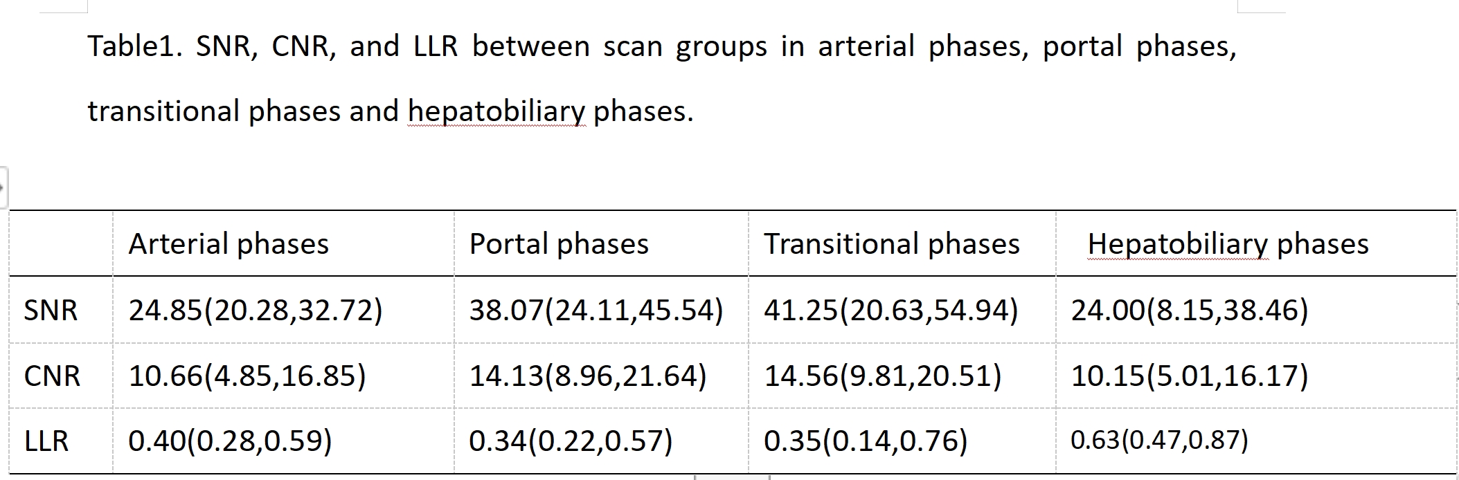
Results: Gd-BOPTA-enhanced MR imaging in the hepatobiliary stage can significantly improve the detection efficiency of metastases, but its SNR and CNR are not as good as those in the transitional stage.
Impact: This experiment demonstrates Gd-BOPTA-enhanced MR imaging in the hepatobiliary stage can significantly improve the detection efficiency of metastases.
Introduce
The early diagnosis of liver metastatic tumors mainly depends on imaging examination. Dynamic enhancement of liver MR Examination has become an effective method for clinical diagnosis and differential diagnosis of liver lesions. The detection, distribution and diagnosis of liver metastases are of great significance for clinical diagnosis and treatment. MRI is a diagnostic technique capable of evaluating the morphological and functional features of the lesion[1], especially the application of liver fine cell specificity to the specific agent gadolinium disodium serate to facilitate the diagnosis of liver metastases. The main objective of this study was to compare the detection effect of multiphase dynamic Gd-BOPTA enhanced magnetic resonance imaging on liver metastases.Purpose
To compare the detection efficiency of Multi-Phase Dynamic Gd-BOPTA Enhanced MR Imaging in liver metastases.Materials and Methods
This study was approved by the local IRB. A total of 21 patients were included in this retrospective study. All patients underwent T1W, T2WI, DWI and Gd-BOPTA-enhanced MRI on a 3.0 T MR scanner (Ingenia CX, Philips Healthcare) using a 16-channel abdominal array coil. The hepatobiliary phase imaging was performed 60 minutes after contrast agent Intravenous bolus, Gd-BOPTA injection. MR raw data were transferred to the workstation (Intellispace Portal v10, Philips Healthcare) for post-processing. Regions of interest (ROIs) were placed in the left lobe, the right anterior and the right posterior lobes of the liver, avoiding the influence of blood vessels and other abnormal liver tissues. ROI was placed on the paraspinal muscles at the same level as the lesion, and the average value of the above parameters was taken for further analysis. Signal intensity (SI), standard deviation (SD), signal-to-noise ratio (SNR), contrast-to-noise ratio (CNR), and liver-to-lesion ratio (LLR) were measured and calculated, and the calculation formule were as follows: SNRLesion=SILesion/SDmuscle, CNRLesion=(SILesion-SImuscle)/SDmuscle, LLR=(SILiver-SILesion)/SILiver. The Mann-Whitney U test was used to compare SNR, CNR, and LLR between scan groups with Dynamic enhancement and hepatobiliary phases with different flip angles and delay times, and the Friedman test was used for pairwise comparison within groups.Results
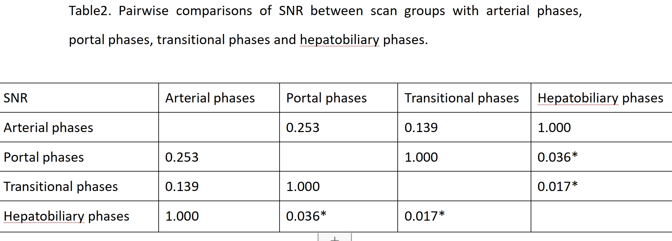
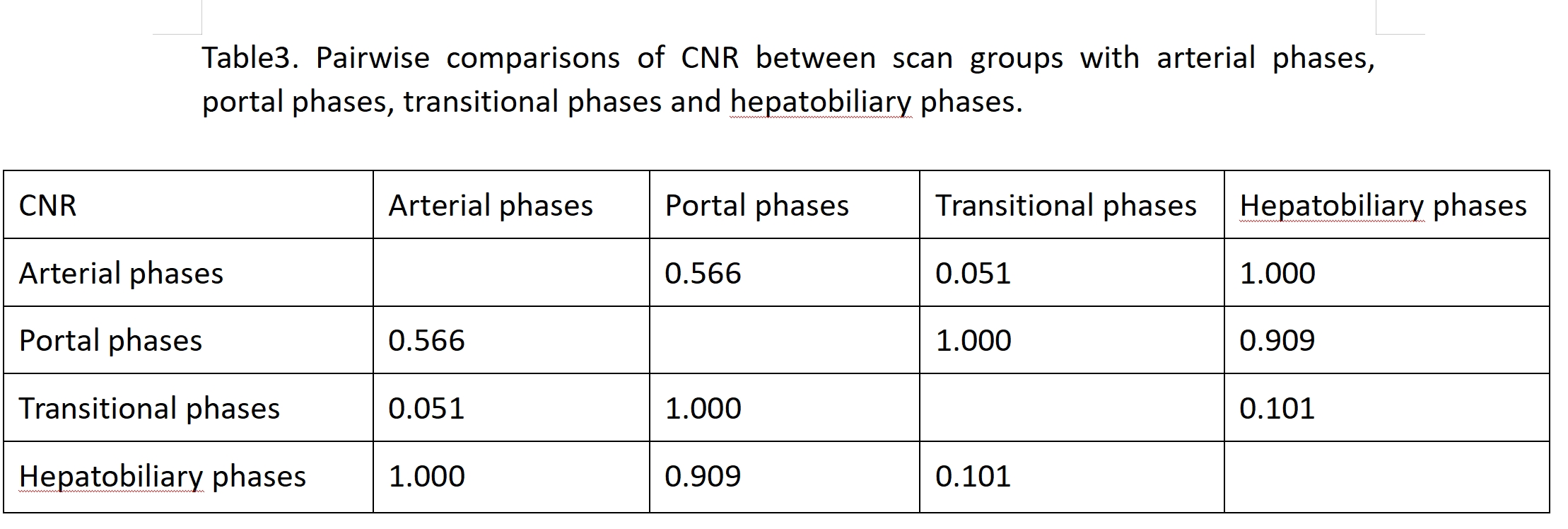
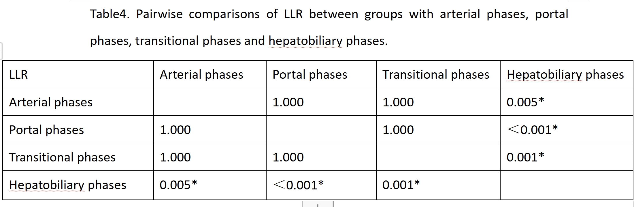
The SNR of the transitional phases was the highest, and there were statistical differences compared with the hepatobiliary phases (p<0.05) (Table1 and 2). The CNR of the transitional phases was also the highest, however, there were no statistical differences compared with others (Table1 and 3). The LLR of the hepatobiliary phases was the highest, and there were statistical differences compared with arterial phases, portal phases, transitional phases and hepatobiliary phases. (both p<0.05) (Table1 and 4).Conclusion
Gd-BOPTA-enhanced MR imaging in the hepatobiliary stage can significantly improve the detection efficiency of metastases, but its SNR and CNR are not as good as those in the transitional stage.Acknowledgements
NoneReferences
NoneFigures