4140
Enhancing liver reserve function prediction in liver cancer through Gd-EOB-DTPA and DWI: Insights on CD34 and glypican-3 expression levels1Department of Radiology, The First People's Hospita of Qinzhou, Qinzhou, China, 2The First People's Hospita of Qinzhou, Qinzhou, China, 3Department of Radiology, The First People's Hospital of QinZhou, Qinzhou, China, 4Siemens Healthineers Ltd, Beijing, China
Synopsis
Keywords: Liver, Cancer, Gd-EOB-DTPA;ADC;CD34;Glypican-3
Motivation: The quantitative magnetic resonance imaging of Gd-EOB-DTPA has been employed to predict liver reserve function, tolerance, and postoperative recovery in patients with primary liver cancer.
Goal(s): To investigate the relationship between quantitative parameters, liver reserve function of primary liver cancer and the expression levels of CD34 and Glypican-3.
Approach: Evaluate the correlation between REHBP, ADC, liver function classification, and immune markers CD34 and Glypican-3
Results: In comparison to patients with benign liver nodules, patients with primary liver cancer exhibited significantly lower REHBP and ADC values.
Impact: The combination DCE-MRI with Gd-EOB-DTPA and DWI has a predictive value for liver reserve function and prognostic molecular marker expression in patients with primary liver cancer. It can also serve as a useful reference index for clinical preoperative planning.
Introduction
Surgical treatment is the commonly used treatment method for primary liver cancer [1]. The accurate and invasive assessment of liver reserve function before surgery is crucial in predicting a patient's tolerance to surgery and the liver's ability to recover post-surgical, promoting surgery safety. In addition, the early prediction of patient prognosis is essential selecting appropriate treatment methods. CD34 (vascular endothelial factor), a type I transmembrane protein, has been found to exhibit significantly increased expression in cancerous liver tissues of patients with liver cancer. High expression of CD34 is significantly related to a poorer prognosis , characterized by early recurrence and metastasis [2]. Additionally, the expression of Glypican-3 is significantly upregulated in liver cancer tissues, and multifactor analysis has indicated that Glypican-3 plays a vital role in the occurrence and development of liver cancer. High expression of Glypican-3 is closely associated with malignancy of liver cancer, therefore serving as a prognostic indicator for patients' poor outcomes [3]. Several studies have demonstrated that Gd-EOB-DTPA-enhanced magnetic resonance imaging (MRI) plays a role in the diagnosis of liver cancer and the evaluation of liver reserve function [4]. Diffusion-weighted imaging (DWI) can quantitatively measure the movement of water molecules within tumors by measuring the apparent diffusion coefficient (ADC), which is of great significance in the diagnosis of liver cancer [5]. This study aims to explore the relationship between the quantitative parameters obtained from Gd-EOB-DTPA-enhanced MRI combined with DWI and the liver reserve function of primary liver cancer, as well as and the expression levels of CD34 and Glypican-3.Methods
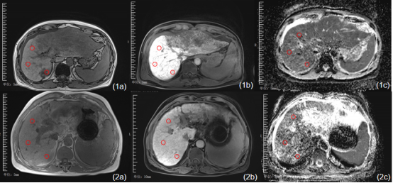
A total of 96 patients with primary liver cancer were included as research subjects, while 50 patients with benign liver nodules were chosen as the control group. All patients underwent liver MRI using a 3T MR scanner (MAGNETOM Skyra, Siemens Healthcare, Erlangen, Germany) with a supine position. A body coil was placed in the position of the subject's sword protrusion to ensure that the liver was positioned at the center of the magnet. Conventional T1-weighted imaging (TIWI), DWI, and dynamic contrast enhancement (DCE)-MRI with Gd-EOB-DTPA- as the contrast agents were performed. Afterwards, the ADC and hepatobiliary phase enhanced-images of all cases were transferred to a workstation (Syngo.via, Siemens Healthcare, Erlangen, Germany) for analysis by two experienced doctors. Three circular regions of interest (ROIs) of equal size were delineated on the liver. The signal intensity of these ROIs before enhancement was denoted as SIpre, whilethe signal intensity during the hepatobiliary phase was represented as SIpost. The relative enhancement at the hepatobiliary phase(REHBP) for each patient was then calculated using the appropriate formula[6]. Furthermore, The average ADC value is measured on the ADC maps (Figure 1-2). Finally, the correlation between these measurements and immune markers (CD34 and Glypican-3) was evaluated.Results
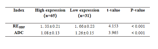
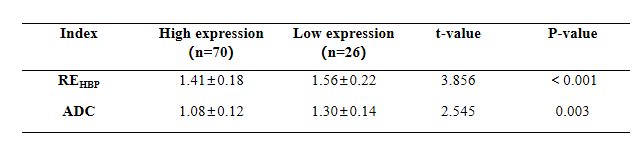
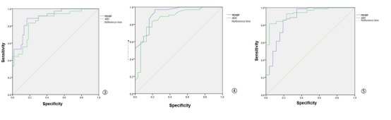
The REHBP and ADC values of patients with primary liver cancer were significantly lower than those of patients with benign liver nodules(P<0.05)(Table1). Additionally, patients with high expression of CD34 and Glypican-3 had significantly lower REHBP and ADC values compared to patients with low expression (P<0.05)(Table2-3).The analysis of the receiver operating characteristic (ROC) curve revealed that the REHBP and ADC values were strong predictors for liver reserve function, with an area under the curve (AUC) of 0.901 and 0.861 respectively. The sensitivity and specificity were 88.9% and 83.3% for REHBP, and 83.3% and 81.7% for ADC (Figure 3). For high CD34 expression, the AUC was 0.923 for REHBP and 0.866 for ADC, and the sensitivity and specificity of 96.9% and 77.4% for REHBP, and 83.1% and 80.6% for ADC (Figure 4). Lastly, for high expression of Glypican-3, the AUC was 0.879 for REHBP and 0.933 for ADC, with sensitivity and specificity of 85.7% and 80.8% for REHBP, and 81.4% and 96.2% for ADC (Figure 5).
Discussion and Conclusion
The results from our study demonstrated that REHBP and ADC values can be used to predict the expression levels of CD34 and Glypican-3. Additionally, for patients with abnormal liver function, the liver's ability to absorb contrast agents is diminished. Therefore, the degree of strengthening can be utilized to determine the amount of Gd-EOB-DTPA update by liver parenchyma. Moreover, there is a correlation between REHBP and ADC values and the expression level of molecular markers of liver cancer prognosis. Preoperative liver reserve function holds significant value as an indicator for assessing the liver's tolerance to surgical interventions, even considered to be a decisive factor in determining the ultimate outcome of a surgery [2]. Consequently, the ability to predict the patient's prognosis is made feasible by analyzing REHBP and ADC values.Acknowledgements
We thank all the participants and the Scientific Cooperation department of Siemens healthineers for their support.References
[1] Abdelraouf A, Hamdy H, El Erian AM, et al. Initial experience of surgical microwave tissue precoagulation in liver resection for hepatocellular carcinoma in cirrhotic liver. J Egypt Soc Parasitol. 2014 Aug;44(2):343-50.
[2] Yao Y, Pan Y, Chen J, et al. Endoglin (CD105) expression in angiogenesis of primary hepatocellular carcinomas: analysis using tissue microarrays and comparisons with CD34 and VEGF. Ann Clin Lab Sci. 2007 Winter;37(1):39-48.
[3] Ning S, BinC, NaH, et al. Glypican- 3, a novel prognostic marker of hepatocellular cancer, is related with postoperative metastasis and recurrence in hepatocellular cancer patients[J] . Mol Biol Rep, 2012, 39(1):351- 357.
[4] Haimerl M, Verloh N, Zeman F, et al. Gd-EOB-DTPA-enhanced MRI for evaluation of liver function: Comparison between signal-intensity-based indices and T1 relaxometry. Sci Rep. 2017 Mar 7;7:43347.
[5] Zhou Y, Zheng J, Yang C, et al. Application of intravoxel incoherent motion diffusion-weighted imaging in hepatocellular carcinoma. World J Gastroenterol. 2022 Jul 21;28(27):3334-3345.
[6]N,Verloh,M,Haimerl,F,Zeman,M,Schlabeck,A,Barreiros,M,Loss,A G,Schreyer,C,Stroszczynski,C,Fellner,P,Wiggermann.Assessing liver function by liver enhancement during the hepatobiliary phase with Gd-EOB-DTPA-enhanced MRI at 3 Tesla.[J].European radiology,2014,24(5):1013-9.DOI:10.1007/s00330-014-3108-y.
Figures