4138
Using Time-Dependent Diffusion MRI for Microstructural Evaluation in Hepatocellular Carcinoma1The department of Radiology, The First Affiliated Hospital of Guangxi Medical University, Nanning, Guangxi, China, 2MR Research Collaboration, Siemens Healthineers, Guangzhou, Guangdong, China, 3MR Research Collaboration, Siemens Healthineers, Erlangen, Germany
Synopsis
Keywords: Liver, Diffusion/other diffusion imaging techniques
Motivation: The purpose of this study is to assess the microstructural parameters of hepatocellular carcinoma by the time-dependent diffusion MRI technique.
Goal(s): Demonstrate the feasibility of TDD-MRI for evaluating hepatocellular cancer cell size with little difference from pathological verification.
Approach: This prospective study will perform TDD-MRI scans for HCC patients before surgery; the cell size of tumor tissue samples obtained from surgery was measured and set as the gold standard.
Results: Data of 13 patients was collected, while there was no remarkable relationship between the TDD-MRI extracted results and histological measurements.
Impact: Although our results show no statistically significant for now, TDD-MRI has still got certain potential values in the application of liver tumors, while the measurement of pathological samples need to be optimized further.
Introduction
Liver cancer is the fifth most common cancer worldwide, especially hepatocellular carcinoma (HCC) within which. The pathological differentiation of HCC can be categorized into low, medium and high grades (classification of WHO 2019 version); or grade I-IV (Edmondson-Steiner classification).1, 2 HCC tumor cells with different degrees of differentiation have different microscopic parameters such as cell size and cell density, which also affect the choice of treatment and prognosis of patients. Diffusion weighted imaging (DWI) with the derived apparent diffusion coefficient (ADC) is an MRI technique which qualitatively reflects microstructural changes of tumor tissues. Recently, time-dependent diffusion MRI (TDD-MRI) has demonstrated value in revealing quantitative characteristics of cell microstructure such as cell diameter and intracellular volume fraction, and has been applied in a series of clinical studies.3-5 This study aimed to investigate the feasibility of TDD-MRI mapping for in-vivo and non-invasive evaluating cellular characteristics of HCC.Method
Patients with a suspected diagnosis of HCC were prospectively recruited at the First Affiliated Hospital of Guangxi Medical University, China, since January 2023, and the patients underwent Gd-EOB-DTPA MRI scanning and TDD-MRI scanning based on an oscillating gradient spin-echo research sequence (OGSE) on a 3T system (MAGNETOM Prisma, Siemens Healthcare, Erlangen, Germany) before surgery. OGSE data were acquired at oscillation frequencies of 25 Hz (effective diffusion time = 9.2 msec, one cycle, b = 0, 250, 500, 750 and 1000sec/mm2) and 50 Hz (effective diffusion time = 4.2 msec, one cycle, b = 0, 100, 200, and 300 sec/mm2). Pulsed gradient spin-echo (PGSE) data were collected with an effective diffusion time of 44.8 msec (b =0, 250, 500, 750, and 1000 sec/mm2). Patients who underwent other preoperative treatments such as TACE, radiofrequency ablation, or lacked MRI images were excluded. To verify the reliability of TDD-MRI data, immunohistochemical staining was performed on histology specimens obtained after surgery and cell diameters were measured under a light microscope.6 Image processing and result estimation were performed with ImageJ. Difference between the tumor cell diameter evaluated by TDD-MRI and the actual cell diameter of pathological specimen was examined by matched T-test; Spearman correlation analysis was performed to evaluate the correlation between TDD-MRI measurements and histology. The significance level was set at P<0.05.Results
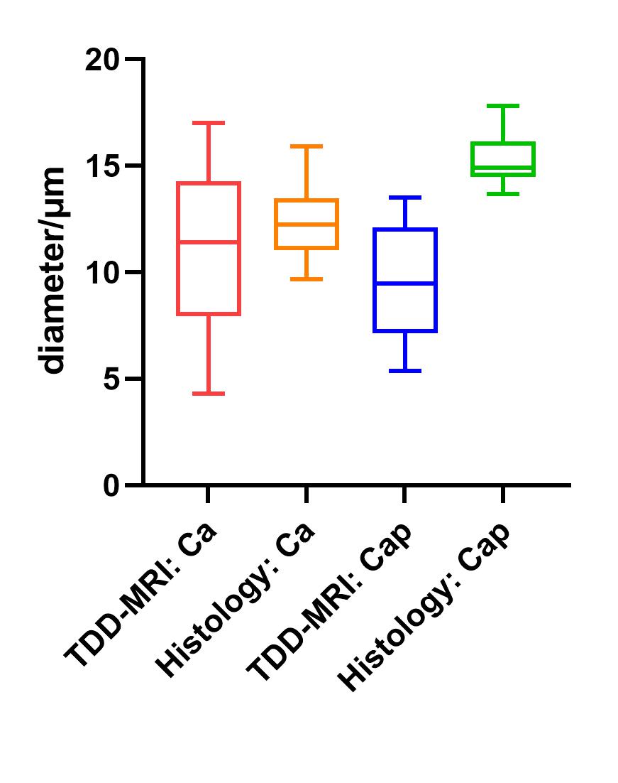
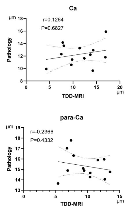
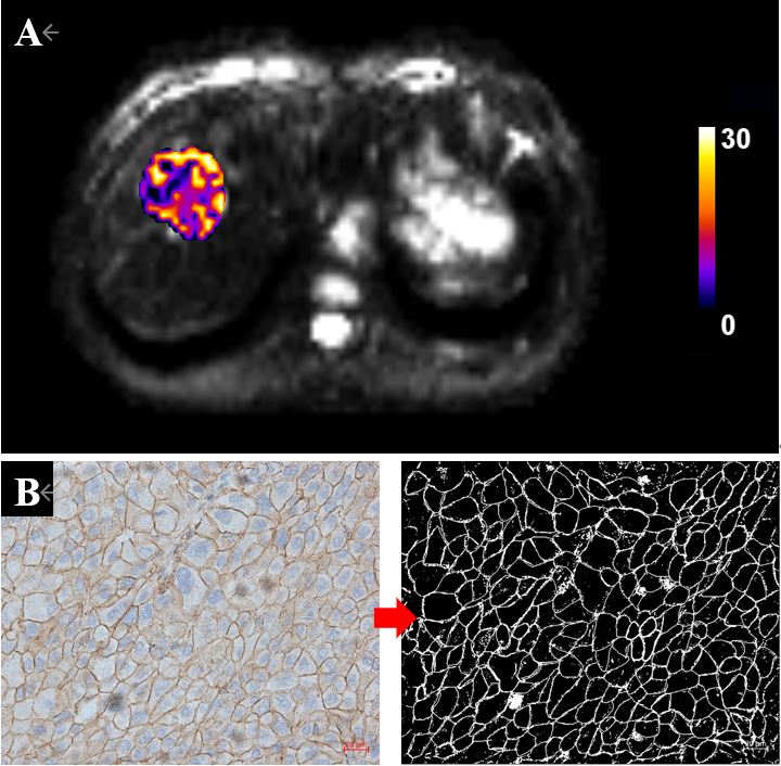
13 patients who underwent TDD-MRI scans followed by surgery were enrolled. There was no significant difference between the cell diameters (d) measured in HCC tissue based on TDD-MRI images (extracted from a Matlab program, based on a two-compartment model with impermeable spheres7) and the cell diameters measured on pathological specimens (P=0.381). Histology showed that para-carcinoma tissue cells are larger than those of cancerous tissue (15±1.2 μm vs. 12±1.7 μm, P<0.05) (Figure 1). There was no correlation between the diameters of cancer tissue cells and para-carcinoma cells derived from TDD-MRI and their measured diameters in pathological specimens (r = 0.1264, P = 0.6827; r = -0.2366, P = 0.4332, respectively) (Figure2). Figure 3A and 3B show an example of a patient.Discussion and Conclusion
This study preliminarily evaluated the feasibility of TDD-MRI for noninvasive assessment of tumor cell size in HCC. It turns out that there was no significant correlation between the measured value of TDD-MRI and the true value measured from histology. This may be related to the high heterogeneity of HCC tumor cells and the image quality of MRI; at the same time, the sample size was too small to reflect the real situation. Further data collection and analysis will be considered in our future study. In conclusion, the application of TDD-MRI in evaluating HCC microstructure requires further discussion.Acknowledgements
We thank those who participated in the study, as well as the radiographers, nurses in the department of Radiology at the first affiliated hospital Guangxi Medical University, for their work, support, and enthusiasm for the study.References
1. Song J, Zhou H, Gu D, Xu Y. Hepatocellular Carcinoma Differentiation: Research Progress in Mechanism and Treatment. Front Oncol. 2021;11:790358.
2. Calderaro J, Ziol M, Paradis V, Zucman-Rossi J. Molecular and histological correlations in liver cancer. Journal of Hepatology. 2019;71(3):616-630.
3. Ba R, Wang X, Zhang Z, et al. Diffusion-time dependent diffusion MRI: effect of diffusion-time on microstructural mapping and prediction of prognostic features in breast cancer. European Radiology. 2023;33(9):6226-6237.
4. Zhang H, Liu K, Ba R, et al. Histological and molecular classifications of pediatric glioma with time-dependent diffusion MRI-based microstructural mapping. Neuro-Oncology. 2023;25(6):1146-1156.
5. Wu D, Jiang K, Li H, et al. Time-Dependent Diffusion MRI for Quantitative Microstructural Mapping of Prostate Cancer. Radiology. 2022;303(3):578-587.
6. Jiang X, Xu J, Gore JC. Mapping hepatocyte size in vivo using temporal diffusion spectroscopy MRI. Magn Reson Med. Nov 2020;84(5):2671-2683.
7. Jiang X, Li H, Xie J, et al. In vivo imaging of cancer cell size and cellularity using temporal diffusion spectroscopy. Magnetic Resonance in Medicine. 2016;78(1):156-164.
Figures