4137
Predicting Liver Metastasis of Pancreatic Ductal Adenocarcinoma Using Radiomics of Whole Pancreas and Tumor from Dynamic Contrast-enhanced MRI1Biomedical Imaging Research Institute, Cedars-Sinai Medical Center, Los Angeles, CA, United States, 2Division of Digestive and Liver Diseases, Cedars-Sinai Medical Center, Los Angeles, CA, United States, 3Bioengineering, University of California, Los Angeles, CA, United States
Synopsis
Keywords: Pancreas, Pancreas, Radiomics, dynamic contrast enhanced MRI, liver metastasis
Motivation: Prediction of subsequent liver metastasis of pancreatic ductal adenocarcinoma (PDAC) is important for patient management and current prediction performance is insufficient.
Goal(s): To improve the prediction of subsequent liver metastasis from PDAC by integrated radiomic analysis of whole pancreas and PDAC using dynamic contrast enhanced (DCE) MRI.
Approach: Radiomic features were extracted from whole pancreas and PDAC on DCE MRI. Logistic regression model was applied to predict likelihood of subsequent liver metastasis.
Results: Combination of radiomics from whole pancreas and tumor improved liver metastasis prediction than that of tumor alone.
Impact: Improved prediction of subsequent liver metastases with PDAC may allow better management of PDAC patients, potentially saving costs and lives.
Introduction
Pancreatic ductal adenocarcinoma (PDAC) is notorious for its aggressive nature and grim prognosis[1]. As the fourth leading cause of cancer death in American, 5-year survival rate is only 12%[1]. Liver metastasis is a strong biomarker associated with serious outcome and shorter overall survival. Accurate prediction of subsequent liver metastasis is critical for the management of PDAC and enhancing patient outcomes. Multiphasic dynamic contrast enhanced (DCE) MRI is clinically used for PDAC diagnosis, staging and follow-up[2]. Radiomic analysis of the tumor has been used to predict PDAC prognosis [3] and future liver metastasis[4] by quantifying image characteristics within the tumor to gain insight into PDAC biological behavior and prognostic factors. The hypothesis of this work is that radiomic features extracted from the whole pancreas in portal venous DCE-MRI can improve the performance of PDAC subsequent liver metastasis prediction.Methods
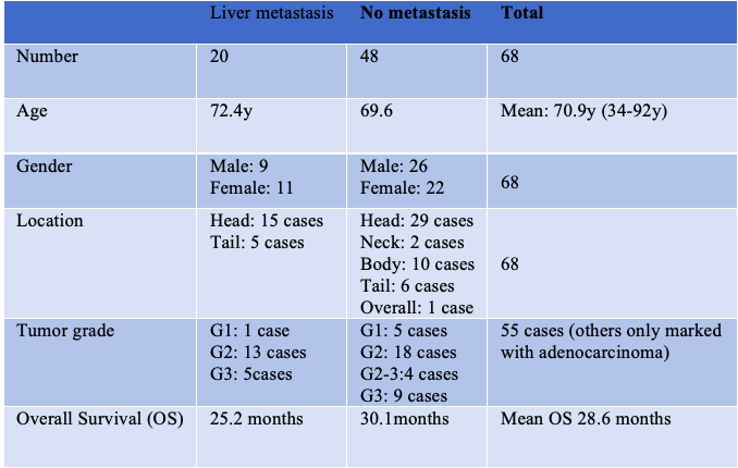
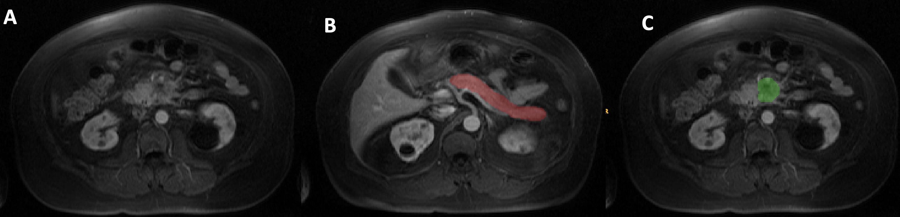
A total of 68 PDAC cases (33 females and 35 males) with a mean age 70.9 years (ranging from 34-92 years) between 2009 and 2021 at Cedars-Sinai Medical Center were selected and constituted as the training and testing dataset. All cases were confirmed PDAC of different grade by surgery or biopsies. DCE MR examination was conducted prior to treatment. The cases with liver metastasis at baseline examination were excluded. A summary of patient’s demographic is listed in table 1. The MRIs of all cases were loaded into ITK-SNAP Software (version 4.0.0-alpha.3). The boundaries of the whole pancreas, tumor, and liver were outlined manually or semi-automatically slice by slice in portal venous phase of DCE MRI (Figure 1) by an experienced radiologist. Radiomic features were extracted using the open-source Python package PyRadiomics in Python3.7. We conducted the Least absolute shrinkage and selection operator (LASSO) to select the maximum of 10 features. Leave one out cross-validation was performed following LASSO. Prediction was carried out using logistic regression model. The area under the receiver operating characteristics (ROC) curve (AUC), accuracy, sensitivity and specificity were calculated as evaluation metrics.Results
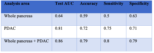
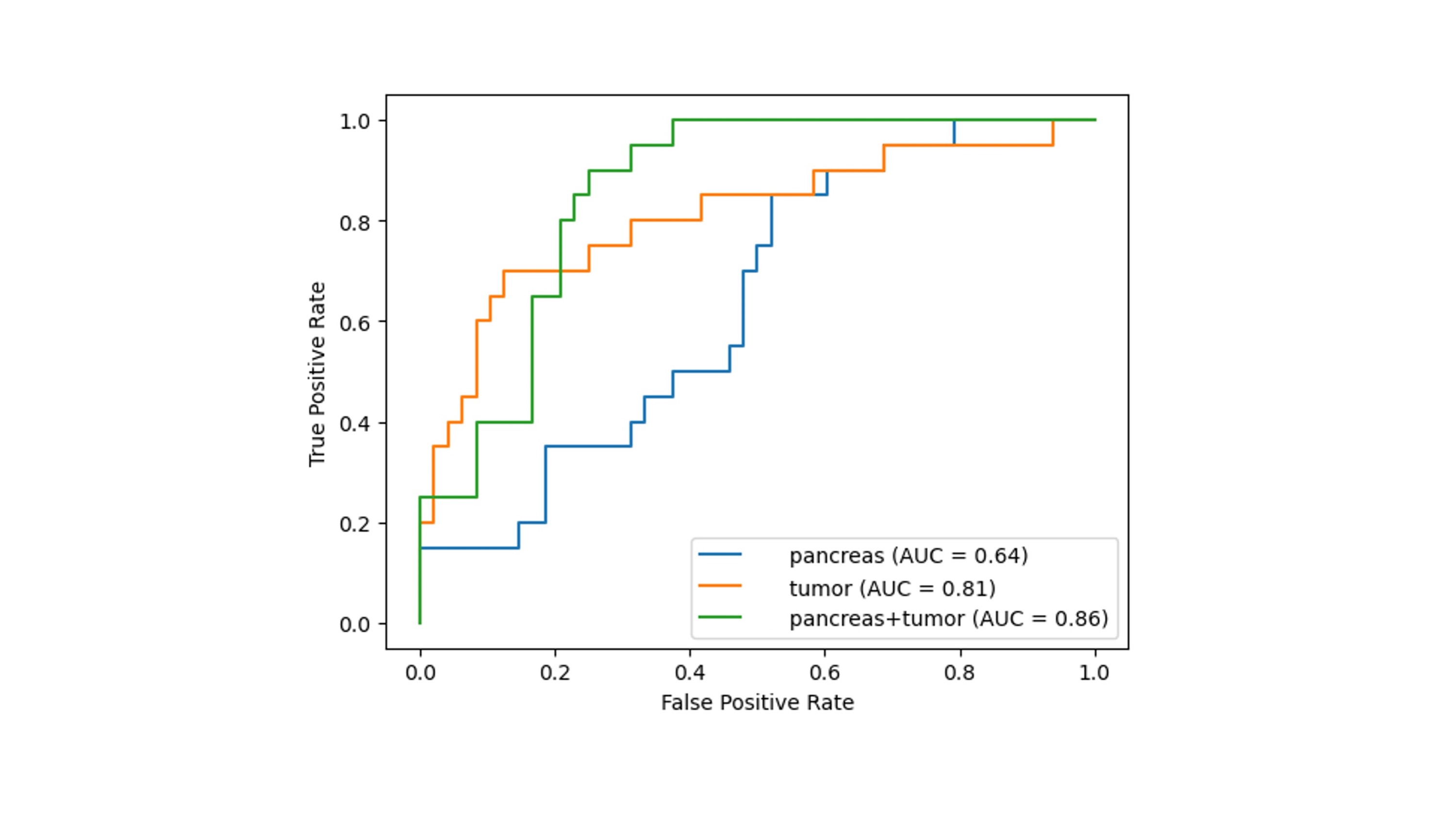
We extracted a total of 107 features separately for the whole pancreas, tumor, and liver. With LASSO feature selection, the maximum of 10 features were selected. The AUC, accuracy, sensitivity, and specificity for prediction of subsequent liver metastasis are given in table 2. Figure 2 shows the ROC curves for the whole pancreas, PDAC region, liver and different combination of regions. Using radiomic features of the tumor alone, the AUC value, accuracy, sensitivity, and specificity were 0.81, 0.72, 0.75 and 0.71, respectively. By adding radiomic features from the whole pancreas, the AUC value, accuracy, sensitivity, and specificity in predicting subsequent liver metastasis improved to 0.86, 0,79, 0.80, and 0.79, respectively.Discussion
Radiomics from PDAC tumor as a quantitative method can reflect tumor characteristic and unique microenvironment, offer essential insight for predicting PDAC subsequent liver metastasis[5, 6] and guiding treatment decisions. As pancreatic inflammatory environment plays an important role in tumor occurrence and progression [7], the background of the whole pancreas can serve as independent prognostic factors in PDAC liver metastasis and a predictor of poor prognosis. The addition of radiomic features from the whole pancreas to tumor features significantly improved the prediction of subsequent liver metastasis. This improvement may be related to factors such as the change of pancreatic anatomy, micro-structure damage and tissue properties due to chronic inflammatory stimulation. For nearly 75% blood supply of liver is from portal vein. Micro-vessel damage and superior mesenteric venous invasion of PDAC will result tumor cell directly deposit to liver through portal vein.Conclusion
Our study demonstrated a substantial improvement in predicting liver metastasis using radiomic features from the whole pancreas and PDAC, in comparison to using the PDAC region alone. This indicates that radiomic features of the whole pancreas should be included in predicting future liver metastasis.Acknowledgements
No acknowledgements found.References
- Siegel, R.L., et al., Cancer statistics, 2023. CA: A Cancer Journal for Clinicians, 2023. 73(1): p. 17-48.2.
- Fukukura, Y., et al., Extracellular volume fraction with MRI: As an alternative predictive biomarker to dynamic contrast-enhanced MRI for chemotherapy response of pancreatic ductal adenocarcinoma. Eur J Radiol, 2021. 145: p. 110036.3.
- Xu, X., et al., Development and validation of an MRI-radiomics nomogram for the prognosis of pancreatic ductal adenocarcinoma. Front Oncol, 2023. 13: p. 1074445.4.
- Yuan, Z., et al., Prediction of postoperative liver metastasis in pancreatic ductal adenocarcinoma based on multiparametric magnetic resonance radiomics combined with serological markers: a cohort study of machine learning. Abdom Radiol (NY), 2023.5.
- Chakraborty, J., et al., Preliminary study of tumor heterogeneity in imaging predicts two year survival in pancreatic cancer patients. PLoS One, 2017. 12(12): p. e0188022.6.
- Huang, Y., et al., Development and validation of a radiomics model of magnetic resonance for predicting liver metastasis in resectable pancreatic ductal adenocarcinoma patients. Radiat Oncol, 2023. 18(1): p. 79.7.
- Hayano, K., et al., Diffusion-weighted MR imaging of pancreatic cancer and inflammation: Prognostic significance of pancreatic inflammation in pancreatic cancer patients. Pancreatology, 2016. 16(1): p. 121-126.
Figures