4135
Habitat imaging’s role in predicting HCC percentage in combined hepatocellular carcinoma-cholangiocarcinoma and its biologic underpinnings1radiology, Zhongshan Hospital Fudan University, Shanghai, China, 2Central Research Institute, United Imaging Healthcare, Shanghai, China
Synopsis
Keywords: Liver, Liver
Motivation: The prognostic value of component percentage in combined hepatocellular carcinoma-cholangiocarcinoma (cHCC-CCA) was verified, but the identification of percentage of each component relies on extensive tumor sampling.
Goal(s): Therefore, the aim of this study was to investigate if MRI-based habitat imaging helps to predict component percentage in cHCC-CCA preoperatively,
Approach: and to further verify the biologic underpinnings of habitat imaging in cHCC-CCA by analyzing RNA sequencing data.
Results: We found that preoperative prediction of HCC percentage in patients with cHCC-CCA can be achieved using an MRI-based habitat imaging model, which was also associated with signaling pathways regulating cell migration and tumor metastasis.
Impact: We achieved prediction of HCC percentage through an MRI-based habitat imaging model, and revealed the biologic underpinnings of habitat imaging, that is, habitat imaging may identify patients at risk of metastasis. These results may guide individual management in cHCC-CCA patients.
Background & Aims
Given the prognostic value of component percentage in combined hepatocellular carcinoma-cholangiocarcinoma (cHCC-CCA), we aimed to construct an MRI-based habitat imaging model to predict component percentage in cHCC-CCA preoperatively, and investigate the biologic underpinnings of habitat imaging.Methods
The study consisted of one retrospective model-building dataset and one prospective validation dataset from two hospitals. All voxels were assigned into different clusters according to the similarity of enhancement pattern by using K-means clustering method, and each habitat’s volume fraction in each lesion was calculated. Least absolute shrinkage and selection operator (LASSO) regression analysis was performed to select optimal predictors among three habitats’ volume fractions, conventional imaging features and clinical findings, and then to establish an MRI-based habitat imaging model. R-squared was calculated to evaluate performance of the prediction models. Performance of the model was verified in the prospective dataset with RNA sequencing data, and Kyoto Encyclopedia of Genes and Genomes (KEGG) pathway enrichment analysis was then applied to investigate the biologic underpinnings of habitat imaging.Results
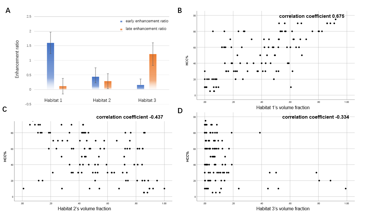
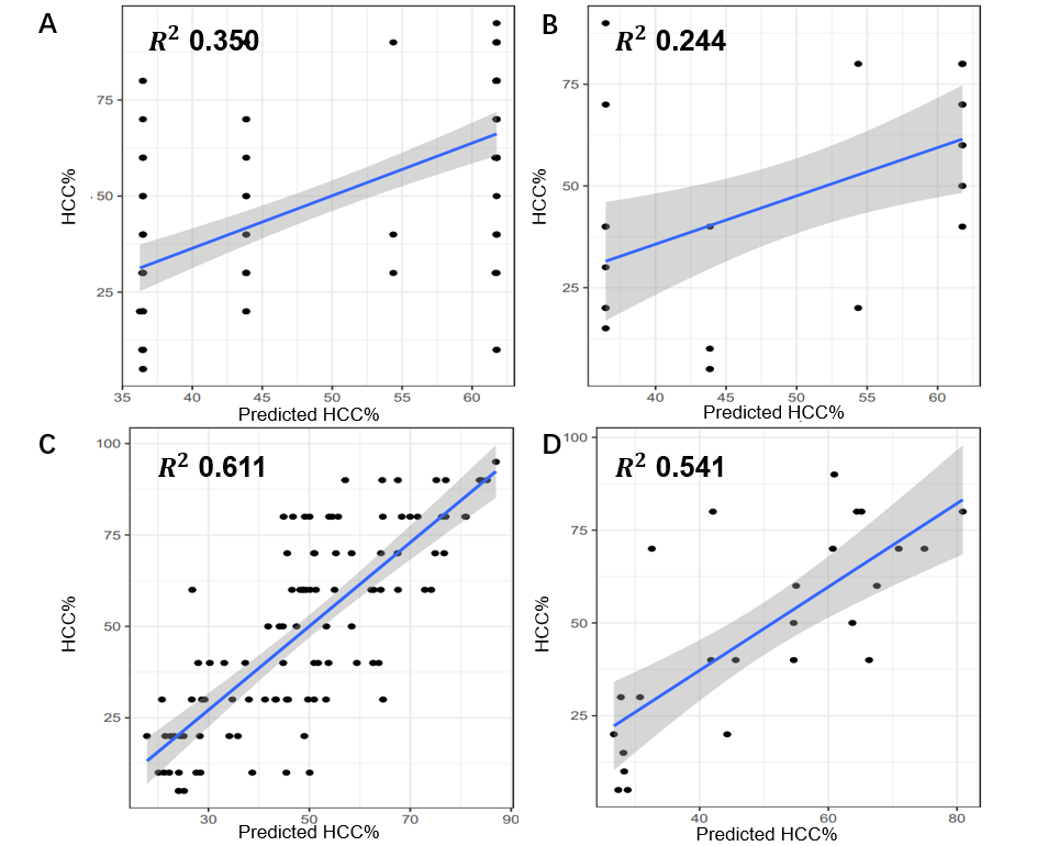
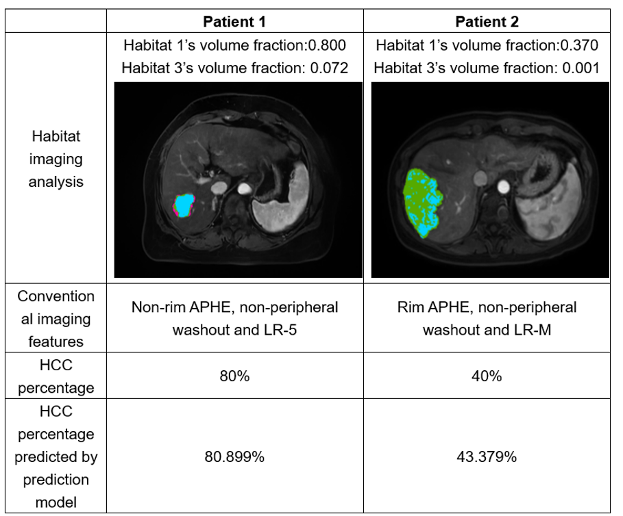
Three habitats, habitat1 (HCC-alike habitat), habitat2 (iCCA-alike habitat) and habitat3 (in-between habitat), were identified. Habitat 1’s volume fraction, habitat 3’s volume fraction, nonrim APHE, nonperipheral washout and LI-RADS categorization were selected to develop an HCC percentage prediction model with R-squared of 0.611 in the model-building set and of 0.543 in the validation set. Habitat 1’s volume fraction was correlated with genes involved in regulation of actin cytoskeleton and Rap1 signaling pathway, which regulate cell migration and tumor metastasis.Conclusion
Preoperative prediction of HCC percentage in patients with cHCC-CCA was achieved using an MRI-based habitat imaging model, which may correlate with signaling pathways regulating cell migration and tumor metastasis.Acknowledgements
no acknowledgements in this paper.References
1. Gatenby RA, Gillies RJ. A microenvironmental model of carcinogenesis. Nat Rev Cancer. 2008;8(1):56-61. doi:10.1038/nrc2255
2. Gatenby RA, Grove O, Gillies RJ. Quantitative imaging in cancer evolution and ecology. Radiology. 2013;269(1):8-15. doi:10.1148/radiol.13122697
3. Napel S, Mu W, Jardim-Perassi BV, Aerts HJWL, Gillies RJ. Quantitative imaging of cancer in the postgenomic era: Radio(geno)mics, deep learning, and habitats. Cancer. 2018;124(24):4633-4649. doi:10.1002/cncr.31630
4. Chang YC, Ackerstaff E, Tschudi Y, et al. Delineation of Tumor Habitats based on Dynamic Contrast Enhanced MRI. Sci Rep. 2017;7(1):9746. Published 2017 Aug 29. doi:10.1038/s41598-017-09932-5
5. Lee DH, Park JE, Kim N, et al. Tumor habitat analysis by magnetic resonance imaging distinguishes tumor progression from radiation necrosis in brain metastases after stereotactic radiosurgery. Eur Radiol. 2022;32(1):497-507. doi:10.1007/s00330-021-08204-1
6. Wei J, Yang G, Hao X, et al. A multi-sequence and habitat-based MRI radiomics signature for preoperative prediction of MGMT promoter methylation in astrocytomas with prognostic implication. Eur Radiol. 2019;29(2):877-888. doi:10.1007/s00330-018-5575-z
7. Beig N, Bera K, Prasanna P, et al. Radiogenomic-Based Survival Risk Stratification of Tumor Habitat on Gd-T1w MRI Is Associated with Biological Processes in Glioblastoma. Clin Cancer Res. 2020;26(8):1866-1876. doi:10.1158/1078-0432.CCR-19-2556
8. Jardim-Perassi BV, Huang S, Dominguez-Viqueira W, et al. Multiparametric MRI and Coregistered Histology Identify Tumor Habitats in Breast Cancer Mouse Models. Cancer Res. 2019;79(15):3952-3964. doi:10.1158/0008-5472.CAN-19-0213
Figures