4133
In vivo 13C MRS of distinct brain region in the mouse brain: how small can we go?1RG Translational Imaging, Central Institute of Mental Health, Mannheim, Germany, 2NeuroImaging, Central Institute of Mental Health, Mannheim, Germany
Synopsis
Keywords: Small Animals, Spectroscopy, X-nuclei, dynamic 13C, mice brain, cerebral metabolism
Motivation: Dynamic 13C MRS is a powerful tool for the investigation of brain metabolism in vivo. Due to its low SNR, it is typically constrained to big voxel sizes covering almost the whole brain in mice. Thus, making region specific research questions difficult.
Goal(s): Investigating the feasibility of acquiring 13C spectra from a distinct brain region (bilateral hippocampi) in the mouse brain at 9.4T.
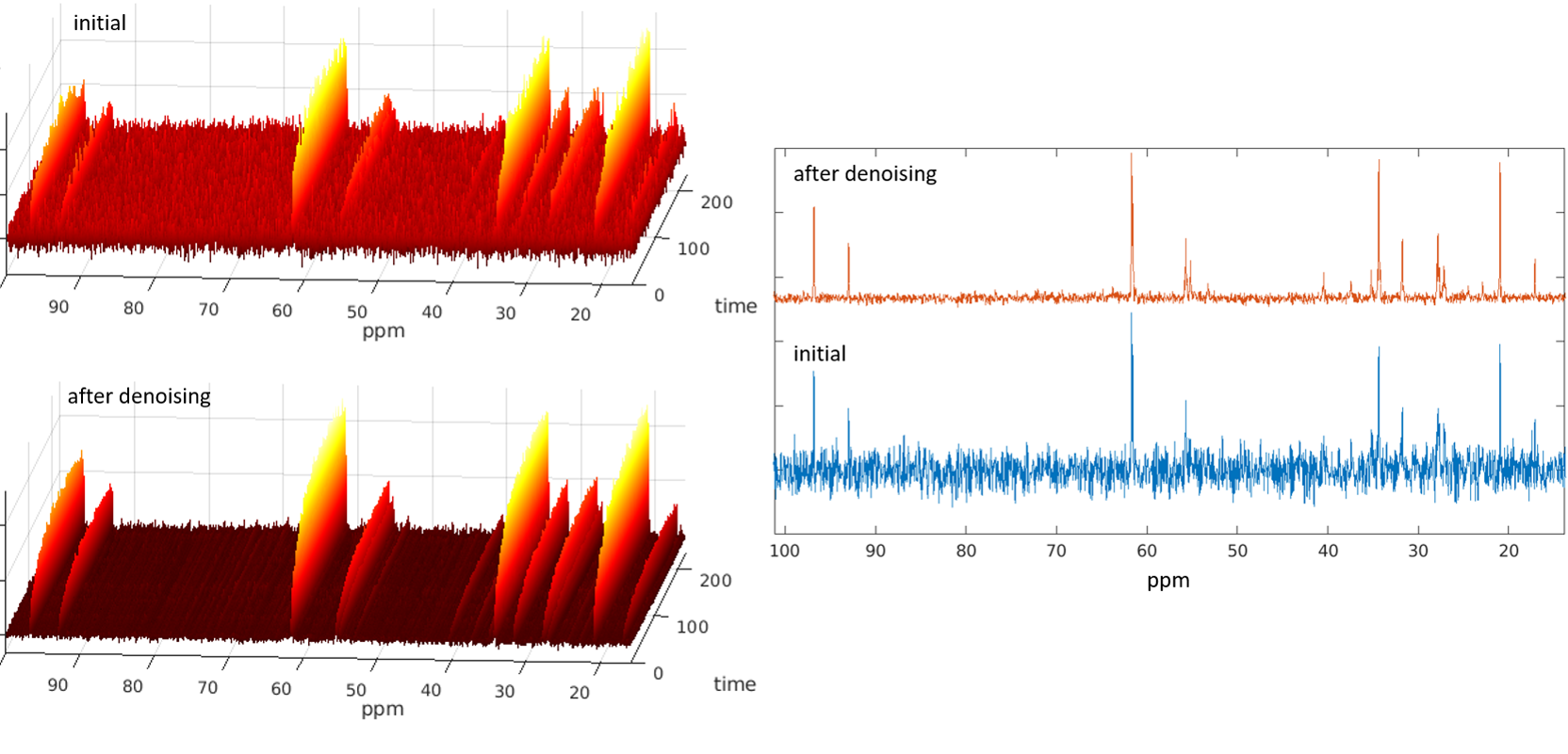
Approach: We applied a combination of polarization transfer, a CryoProbe, frequency correction and ‘SVD denoising’ for the acquisition of 13C spectra.
Results: We achieved sufficient spectral quality to quantify even small metabolite pools like GABA in the bilateral hippocampus.
Impact: 13C MRS of distinct brain regions in the mouse brain paves the way for more specific research questions.
Introduction
Dynamic 13C MRS is a powerful tool for the investigation of brain metabolism in vivo. In conjunction with the administration of 13C-enriched substrates, the 13C label incorporation into different carbon positions of many important brain metabolites, i.e. glutamate and glutamine, can be measured 1-3 and allows for the calculation of metabolic flux rates. Due to its low natural abundance and low gyromagnetic ratio, 13C MRS is challenging, especially in tiny mouse brains. Despite the use of polarization transfer techniques and administration of double-labeled substances, to obtain sufficient SNR in vivo experiments are conducted typically with large voxel sizes covering almost the whole brain or at least several brain regions 2, 4. This limits the application of 13C MRS for certain research questions in which the investigation of distinct brain regions is necessary or mandatory. We present preliminary data of dynamic in vivo 13C MRS in mice.Methods
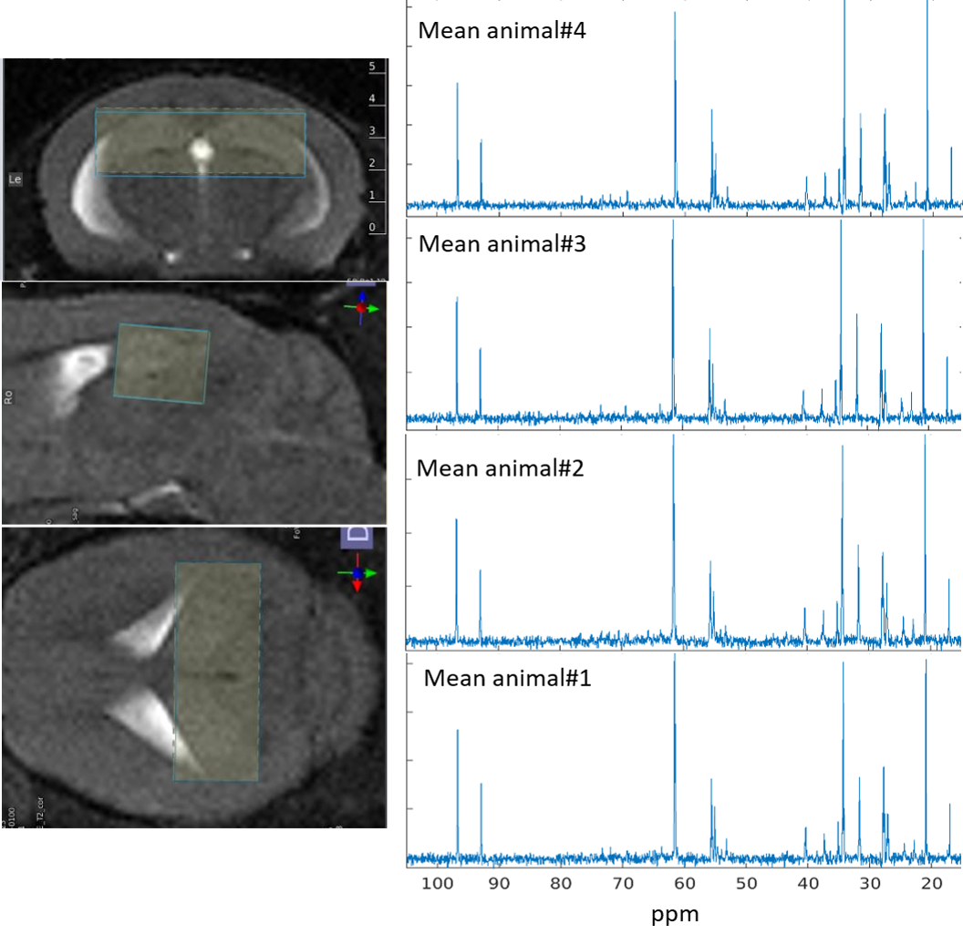
In total, 5 mice (C57/Bl6) were investigated (of which one experiment had to be discarded due to technical issues). MRI experiments were conducted on a 9.4T MR-scanner (Bruker, Ettlingen, Germany) with a cryogenically cooled 13C coil (CryoProbe). The CryoProbe increases the available SNR by a factor of ~4 compared to a similar shaped room temperature coil (5). The freely breathing mice were anesthetized using isoflurane. With onset of [1,6-13C2]-enriched glucose administration (5min bolus of 20% (w/v) 99%-enriched glucose solution followed by continues infusion of 70%-enriched glucose solution at a rate of 9.96 mL/kg/h via the tail vein), 13C acquisition began using an ISIS-DEPT sequence. The ISIS-DEPT (TR=2.5s; 16 ISIS cycles, 128 averages; duration 5.3min; plus 1 dummy cycle; interpulse delay of 3.45ms / JCH = 145Hz) was set with a theta-pulse flip angle of 45°, 0° adiabatic segmented BIR-4 pulse on 13C channel, WALTZ decoupling, and outer volume suppression. The voxel (6.5x2.5x2 mm³) was placed in the hippocampal area (see Fig. 1). Total duration of glucose administration and 13C acquisition was about 4h. The acquired spectra were frequency corrected, SVD denoised using in-house MATLAB scripts 6, and finally pairwise summed (resulting in a time resolution of ~11.3min) and quantified via AMARES (jMRUI v7).Results
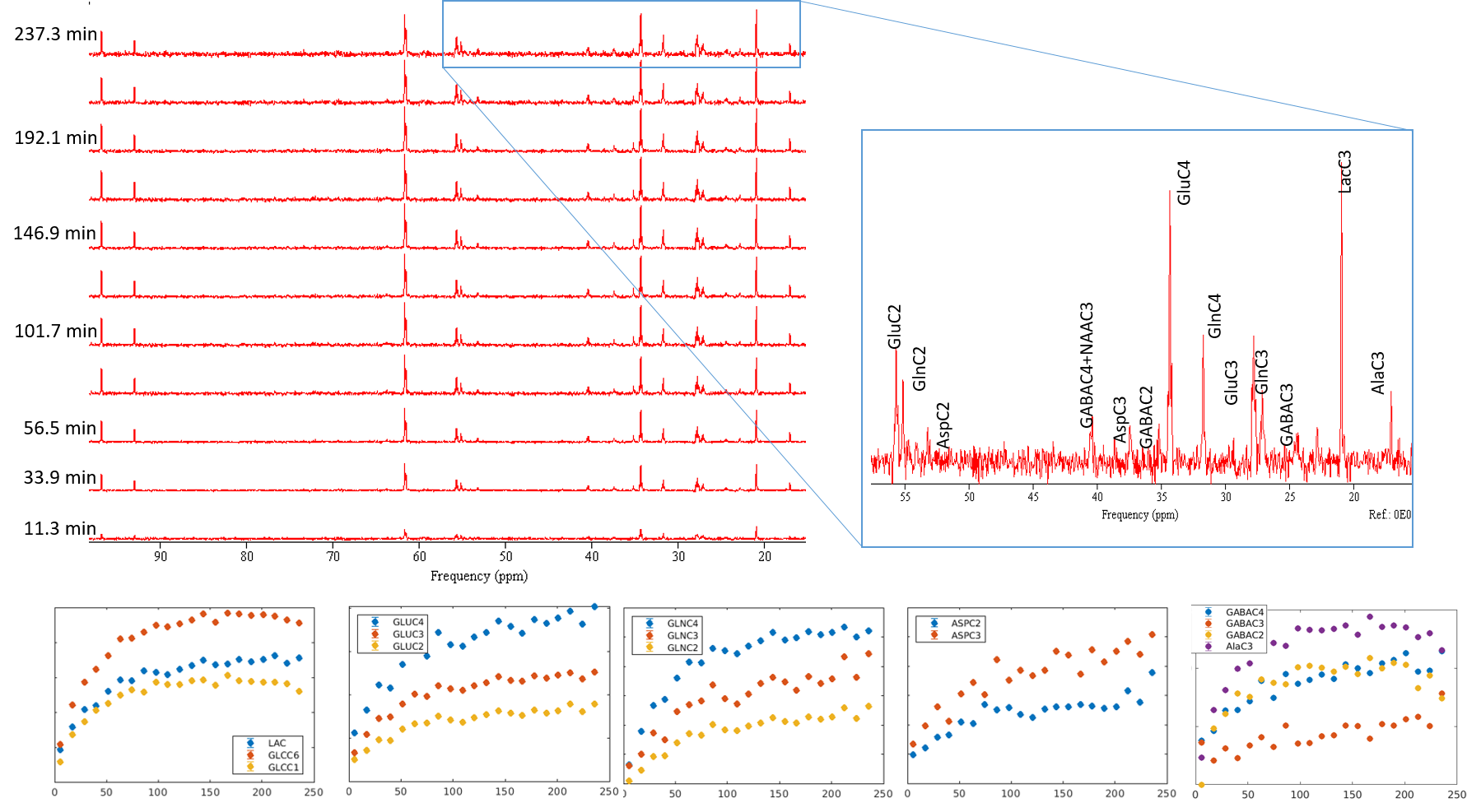
Fig. 1 depicts individual mean spectra demonstrating the consistent spectral quality achieved in the experiments. The combined use of SNR-enhancing techniques (polarization transfer, CryoProbe, frequency correction and ‘SVD denoising’) allowed for the quantification of even small metabolite pools like Asp and GABA (Fig. 2 and 3) from a voxel, which covered mainly the bilateral hippocampal area.Discussion
Despite the small voxel size, the obtained spectral quality promises the chance to even go smaller and maybe investigate the unilateral hippocampus. Especially SVD denoising has a huge impact on single spectra, and thus, also on the found evolution of label incorporation. To our knowledge, this technique has not been used yet for calculation of metabolic flux rates in continues labeled substrate infusion experiments and needs further investigation in this regard.Acknowledgements
We thank Felix Hörner and Claudia Falfan-Melgoza for the skillfully conducted MR-measurements.References
1. Gruetter R, Adriany G, Choi IY, Henry PG, Lei H, Oz G. Localized in vivo 13C NMR spectroscopy of the brain. NMR Biomed. 2003;16(6-7):313-38.
2. Nabuurs CI, Klomp DW, Veltien A, Kan HE, Heerschap A. Localized sensitivity enhanced in vivo 13C MRS to detect glucose metabolism in the mouse brain. Magn Reson Med. 2008;59(3):626-30.
3. Valette J, Tiret B, Boumezbeur F. Experimental strategies for in vivo13C NMR spectroscopy. Anal Biochem. 2016.
4. Lai M, Lanz B, Poitry-Yamate C, Romero JF, Berset CM, Cudalbu C, et al. In vivo 13C MRS in the mouse brain at 14.1 Tesla and metabolic flux quantification under infusion of [1,6-13C2]glucose. J Cereb Blood Flow Metab. 2017:271678X17734101.
5. Sack M, Wetterling F, Sartorius A, Ende G, Weber-Fahr W. Signal-to-noise ratio of a mouse brain (13) C CryoProbe system in comparison with room temperature coils: spectroscopic phantom and in vivo results. NMR Biomed. 2014;27(6):709-15.
6. Brender JR, Kishimoto S, Merkle H, Reed G, Hurd RE, Chen AP, et al. Dynamic Imaging of Glucose and Lactate Metabolism by (13)C-MRS without Hyperpolarization. Sci Rep. 2019;9(1):3410.Figures