4129
Impact of Fasting in the Brain Energy Metabolism in Mice1NMR Microimaging and Spectroscopy, CSIR-Centre for Cellular and Molecular Biology, Hyderabad, India, 22Academy of Scientific & Innovative Research (AcSIR), Ghaziabad, India
Synopsis
Keywords: Neurotransmission, Metabolism, Glucose, glutamate, GABA, Neurons, neurotransmission
Motivation: Fasting is used in weight loss but its impact on brain activity is not well understood.
Goal(s): To assess the impact of prolonged fasting on the excitatory and inhibitory neurotransmission in the brain.
Approach: Infuse [1,6-13C2]glucose in mice fasted for 4h to 60h, and measure 13C labeling of brain metabolites by 1H-[13C]-NMR spectroscopy.
Results: The glutamatergic neurometabolic activity was reduced in the cerebral cortex of mice fasted for 60 h.
Impact: These findings can be used for modifying the fasting paradigm in different interventions involving fasting.
Methods: All animal experiments were approved by the Animal Ethics Committee of CSIR-CCMB, Hyderabad. C57BL6 male mice (7-month-old) were randomly assigned to three groups: Group (i) 4 h (n=12), Group (ii) 24 h (n=12), and Group (iii) 60 h (n=12) fasting. During the fasting, mice had free access to water. Mice were infused with either [1,6-13C2]glucose (0.225 M) or [2-13C]acetate (1M) via the tail vein for 2 min using a bolus variable infusion protocol2 after the designated fasting period. The brain metabolism was arrested using focused beam microwave irradiation (3 kW, 1.2 s) at 6 and 10 min for glucose and acetate, respectively. The 13C Labeling of neurometabolites were measured in 1H-[13C]-NMR spectra of the brain tissue extracts acquired at 600 MHz NMR spectosocpy3. The rates of glucose and acetate oxidation were calculated from the 13C labeled trapped into different amino acids4. One-way ANOVA analysis was carried out using GraphPad prism to find the statistical significance of differences among three groups
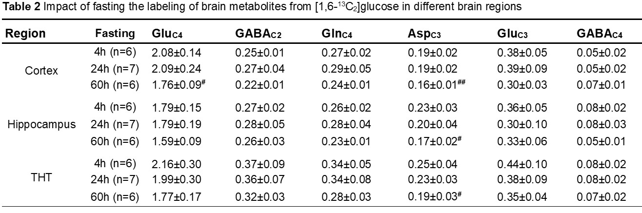
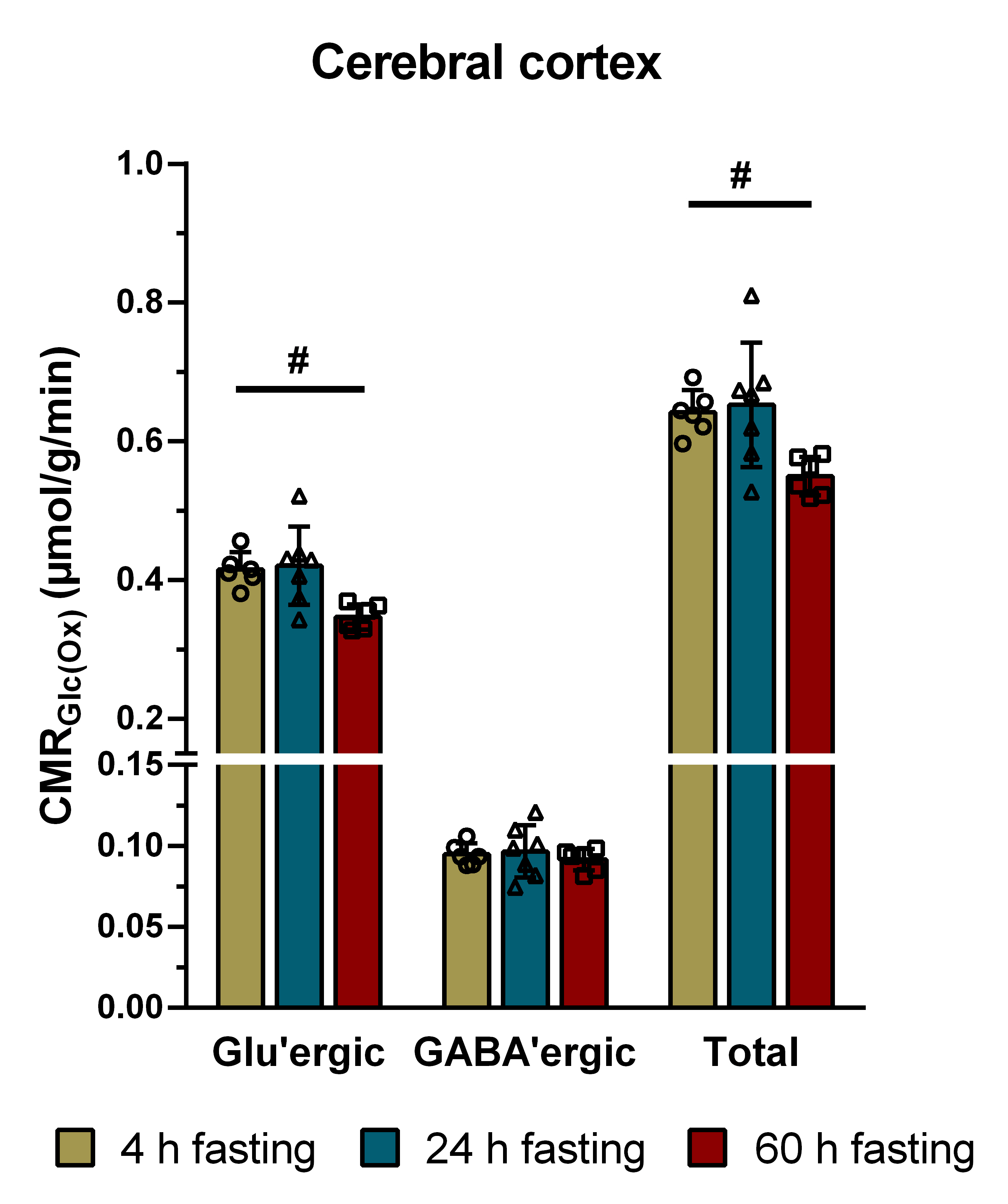
Results: A representative 1H-[13C]-NMR spectra cortical extract from mice infused with [1,6-13C]glucose is presented in Fig. 1. The levels of cortical glutamate (p=0.0286), GABA (p=0.0121), glutamine (p=0.0063) and aspartate (p=0.0348) were reduced significantly in 24 h fasted mice when compared to 4h controls (Table 1). The hippocampus revealed a reduction in the levels of glutamate and glutamine only (Table 1). The cerebral cortex and hippocampus also showed a reduction in glutamate levels after 60h of fasting. Notably, the levels of total choline were reduced across the brain at 24 h as well as 60 h of fasting. The concentrations of 13C labeled GluC4 (60h 1.76±0.09 µmol/g; 4 h 2.08±0.14 µmol/g, p<0.05) and AspC3 (60h 0.16±0.01 µmol/g; 4h 0.19±0.02 µmol/g, p<0.05) from [1,6-13C2]glucose were decreased in mice after 60h of fasting when compared with 4h fasted controls (Table 2). The labeling of AspC3 was decreased in the hippocampus and thalamus. There was not much impact of 24h fasting on the labeling of neurometabolites across the brain (Table 2). The cerebral metabolic rates thus estimated from the 13C labeling of metabolites revealed a significant reduction (p=0.019) in the glucose oxidation in the glutamatergic neurons following 60 h of fasting (0.314±0.016 µmol/g/min) when compared with 4 h fastest mice (0.410±0.026 µmol/g/min) (Fig. 2). Fasting did not have any impact on the glucose oxidation in the GABAergic neurons (60h 0.090±0.006 µmol/g/min, 4h 0.094±0.006 µmol/g/min, p=0.87). The neurometabolic activity was unperturbed in other brain regions irrespective of the extent of the fasting (24 or 60 h).
Discussion As per our knowledge, this is the first quantitative study to understand the impact of fasting on Brain Energy metabolism. The neurotransmitter cycling is stoichiometrically coupled with neuronal glucose oxidation5. Hence, the finding of decreased neuronal glucose oxidation is suggestive of reduced excitatory neurotransmission in the cerebral cortex after 60 h of fasting.
Acknowledgements
This work was supported by the Council for Scientific and Industrial Research (CSIR), Government of India (Health Care Theme NCP/MLP0139).References
1. Longo VD, Mattson MP (2014) Fasting: molecular mechanisms and clinical applications. Cell Metab 4;19(2):181-92
2. Soni ND, Ramesh A, Roy D, Patel AB (2021) Brain energy metabolism in intracerebroventricularly administered streptozotocin mouse model of Alzheimer's disease: A 1H-[13C]-NMR study J Cereb Blood Flow Metab 41(9):2344-2355
3. de Graaf RA, Brown PB, Mason GF, Rothman DL, Behar KL (2003) Detection of [1,6-13C2]-glucose metabolism in rat the Brain by in vivo 1H-[13C]-NMR spectroscopy. Magn Reson Med 49:37-46
4. Mishra PK, Kumar A, Behar KL, Patel AB (2018) Subanesthetic ketamine reverses neuronal and astroglial metabolic activity deficits in a social defeat model of depression. J Neurochem. 146(6):722-734.
5. Sibson NR, Dhankhar A, Mason GF, Rothman DL, Behar KL, Shulman RG (1998) Stoichiometric coupling of brain glucose metabolism and glutamatergic neuronal activity. Proc Natl Acad Sci US A. 6;95(1):316-21
Figures
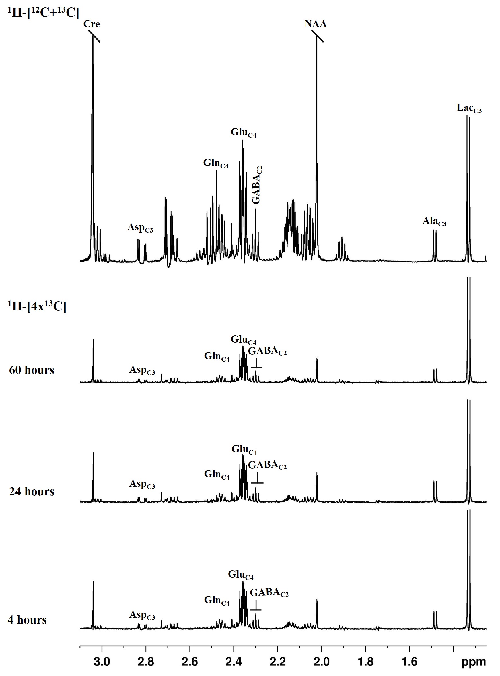
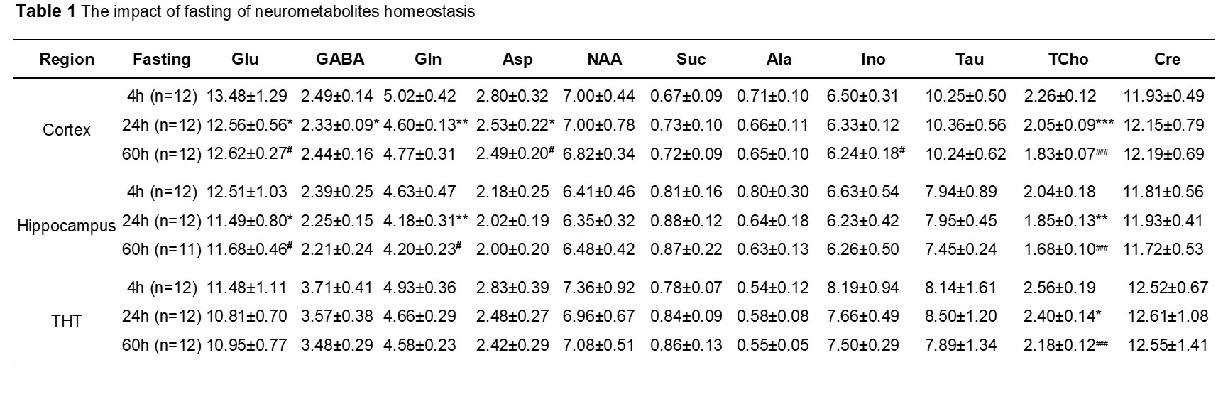
The concentrations (µmol/g) of neurometabolites were measured in the unedited 1H-[12C+13C]-NMR spectrum using [2-13C]glycine as a reference. Values are presented as mean±SD; *or# p<0.05, **or## p<0.01, ***or###p<0.001. Abbreviations: Ala: alanine; Asp: aspartate; Cho: Choline; PC: phosphocholine; GABA: g-aminobutyric acid; Glu: glutamate; Gln: glutamine; NAA: N-acetyl aspartate; Lac: lactate