4123
Assessment of arterial and venous sinuses cerebral blood flows in rats with phase contrast MRI1University Centre for Health Research (CURS, PIRMPA), University of Picardy Jules Verne, Amiens, France, 2Facing Faces Institute/CHIMERE EA 7516, University of Picardy Jules Verne, Amiens, France
Synopsis
Keywords: Small Animals, Velocity & Flow, Magnetic resonance velocity mapping; Phase contrast; Velocity; Rat.
Motivation: Several human cerebral diseases are associated to neurofluids dynamics alterations and continue to require preclinical research studies. Rat models were widely used to understand these diseases but its cerebral hemodynamic is not fully elucidated. Phase contrast (PCMRI) allows noninvasive quantification of blood flow dynamics.
Goal(s): Our goal was to investigate both cerebral arterial and venous sinuses flows in rats.
Approach: Twelve Sprague-Dawley rats underwent a 7T preclinical MRI. PCMRI quantified blood flows dynamics in the internal/external carotid arteries, basilar artery and transverse sinuses during cardiac cycle.
Results: We obtained physiological references of cerebral blood flows parameters for these main cerebral vessels in rats.
Impact: This study highlights the potential of PCMRI to investigate cerebral blood flows dynamics in rats. These findings may be important to study neurofluids dynamics interactions, pressure and compliance, to better understand human idiopathic pathologies such as hydrocephalus and intracranial hypertension.
INTRODUCTION
Based on currently knowledge on cerebral blood hydrodynamics in experimental animal, only a few studies1,2 were focused on the quantification of blood flows of cerebral arterial vessels in rats. Moreover, comparable flow measurements for arterial and sinus systems have not been yet performed in rats. The aim of this study was to quantify cerebral blood flow dynamics in the major arterial and venous vessels in rats and to establish reference physiological values based on PCMRI.METHODS
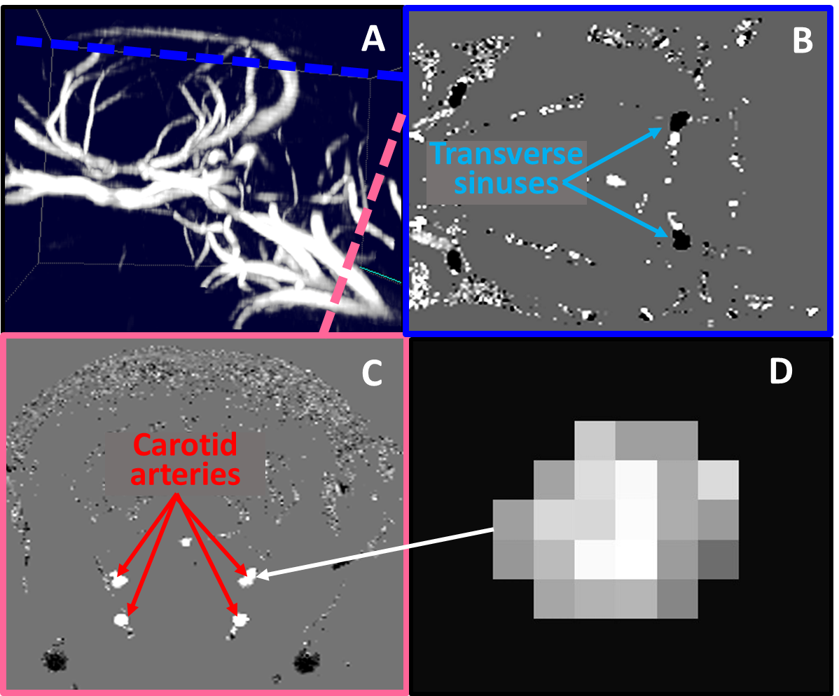
Twelve Sprague-Dawley rats weighing 378 ± 100 g were used in this study. The experimental protocol was approved by our Regional Ethics Committee. Anesthesia was maintained during the images acquisition by using 2% vaporized isoflurane. Blood velocities measurements were performed using a 7T system (Biospec 70/20, Bruker, Ettlingen), with a 72-mm volume resonator for RF transmission and a rat head surface coil for RF reception. We first used a standard 3D-PC angiography sequence to perform vascular imaging which was used as anatomical reference to position the PC imaging planes perpendicularly to the vessels of interest. A prospectively-gated 2D flow-sensitive PC-MRI sequence was used to assess blood velocities with a velocity encoding sensitization (VENC) of 60 cm/s for the carotid and basilar arteries, while, the venous sinus system blood flow was measured with VENC = 12 cm/s. The other parameters of the flow sequence were: TR/TE = 16/2.8 ms, FA=30°, spatial resolution = 0.17x0.17x1mm3, matrix = 256x256, number of averages=3, acquisition time ≈ 3.2 mn depending on cardiac frequency.The slices location of velocity measurements is indicated in the figure1. Arterial flows were measured in the major cerebral arteries (left/right internal carotid arteries (ICA), left/right external carotid arteries (ECA) and basilar artery). While, cerebral blood drainage was calculated by summation of the left and right transverse sinuses flows. Flow data were post-processed using a dedicated semi-automatic software3 allowing segmentation of vessels, reconstruction of blood flows curves through the cardiac cycle as well as calculation of peaks flows, mean flows and pulsatility index.
RESULTS AND DISCUSSION
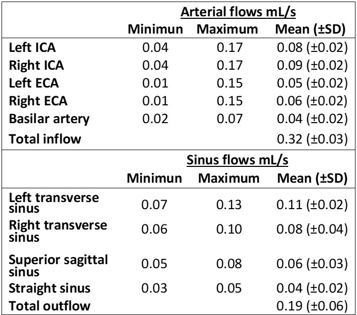
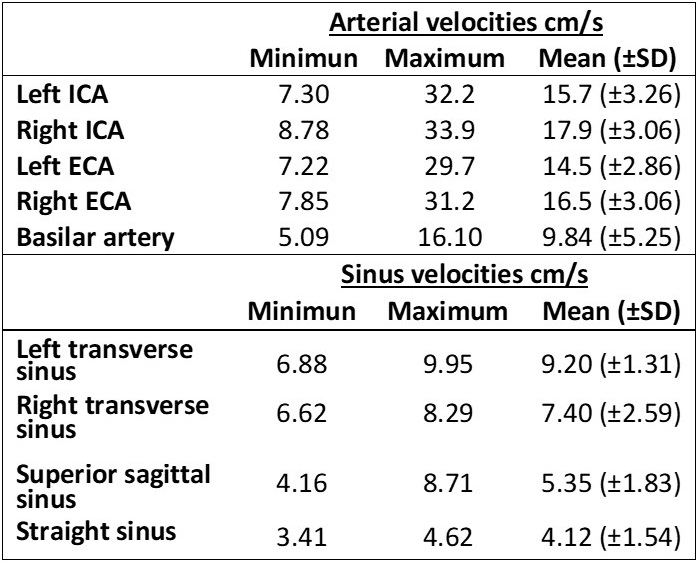
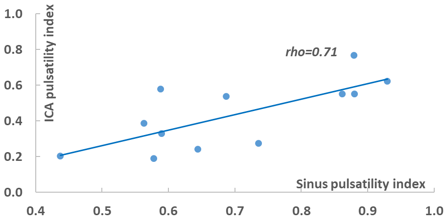
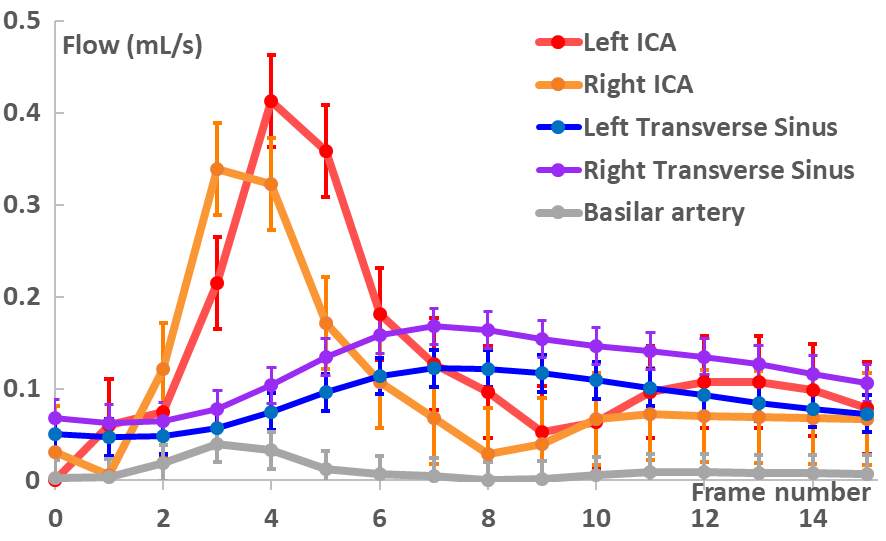
Key flows parameters are reported in the tables. All animals showed bilateral flows in their ICA (mean left ICA flow= 0.08 ± 0.02 mL/s vs right ICA flow= 0.09 ± 0.02 mL/s; mean left ECA flow= 0.05 ± 0.02 mL/s vs right ECA flow= 0.06 ± 0.02 mL/s). However, two animals exhibited unilateral transverse sinus flow in the group (mean left outflow= 0.11 ± 0.02 mL/s vs right outflow= 0.08 ± 0.04 mL/s). This unilateral blood circulation may be related to differences in anatomy and reflects the complexity and variability of the venous sinus system compared to that of arterial system, as previously observed in human4. Moreover, this study revealed that measured total mean flow in the sinuses are lower than that measured in the arteries (total arterial flow = 0.32 ± 0.03 mL/s vs 0.19 ± 0.06 mL/s), resulting of the presence of secondary drainage pathways in the rat brain. The pulsatility index of the arterial flows were significantly correlated with that of the sinus system (Spearman's rank correlation rho= 0.71, p-value<0.01, figure2), reflecting an interaction between these two compartments. Furthermore, pulsatility of the sinus flow waveforms was lower compared to that of the arterial system (figure3), as previously observed in human5.CONCLUSION
PCMRI quantified blood flows dynamics in the ICA, ECA, basilar artery, sagittal, lateral and straight sinuses during cardiac cycle. We obtained physiological references of the main cerebral blood flows parameters from the main cerebral vessels in rats. These new results about cerebral blood flow dynamics in rats open new possibilities one research studies concerning cerebral diseases in animal models.Acknowledgements
We would like to thank Julie Le-Ber and Anais Jovelet for their help in animals’ preparation. We would like to thank also Dr. Jerome Voiron at Bruker technical support service for assistance.References
(1) Chiu, S.-C.; Hsu, S.-T.; Huang, C.-W.; Shen, W.-C.; Peng, S.-L. Phase Contrast Magnetic Resonance Imaging in the Rat Common Carotid Artery. J. Vis. Exp. JoVE 2018, No. 139.
(2) Peng, S.-L.; Shih, C.-T.; Huang, C.-W.; Chiu, S.-C.; Shen, W.-C. Optimized Analysis of Blood Flow and Wall Shear Stress in the Common Carotid Artery of Rat Model by Phase-Contrast MRI. Sci. Rep. 2017, 7 (1), 525.
(3) Balédent, O.; Henry-Feugeas, M. C.; Idy-Peretti, I. Cerebrospinal Fluid Dynamics and Relation with Blood Flow: A Magnetic Resonance Study with Semiautomated Cerebrospinal Fluid Segmentation. Invest. Radiol. 2001, 36 (7), 368–377.
(4) Durgun, B.; Ilglt, E. T.; Cizmeli, M. O.; Atasever, A. Evaluation by Angiography of the Lateral Dominance of the Drainage of the Dural Venous Sinuses. Surg. Radiol. Anat. SRA 1993, 15 (2), 125–130.
(5) Stoquart-Elsankari, S.; Lehmann, P.; Villette, A.; Czosnyka, M.; Meyer, M.-E.; Deramond, H.; Balédent, O. A Phase-Contrast MRI Study of Physiologic Cerebral Venous Flow. J. Cereb. Blood Flow Metab. Off. J. Int. Soc. Cereb. Blood Flow Metab. 2009, 29 (6), 1208–1215.
Figures