4122
Glymphatic Diffusion-Weighted MRI in Awake Mice: Comparison to Anesthetized Brain States.1Center for Translational Neuromedicine, University of Copenhagen, Copenhagen, Denmark, 2Center for Translational Neuromedicine, University of Rochester Medical Center, Rochester, NY, United States
Synopsis
Keywords: Neurofluids, Brain, Glymphatic System
Motivation: This study investigates the glymphatic function in awake vs. anesthetized mice by means of diffusion-weighted (DW) MRI.
Goal(s): Application of developed ‘silent’ DW-MRI protocol is presented for comparison of awake and anesthetized mice under ketamine-xylazine (K/X), and isoflurane.
Approach: In total, n=17 C57BL/6 mice were habituated to awake MRI setup, and successfully underwent DW-MRI with monitored body temperature and respiratory rate.
Results: Results showed that brain water mobility in awake mice was consistent, while K/X and isoflurane affected both slow and fast MR diffusivity measures differently. Awake DW-MRI offers a promising alternative to MRI under anesthesia, promoting increased biological relevance and translational potential.
Impact: This research on glymphatic function using DW-MRI underscores the importance of non-invasive MRI in awake mice, emphasizing anesthesia's impact on physiological stability. It highlights the translational potential of awake DW-MRI and is of importance to neuroscientific and biomedical research communities.
Introduction
The glymphatic system is formed of a network of perivascular spaces enclosed by astrocytic endfeet ensheathing the vascular walls [1, 2]. It serves a vital role of a brain waste clearance pathway facilitated by exchange of cerebrospinal fluid (CSF) with interstitial fluid (ISF). The system undergoes significant variations during natural sleep and under anesthesia, attributed to the modulation of the brain's ISF space and perivascular flow albeit the functional changes within the vascular bed [3-7]. While invasive dynamic contrast-enhanced MRI is a common method for glymphatic assessment, both low-pressure CSF and ISF flow can also be substantially influenced by contrast injections. This limits the translational relevance of preclinical findings, especially when utilizing diffusion-weighted (DW) MRI to evaluate water mobility in the entire brain is considered.Thus, we have recently successfully developed and validated a fully non-invasive glymphatic MRI protocol for evaluating brain water mobility in anesthetized mice [8]. In this study, we present further progress on glymphatic diffusion-weighted MRI in an awake murine model, comparing it to two common anesthetic states of ketamine-xylazine and isoflurane.
Methods
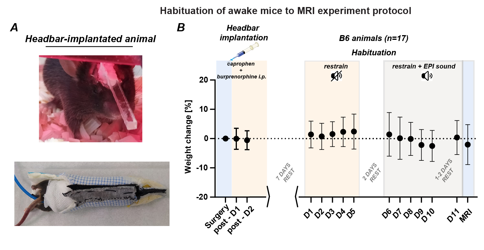
Experiments were performed in n=17 12-16 weeks C57BL/6 mice, group-housed with food and water ad-libitum, temperature and humidity-controlled environment, and 12/12h reverse light/dark cycle. Each group of randomly subdivided animals underwent implantation of ~25x3x3mm dimensions acrylic headbar to the surface of the skull along the cranio-caudal direction, for the head fixation during habituation and MRI (Figure 1A top). The habituation aimed adapting the animal to DW-MRI and lasted 12 days in 2 phases. During each phase, continued over 5 consecutive days and separated one from another by 2 days of rest (Figure 1B), each animal was placed in an in-house designed restrain coat and a mock MR bed (Figure 1A, bottom). After successful habituation, animals underwent DW-MRI using a 9.4T MR system (BioSpec 94/30USR, Bruker BioSpin) in awake state and under subsequent anesthesia (ketamine-xylazine, K/X: i.p. 100/10mg/kg; isoflurane (ISO) low dose: 0.5-1%; ISO high dose: 1-1.5%). During DWI, all animals were securely fixed to the MR holder with an implanted headbar and confined using the designed restrain coat (Figure 1A, bottom). Their body temperature was maintained at 37±1°C, and both body temperature and respiratory rate were continuously monitored. To evaluate differences in brain water mobility in awake mice, a ‘silent’ version of a previously employed [8] respiratory-gated echo-planar-imaging (EPI) sequence was used (EPI: TR/TE=3600/30ms; FA=85deg.; repetitions=3; segment=3; diffusion encoding directions=3; voxel dimension=0.15x0.15x0.5mm; gradient shape=sine; gradient duration time=11ms; gradient separation time=3.5ms). A total of 17 b-values were measured, ranging from 40 to 3081s/mm². Data analysis was conducted using a voxel-wise basis and both monoexponential model (apparent diffusion coefficient, ADC) and biexponential intravoxel-incoherent motion (IVIM) model with a segmented approach [8], and evaluated within 21 parenchymal ROI set-based on the Allen Brain Atlas.Results
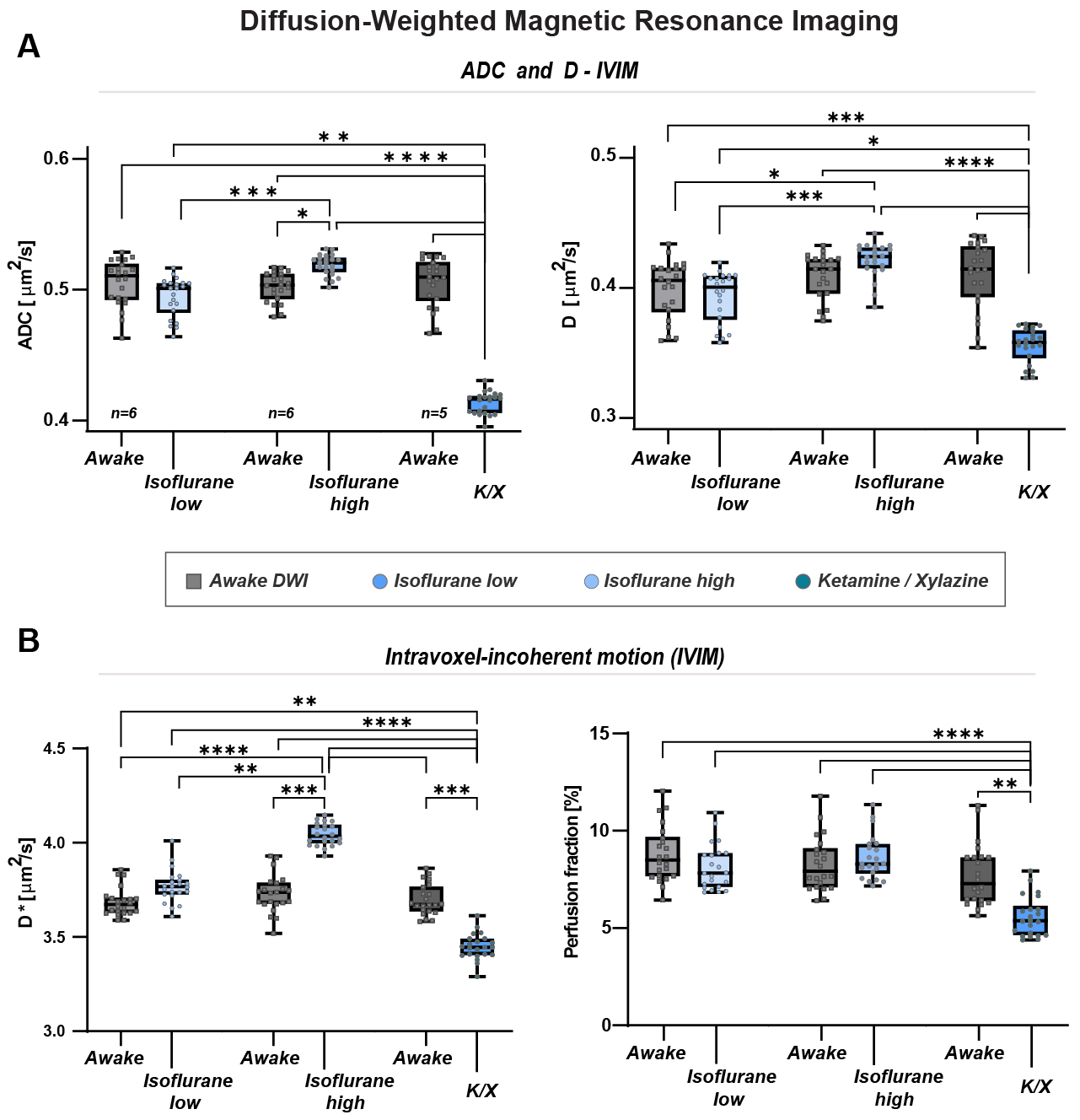
All n=17 animals underwent successful habituation followed by DW-MRI (K/X: n=5; low dose ISO: n=6; high dose ISO: n=6). Using the 'silent' DW-MRI approach, both slow diffusivity measures using a standard ADC and ‘pure’ slow diffusivity (D) using IVIM were found similar among all parenchymal ROI in all awake animals (ADC=0.504±0.016 and D=0.408±0.021µm2/ms; Figure 2A). Low dose ISO resulted in a non-significant decrease, while high dose ISO resulted in a similar increase in ADC and D, compared to awake conditions. As expected, K/X resulted in a significant ~20% decrease in both slow diffusivity measures (p<0.0001). Fast MR diffusivity measures expressed by pseudodiffusion (fast diffusivity, D*) were not significantly increased by low dose ISO, while high dose ISO and K/X resulted in similar in magnitude but contradictory effects (increase for ISO and decrease for K/X, p<0.001 for both; Figure 2B left) which might be attributed to their contradictory effect on circulatory system [9, 10]. Still, using 'silent' DWI approach blood/fluid volume changes assessed using perfusion fraction IVIM measure were found altered and decreased by ~25-30% only after K/X (p<0.01; Figure 2B right).Conclusion
Our study highlights the feasibility and advantage of awake diffusion-weighted MRI in mice as an alternative to traditional anesthetic methods. Our findings also suggest that the choice of anesthesia or the awake state should be carefully considered in preclinical research to ensure the reliability and translational relevance of MRI data, as both ISO and K/X are known to possess contradictory effect on vascular pulsatility and the volume of vascular bed. Since awake mice exhibit closer-to-baseline steady state conditions, adoption of awake MRI in murine studies ultimately provides more biologically relevant approach to study the brain physiology in more natural non-anesthetized state, and improves the translational potential of these studies to human conditions.Acknowledgements
No acknowledgement found.References
1. Iliff, J.J., et al., A paravascular pathway facilitates CSF flow through the brain parenchyma and the clearance of interstitial solutes, including amyloid beta. Sci Transl Med, 2012. 4(147): p. 147ra111.
2. Iliff, J.J., et al., Brain-wide pathway for waste clearance captured by contrast-enhanced MRI. J Clin Invest, 2013. 123(3): p. 1299-309.
3. Hablitz, L.M., et al., Circadian control of brain glymphatic and lymphatic fluid flow. Nat Commun, 2020. 11(1): p. 4411.
4. Xie, L., et al., Sleep drives metabolite clearance from the adult brain. Science, 2013. 342(6156): p. 373-7.
5. Slupe, A.M. and J.R. Kirsch, Effects of anesthesia on cerebral blood flow, metabolism, and neuroprotection. J Cereb Blood Flow Metab, 2018. 38(12): p. 2192-2208.
6. Mestre, H., Y. Mori, and M. Nedergaard, The Brain’s Glymphatic System: Current Controversies. Trends in Neurosciences, 2020.
7. Ozturk, B.O., et al., Disparate volumetric fluid shifts across cerebral tissue compartments with two different anesthetics. Fluids Barriers CNS, 2021. 18(1): p. 1.
8. Gomolka, R.S., et al., Loss of aquaporin-4 results in glymphatic system dysfunction via brain-wide interstitial fluid stagnation. Elife, 2023. 12.
9. Iida, H., et al., Isoflurane and sevoflurane induce vasodilation of cerebral vessels via ATP-sensitive K+ channel activation. Anesthesiology, 1998. 89(4): p. 954-60.
10. Peltoniemi, M.A., et al., Ketamine: A Review of Clinical Pharmacokinetics and Pharmacodynamics in Anesthesia and Pain Therapy. Clin Pharmacokinet, 2016. 55(9): p. 1059-77.
Figures