4121
A fMRI study of the face-processing regions in Dog’s brain1State Key Laboratory of Brain and Cognitive Science, Institute of Biophysics, Chinese Academy of Sciences, BeiJing, China, 2University of Chinese Academy of Sciences, Beijing, China, 3Beijing Municipal Public Security Bureau Public Transport Safety and Security Corps Canine Unit, BeiJing, China
Synopsis
Keywords: Large Animals, Nonhuman Primates, Preclinical, animal models
Motivation: Functional magnetic resonance imaging is increasingly used to study brain function and cognition in domestic dogs.
Goal(s): The objective of this study was to acquire the high-quality fMRI data which dogs can be trained to remain awake and still inside MRI scanners and detect the pathway for dog’s face processing.
Approach: We use a combination of simulated and real MRI environments to train awake dogs. A visual stimulation paradigm with block design was used to compare activity elicited by human faces against objects.
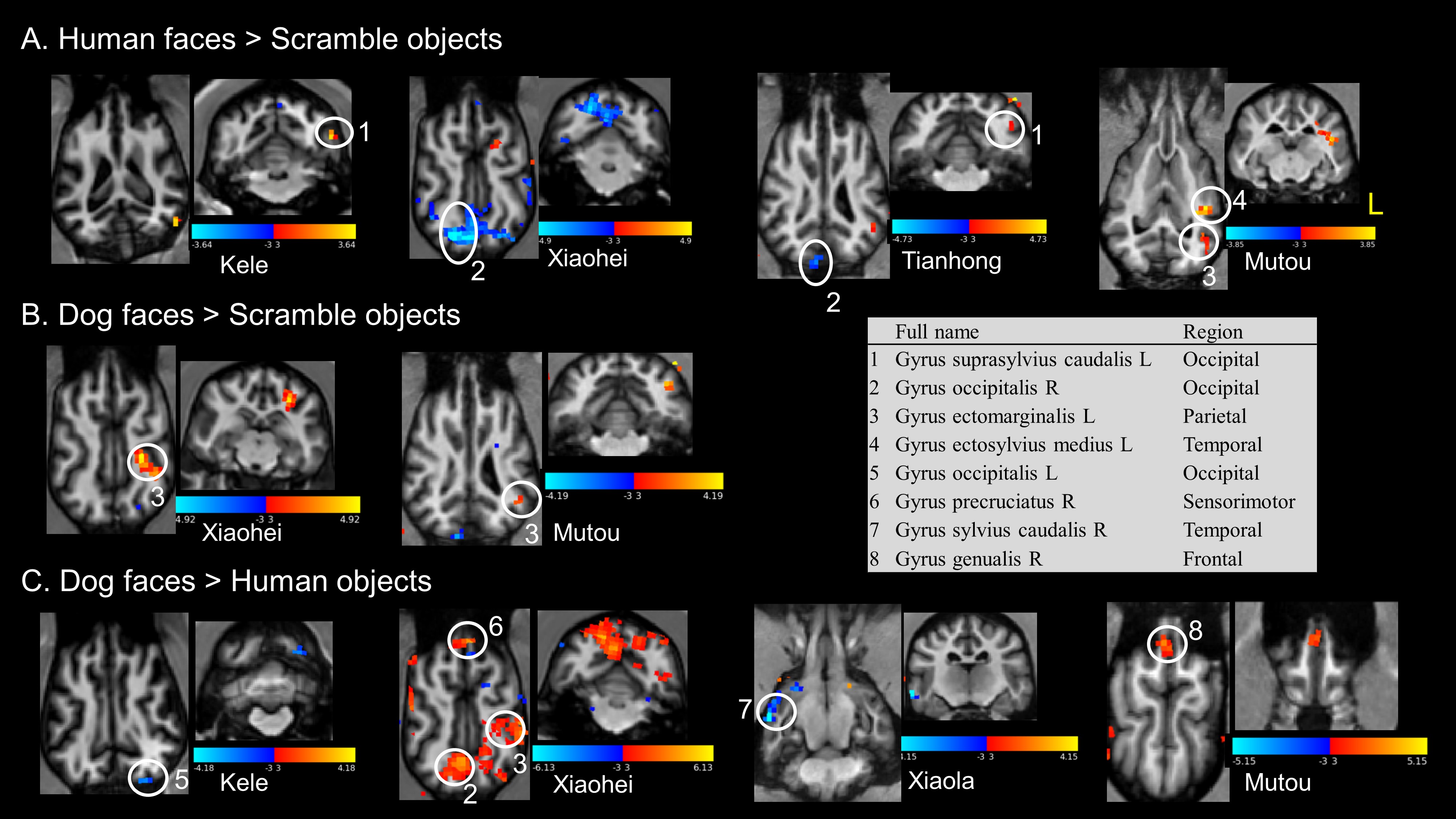
Results: We successfully detect the activation of human faces against scramble objects in occipitalis, ectomarginalis, and ectosylvius medius.
Impact: This study provides a process for training dogs for fMRI acquisition while awake and introduces the temporal cortex as candidate to process human faces and dog faces.
Introduction
In recent years, dogs have become one of the experimental subjects of cognitive science research. Dogs have strong learning abilities and are easy to train, coupled with the safety and non-invasive nature of fMRI, making dogs a promising animal model for studying brain diseases and social disorders related to social cognition1. Many studies on basic visual graphics, facial emotion recognition, and wake-up dog language have been carried out internationally. The difficulty of conducting fMRI experiments on awake dogs mainly lies in the need for the dog to keep its head and body still and actively participate in the experimental task2-4. We will use a combination of simulated and real MRI environments to train awake dogs, which will save the cost of training in a real MRI environment and achieve the goal of dogs completing fMRI experiments "freely and autonomously" in an awake state.Methods
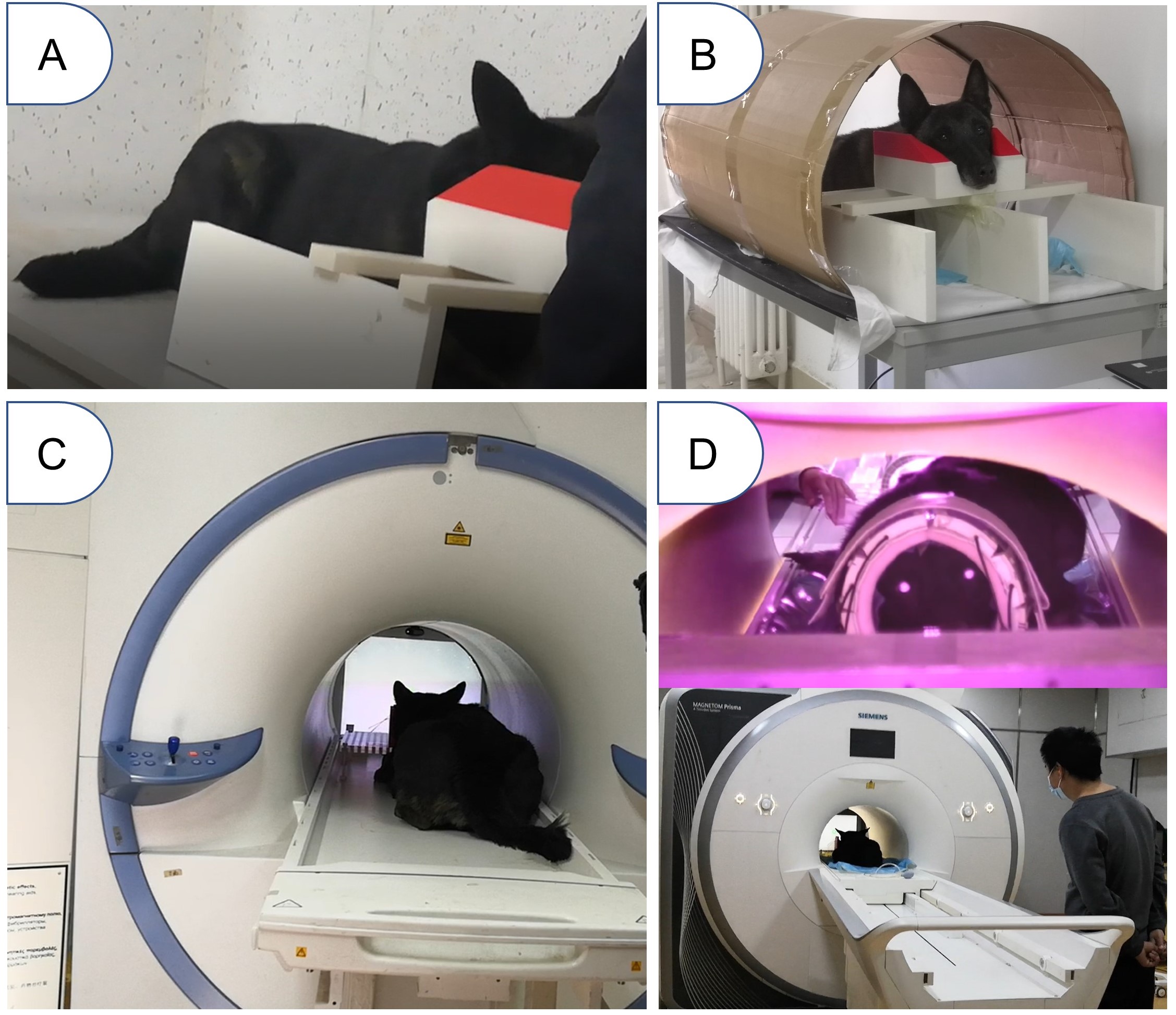
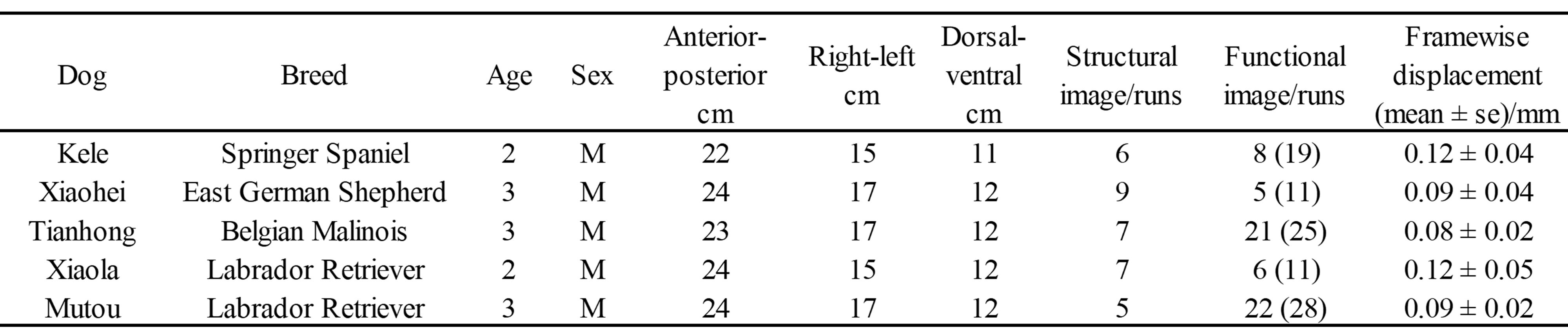
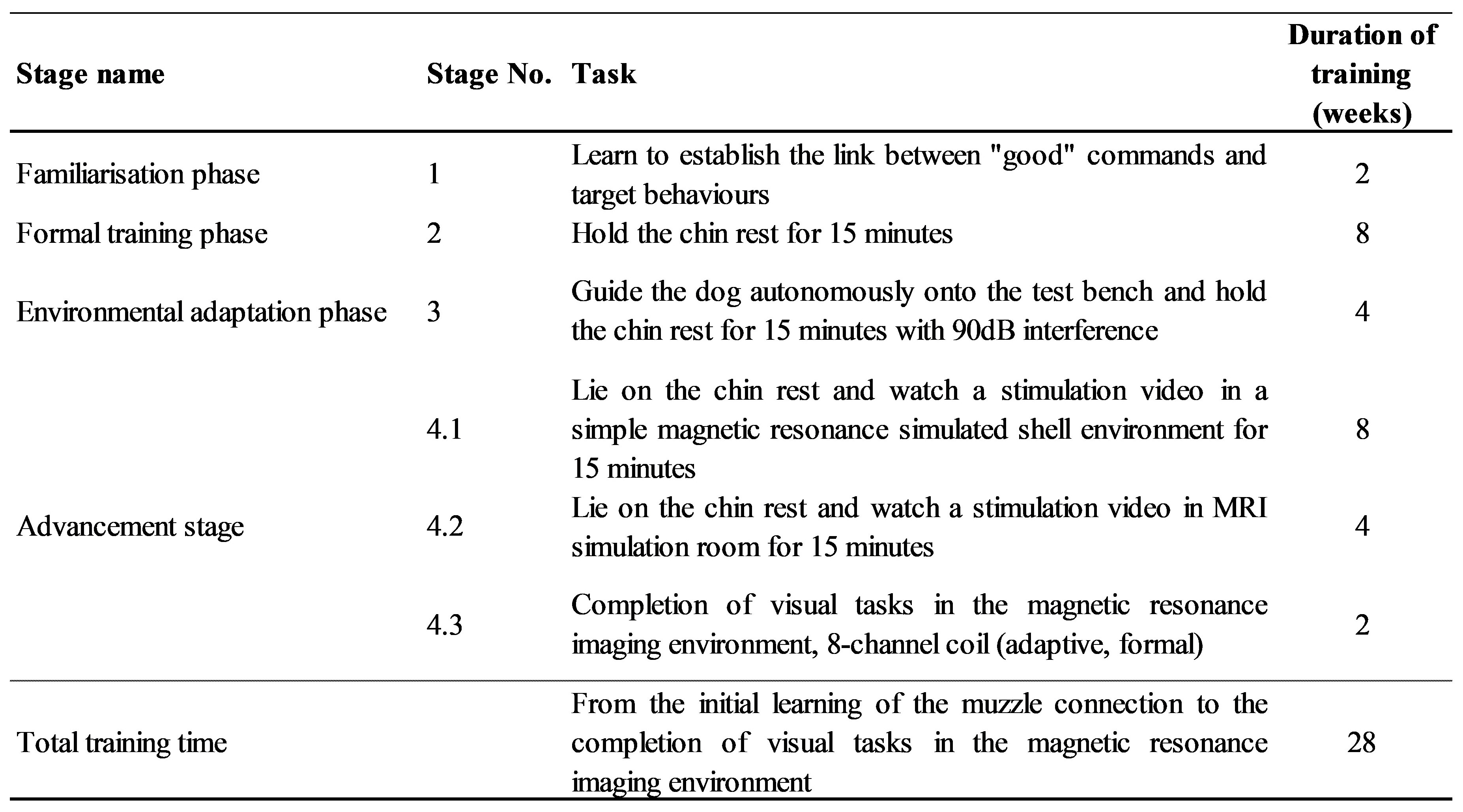
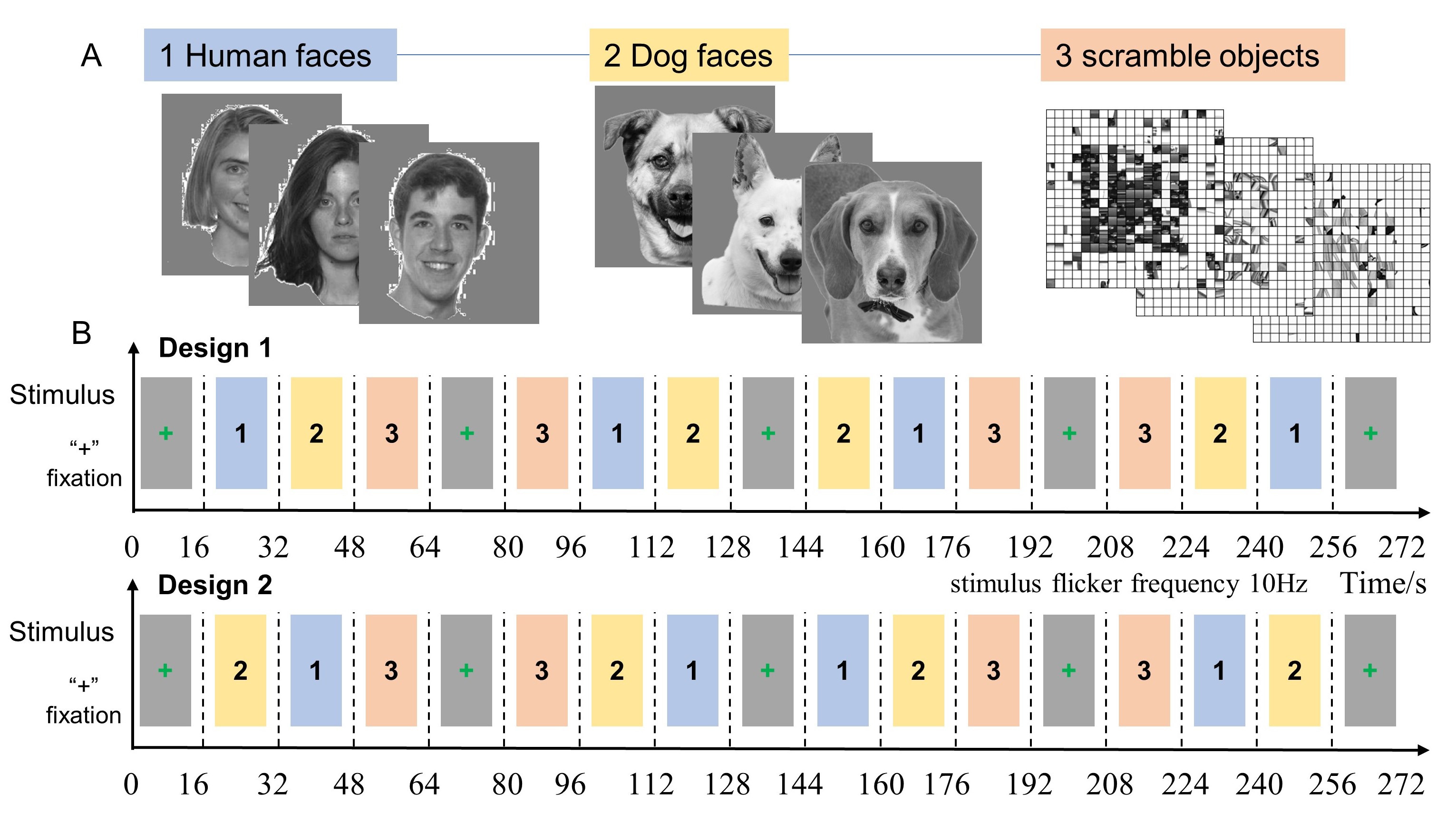
Training: To allow dogs to "freely and autonomously" enter the MRI scanner while awake, all dogs complete four stages of training (affinity, simulation training, environmental adaptation, and improvement) prior to the scanning, and ensure that they lie still and watch images during the fMRI acquisition (Figure 1). Each training session lasted 5-20 minutes, with 5 sessions conducted daily (Figure 2). A total of 11 dogs completed the training, and their basic information is provided in Figure 3.Data Acquisition: MR scans of all dogs were performed on a Siemens Prismafit 3.0T MR scanner at the Institute of Biophysics using a homemade 8-channel Tx/Rx RF coil. Structural images using MPRAGE sequence: FOV = 180×180 mm2; TE/TR/TI = 3.37/2200/800 ms; FA = 8°; slice thickness = 0.7 mm, acquisition data matrix size 256×256, TA = 5min18s. Functional images covered the whole brain with 30 contiguous slices acquired with a gradient-echo EPI sequence: FOV = 119 × 119 mm2; TE/TR = 29/2000 ms; FA = 90°; slice thickness = 1.8 mm, acquisition data matrix size 66 × 66, measurements = 136, TA = 4min16s. The visual stimulation paradigm had a block design including 3 types of blocks: human faces, dog faces and scramble objects (Figure 4). To ensure optimal data quality, three runs were acquired per imaging session, with 2-3 sessions per MRI day separated by 1-2 weeks, until a total of 8-12 runs were successfully acquired, with each run showing the average framewise displacement of less than 0.5 mm.
Image analysis: T1w images in individual spaces were noise-reduced and corrected for offset fields, and then realigned to the one with the best SNR and contrast. The average T1w image for each dog was generated using Serial Longitudinal Registration on SPM12. The functional images were reoriented and corrected for eddy current distortion after slice timing and realignment, then detrended and co-registered to the average T1w for each dog, and finally smoothed with 4 mm FWHM. Potential outlier scans were identified using ART as acquisitions with framewise displacement above 0.5 mm. Statistical analysis of fMRI data was performed using a General Linear Model in SPM. The resulting statistical parametric maps were threshold at z > 3 and pcluster < 0.001 uncorrected.
Results
After 28 weeks of training, 5 dogs were able to achieve an average FD of less than 0.5 mm in the fMRI experiment and successfully completed the visual stimulation experiment. There were significant differences in occipitalis, ectomarginalis, and ectosylvius medius for human faces against scramble objects, extended to Occipital, partial, and temporal cortex. The precruciatus, sylvius caudalis and gyrus genualis might distinguish dog and human faces (Figure 5).Discussion and Conclusion
In preliminary exploration, we achieved the goal of dogs completing fMRI experiments "freely and autonomously", indicating that the prospects in this direction are very optimistic. Based on the awake dog fMRI research platform, it is a very promising direction to conduct a series of studies on the neural mechanisms of dog socialization and emotion5. Combining behavioral indicators with changes in structure and function during brain development can establish a set of selection criteria for working dogs. Additionally, expanding research into canine olfaction, exploring canine olfactory mechanisms, and delving into studies on canine pain empathy and social empathy can shed light on the establishment and dependence of canine-human emotions, as well as the emotional connection between humans and dogs.Acknowledgements
This work was supported in part by the Ministry of Science and Technology of China grant (2022ZD0211901, 2019YFA0707103, 2020AAA0105601), and the Chinese Academy of Sciences grants (ZDBS-LY-SM028).References
1. Sriganesh K, Balachandar R, Bagepally BS, et al. Effect of propofol anesthesia on resting-state brain functional connectivity in Indian population with chronic back pain. Neurol India, 2017, 65(2):286-292.
2. Karl, S., Boch, M., Virányi, Z. et al. Training pet dogs for eye-tracking and awake fMRI. Behav Res, 2020, 52, 838–856.
3. Wallis, L.J., Virányi, et al. Aging effects on discrimination learning, logical reasoning and memory in pet dogs.Age, 2016, 38, 6.
4. Johnson, P. J., Luh, et al. Stereotactic Cortical Atlas of the Domestic Canine Brain.Scientific reports, 2020, 10(1), 4781.
5. Boch M, Karl S, Sladky R, et al. Tailored haemodynamic response function increases detection power of fMRI in awake dogs (Canis familiaris). NeuroImage, 2021, 224:117414
Figures