4119
Hemodynamic response to activation of excitatory and inhibitory neurons under awake and different anesthetic conditions1Center for Neuroscience Imaging Research, Institute for Basic Science (IBS), Suwon, Korea, Republic of, 2Department of Biomedical Engineering, Sungkyunkwan University, Suwon, Korea, Republic of, 3Department of Intelligent Precision Healthcare Convergence, Sungkyunkwan University, Suwon, Korea, Republic of
Synopsis
Keywords: Small Animals, Neuroscience, Neurovascular coupling;Excitatory neurons;Inhibitory neurons;Anesthesia
Motivation: Understanding hemodynamic activity is essential for blood-based brain mapping techniques like fMRI.
Goal(s): Many studies have demonstrated that excitatory activity leads to an increase in hemodynamic response. However, how inhibitory neurons regulate the brain blood’s supply is less understood. Also, how hemodynamic response is modulated by brain status induced by different anesthetic is still unclear.
Approach: To investigate that, we used optical intrinsic signal imaging to investigate the roles of excitatory and inhibitory activity under awake and different anesthetic conditions.
Results: Our findings revealed that different conditions not only shape the response time and peak change but also causes different hemodynamic response.
Impact: We investigated the hemodynamic response of excitatory and inhibitory neurons by optogenetic stimulation under awake and 3 commonly used anesthetics. Our study may have an impact on investigating neurovascular coupling in different brain conditions.
Introduction
Understanding hemodynamic activity is essential for blood-based brain mapping techniques including fMRI, which indirectly measures changes in blood flow and oxygenation associated with neuronal activity. Many studies have demonstrated that excitatory activity leads to an increase in cerebral perfusion and cerebral blood volume (CBV)1,2. However, how inhibitory neurons regulate the brain blood’s supply is less understood2. Hemodynamic responses to both excitatory and inhibitory neuronal activities can be highly affected by the brain status induced by anesthetic. To address how different anesthetics modulate hemodynamic responses, we performed hemoglobin-based optical intrinsic signal imaging studies to optogenetic stimulation of excitatory and inhibitory neurons under awake and three commonly used anesthetics in optics and MRI communities: Ketamine, dexmedetomidine and isoflurane3 4. As a control, we also used electrical forepaw stimulation under different conditions. We observed positive hemodynamic response of excitatory activity and biphasic hemodynamic response of inhibitory activity. Also, we found that different conditions shape the response time and peak change slightly different.Methods
Animals:Thy1-ChR2 (n=12) and VGAT-ChR2(n=20) mice with thinned-skull on the entire brain.
Anesthetic:
Dex-ISO: An initial mixture of Dexmedetomidine (Dex:50μg/kg/h) and a supplementary dose (Dex:50-63μg/kg) with 0.3% of isoflurane3.
Isoflurane: 1.5% isoflurane.
Ket-Xyl: An initial mixture of ketamine (Ket:100mg/kg) and xylazine (Xyl:10mg/kg), and a supplementary dose (25mg/kg Ket and 1.25mg/kg Xyl) 5.
Stimulation:
Optogenetic stim: 5-s and 20-s photostimulation duration with 20Hz and 1Hz frequency, 20% duty cycle, power of 3mW (3 and 5.7mW) for Thy1-ChR2 (VGAT-ChR2) mice.
Forepaw electric stimulation as a control: 4Hz, 0.5 mA, and 0.5 ms.
Optical intrinsic signal imaging: OISI was performed at 530 nm (CBV-like) and 625 nm (BOLD-like). Total hemoglobin concentration (HbT) and CMRO2 changes were calculated6.
Results
We first performed optogenetic stimulation in the left somatosensory area of awake Thy1 and VGAT mice. We observed increased CBV response of excitatory activity and biphasic CBV response of inhibitory activity at the stimulation site (Figure 1C and 1D). Excitatory activity increases CBV, while inhibitory activity induces initial CBV increase followed by negative CBV response.The magnitude of hemodynamic response depended on frequency and laser power of photostimulation (Figure 2). For excitatory Thy1 mice, increasing laser power at a fixed 20Hz frequency results in a gradual increase in the peak value of total hemoglobin (Figure 2A). For inhibitory VGAT mice, except the increase in peak response, high laser power also induces post-vasodilation which did not show in other lower laser power group (Figure 2B). One possibility is high power may activate interneurons in deeper layer which may induce later responses. 1Hz stimulation changed the polarity of CBV response in the VGAT group (Figure 2C and 2D), which can be explained by the relative contribution of vasodilative inhibitory activity and vasoconstrictive excitatory suppression1.
Our next question is whether CBV response is modulated by different anesthetic which can significantly modulate synaptic activity and neuronal response properties by changing the balance between excitatory and inhibitory activities7 8 9. For Thy1 mice, we found Ket-Xyl condition induced the highest hemodynamic and oxygen consumption increase, but not much difference in the peak change between other 3 conditions (Figure 3A 3C and 3E). Under anesthesia, the slow return to baseline after stimulus offset was observed. For VGAT mice, response patterns were quite similar, except the initial vasodilation under awake condition and post-stimulus dilation only occurred under Dex-Iso (Figure 3B 3D and 3F).
As a control, we also did forepaw stimulation under different anesthetic, we found that the Ket-Xyl condition induced higher HbT response than Dex-Iso, which is consistent of our optogenetic study (Figure 4A). We also did experiment under multiple doses of isoflurane, but did not observe significant response as previous study 10.
Discussion & Conclusion
In summary, we investigated the hemodynamic response of excitatory and inhibitory neurons by optogenetic stimulation under awake and anesthetic conditions. We found that activation of excitatory neurons induces vasodilation regardless of the brain status. Similarly, activation of inhibitory neurons induces similar negative CBV response due to inhibition of excitatory neurons, regardless of anesthetics. Also, initial dilation was observed with post-stimulus dilation, which magnitude is dependent on awake and anesthesia condition. The initial dilation could be due to activation of nNOS neurons, and later post-stimulus vasodilation may be due to inhibitory neuron-induced slow astrocytic activity. Our study may provide valuable insights of anesthetics for investigating neurovascular coupling.Acknowledgements
This research was supported by the Institute of Basic Science (IBS-R015-D1).References
1. Moon, H. S. et al. Contribution of excitatory and inhibitory neuronal activity to BOLD fMRI. Cerebral Cortex 31, 4053-4067 (2021).
2. Lee, J., et al. Opposed hemodynamic responses following increased excitation and parvalbumin-based inhibition. Journal of Cerebral Blood Flow & Metabolism 41, 841-856 (2021).
3. You, T., et al. Characterization of brain-wide somatosensory BOLD fMRI in mice under dexmedetomidine/isoflurane and ketamine/xylazine. Scientific reports 11, 13110 (2021).
4. Vazquez, A. L., et al. Inhibitory neuron activity contributions to hemodynamic responses and metabolic load examined using an inhibitory optogenetic mouse model. Cerebral cortex 28, 4105-4119. (2018).
5. Shim, H.-J., et al. Mouse fMRI under ketamine and xylazine anesthesia: Robust contralateral somatosensory cortex activation in response to forepaw stimulation. Neuroimage 177, 30-44 (2018).
6. Dunn, A. K., et al. Spatial extent of oxygen metabolism and hemodynamic changes during functional activation of the rat somatosensory cortex. Neuroimage 27, 279-290 (2005).
7. Reimann, H. M. and T. Niendorf The (un) conscious mouse as a model for human brain functions: key principles of anesthesia and their impact on translational neuroimaging. Frontiers in systems neuroscience 14, 8. (2020).
8. Brown, E. N., et al. General anesthesia and altered states of arousal: a systems neuroscience analysis. Annual review of neuroscience 34, 601-628 (2011).
9. Kim, S., et al. Whole-brain mapping of effective connectivity by fMRI with cortex-wide patterned optogenetics. Neuron. (2023).
10. Shim, H. J., et al. BOLD fMRI and hemodynamic responses to somatosensory stimulation in anesthetized mice: spontaneous breathing vs. mechanical ventilation. NMR in biomedicine 33(7), e4311 (2020).
Figures
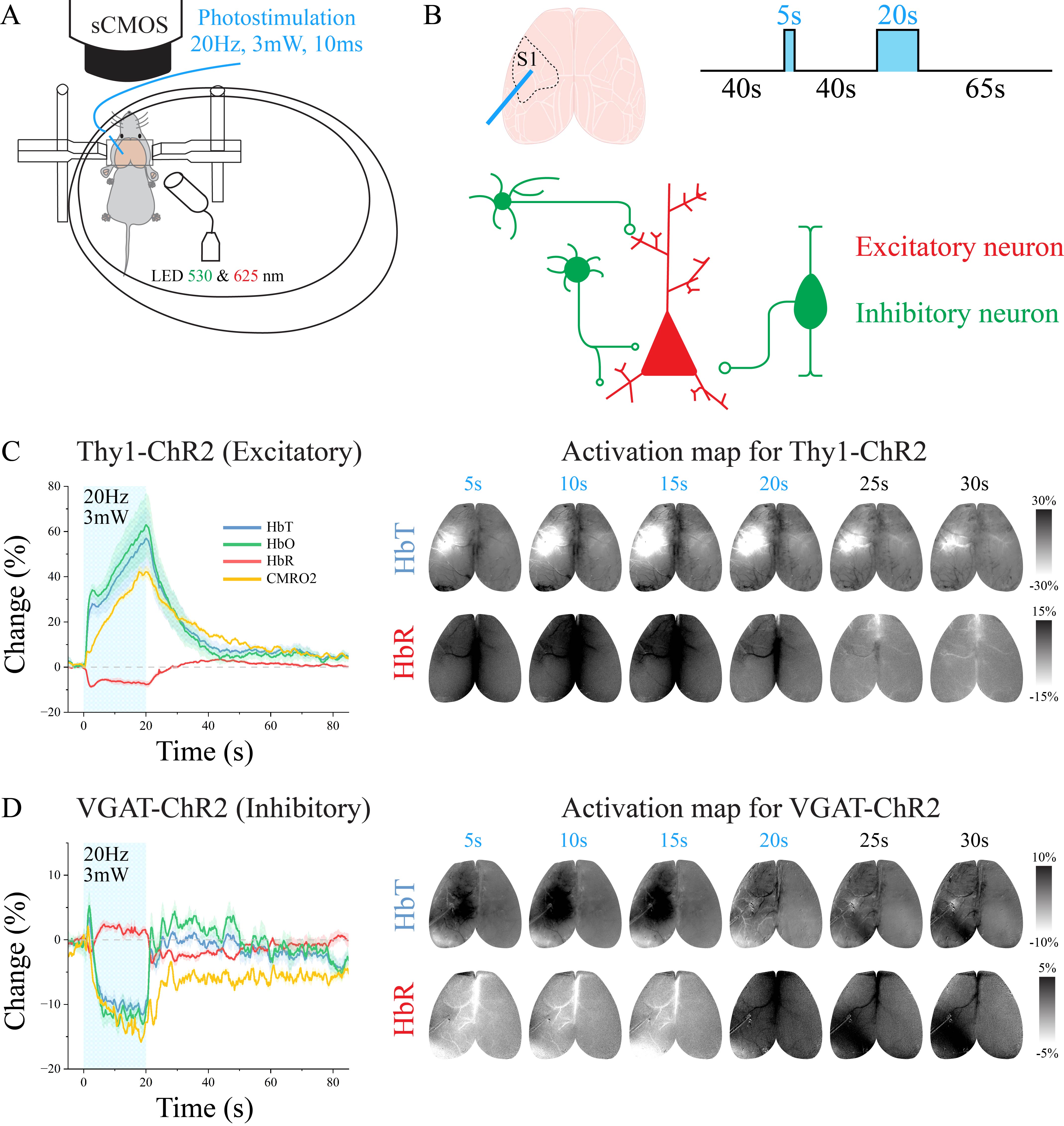
Figure 1. Hemodynamic and metabolic responses to optogenetic stimulation of excitatory and inhibitory neurons under awake condition. (A) Schematic of the experiment. (B) Stimulation paradigm, classification and interaction of excitatory and inhibitory neurons. (C)(D) Time course of total hemoglobin (HbT), deoxyhemoglobin (HbR), oxyhemoglobin (HbO) and CMRO2 changes for 20s stimulation of excitatory neurons and inhibitory neurons(left). Activation timeseries maps for HbT (CBV) and HbR (right). Dark and bright indicate negative and positive change, separately.
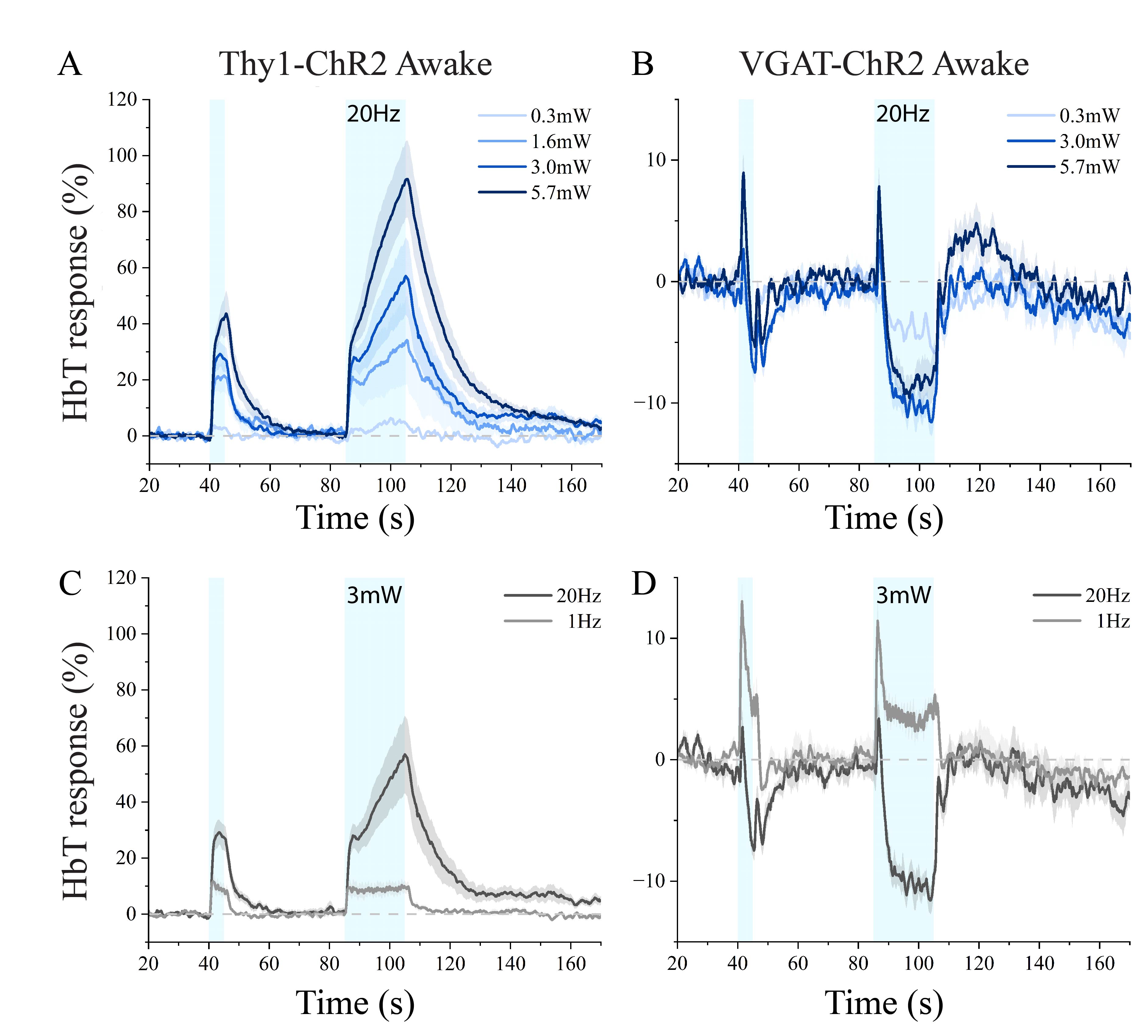
Figure 2. Power and frequency dependent HbT response of excitatory and inhibitory neurons under awake condition. (A)(B) HbT responses under different laser power of excitatory and inhibitory neurons (20Hz stimulation). (C)(D) HbT response under different frequency of excitatory and inhibitory neurons. (3mW stimulation).
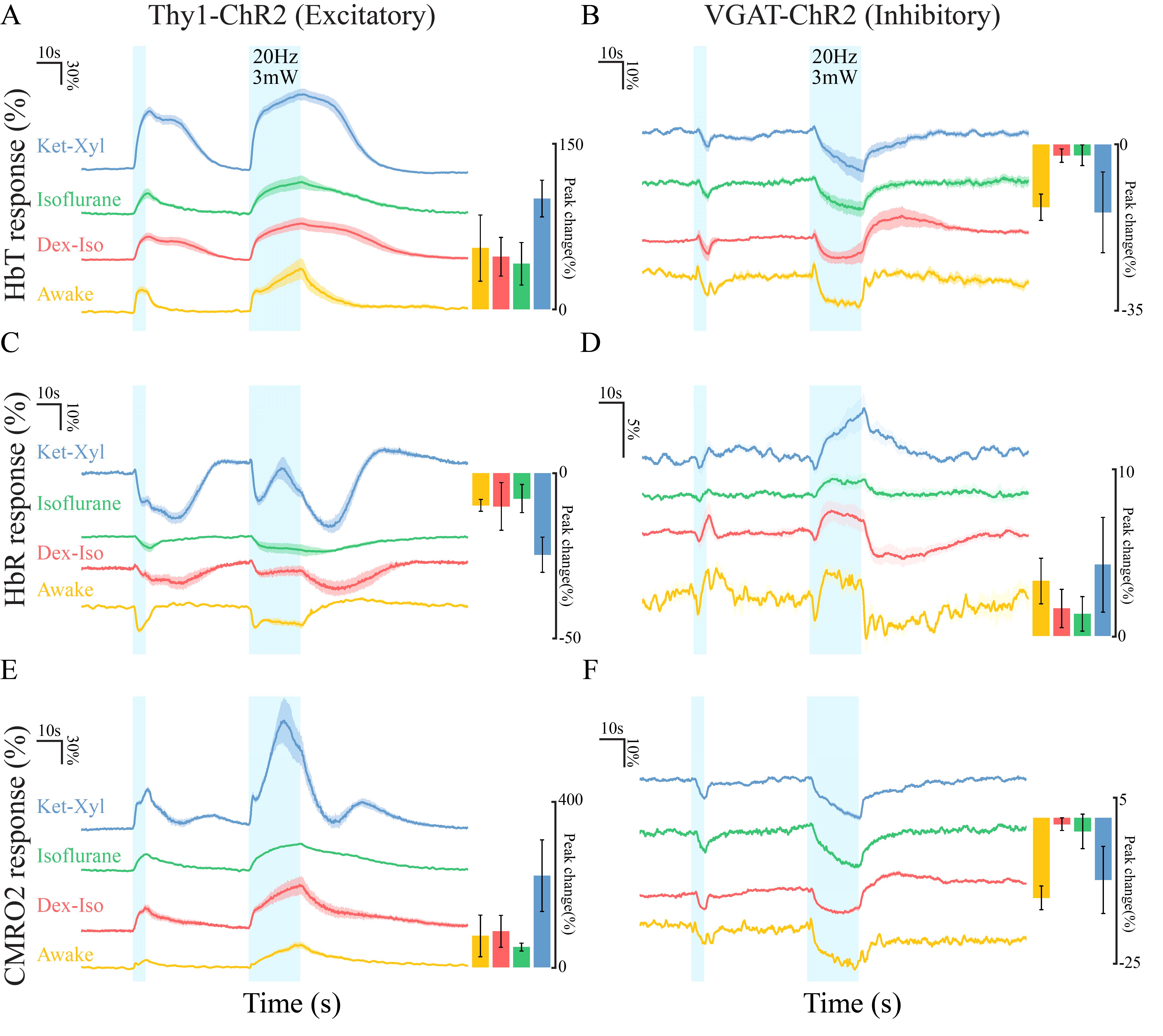
Figure 3. Hemodynamic and metabolic responses to optogenetic stimulation of excitatory and inhibitory neurons under awake and different anesthetic conditions. (A)(C)(E) Time courses and peak amplitudes (20s-stimulation) of HbT, HbR, and CMRO2 response for excitatory neurons under awake, Dex-Iso, Isoflurane and Ket-Xyl conditions. (B)(D)(F) Time courses and peak amplitudes (20s-stimulation) of HbT, HbR, and CMRO2 response for inhibitory neurons under awake, Dex-Iso, Isoflurane and Ket-Xyl conditions.
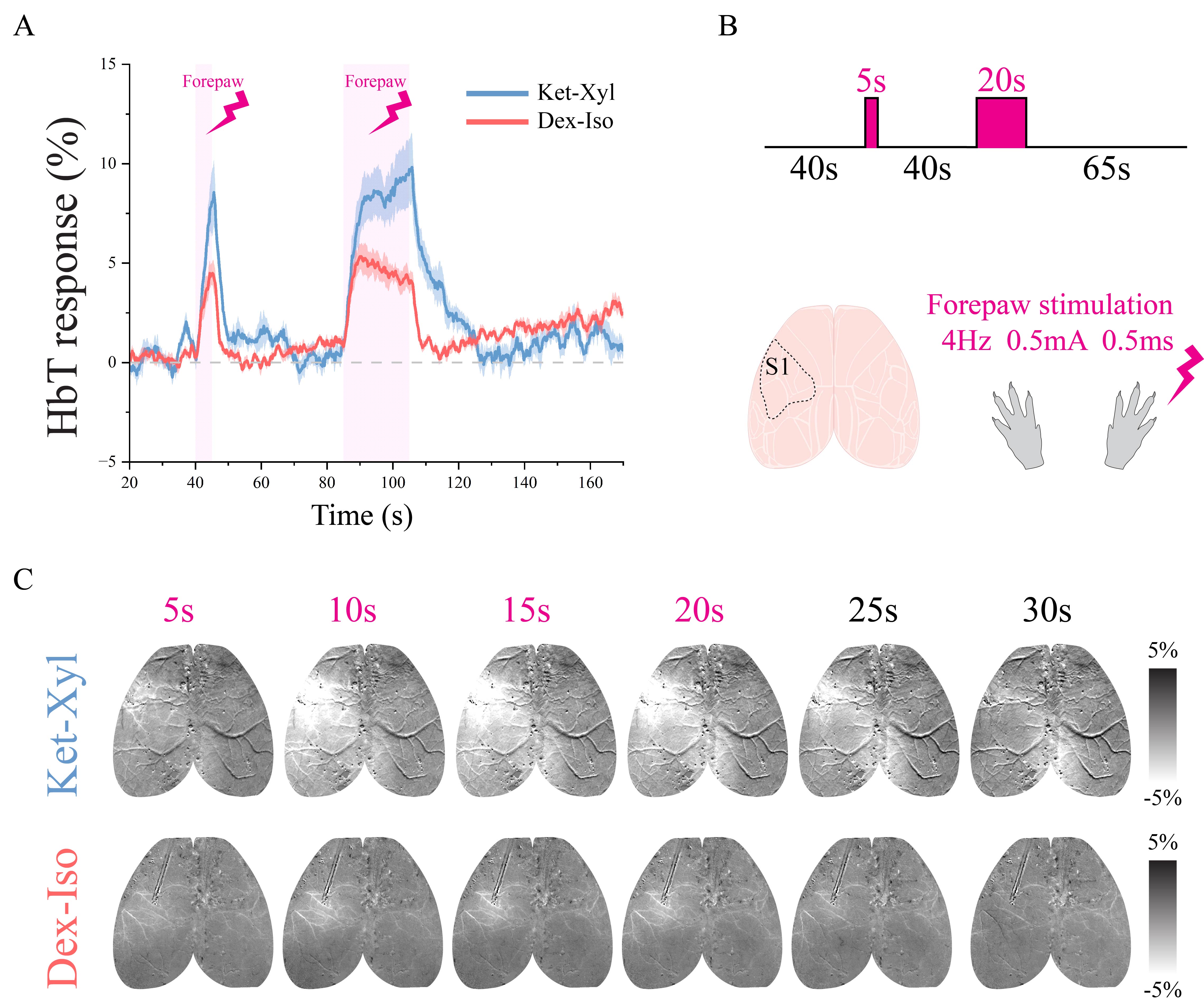
Figure 4. CBV responses to forepaw stimulation under different anesthetics. (A) Time course of HbT response under Dex-Iso and Ket-Xyl. (B) Stimulation paradigm. (C) Activation map for HbT response of 20s-stimulation.