4118
Characterization of atlas based volumetric changes in unilateral 6-OHDA-induced Parkinson’s rat model1Department of Computer Science, University of Delhi, Delhi, India, 2Department of NMR, All India Institute of Medical Sciences, New Delhi, India, 3Department of Physiology, All India Institute of Medical Sciences, New Delhi, India, 4Department of Psychiatry, Yale School of Medicine, New Haven, CT, United States
Synopsis
Keywords: Preclinical Image Analysis, Segmentation, Parcellation, atlas based volume extraction
Motivation: Manual characterization of brain regions is studied without prior knowledge to identify changes in GM/WM volume in PD.
Goal(s): Characterisation of GM and WM changes in 6-OHDA PD model as compared to sham control
Approach: Parcellated the rat brain into predefined regions using a rat atlas and performed the Wilcoxon rank signed rank test to identify significant changes in GM/WM volume.
Results: Significant loss in GM volume of a few brain regions in PD and Sham observed in 3rd and 7th week. Increase in WM volume for 3rd week is observed.
Impact: We identified GM loss and WM gain in a few brain regions without manual intervention which helps in understanding the structural changes in the whole brain in PD and Sham.
Introduction
Parkinson’s disease (PD) is a neurodegenerative disorder wherein slow degeneration of dopaminergic neurons in the substantia nigra pars compacta is reported1. We attempted to identify the significant volume changes in the brain regions in the unilaterally-lesioned 6-hydroxydopamine (6-OHDA) model.Methods
The adult male Wistar rats (n = 12, weighing 250–300 g) study was conducted after taking ethical approval from Institute Animal Ethics Committee. The adult male Wistar rats were divided into PD and Sham groups (n=6 in each). Two injections of 2 µL of 6-OHDA solution (7.5 µg/µL 6-OHDA dissolved in 0.2% ascorbate saline) were given in the right hemispheric striatum in the coordinates with reference to the bregma2. The sham-lesioned group went through the same procedure with the injection of saline (instead of 6-OHDA). Necessary postoperative care was provided and all procedures were performed as per the guidelines of the Committee for Control and Supervision of Experiments on Animals (CCSEA).The extent of the 6-OHDA lesioning was confirmed by the apomorphine-induced rotational test.High-resolution T1- weighted 3D gradient-echo MRI sequence (RF-spoiled fast low-angle shot (FLASH), repetition time (TR)/ echo time (TE) = 317.89/4.5ms, flip angle = 30°; voxel size = 0.0684 x 0.0684x0.8mm3; number of averages = 3. The data was acquired at baseline, after three weeks and after seven weeks.Each image was reconstructed using Bruker’s Paravision software, exported in DICOM format, and converted to NIfTI format. Preprocessing was carried out in SPM12. NifTI files were resized by a factor of 10 to account for the brain size difference between rodents and humans; followed by manual rotation to match the standard stereotaxic space in rat3. By registering each image to the template image using “Coregister” tool, the images were aligned to the stereotaxic space and was resampled into 0.125-mm isotropic voxels and then segmented into probability maps of gray matter (GM), white matter (WM), and cerebrospinal fluid with rat tissue priors3. These GM and WM maps were co-registered with the Wistar Rat Atlas. Thereafter, using in-house MATLAB code, the volume of each brain region defined by Wistar Rat Atlas is obtained. The volumetric changes in each brain region are evaluated using Wilcoxon signed rank test by keeping level of significance (α)=0.05. These tests are conducted for baseline vs 3rd week, baseline vs 7th week and 3rd week vs 7th week for PD and Sham controls.Results
Using Wilcoxon signed rank test with α=0.05, a few regions were found significant in GM, namely Left frontal assocn cortex, Left agranular insular cortex, posterior part (AIP) and Right anterior commissure (Table 1, Figure 1) and the change in volume in significant brain areas are shown in (Figure 2). In PD, a decrease in GM volume in Left frontal assocn cortex (FrA), Left agranular insular cortex, posterior part (AIP), and right anterior commissure is observed while WM gain is observed in Left parietal cortex, posterior area, rostral part (PtPR) except Left pallidum. In Sham controls, GM volume loss is observed while gain in WM is observed. No significant change in volume is observed in acquired data during baseline and week 7th. In Sham controls, no significant changes were observed in acquired data during weeks 3 and 7.Discussion
The results confirm the PD lesion at 3rd week, and since 6-OHDA has the maximum effect for a couple of weeks, the restoration of normality is observed at 7th weeks. The reduction in volume corresponds to the motor impairment observed in the rats.Conclusion
Using Wilcoxon signed rank test, significant changes in GM and WM volume could be detected in PD experimental model. Atlas based automated volume extraction may help in unbiased statistical estimation of the affected regions in an experimental 6-OHDA model.Acknowledgements
No acknowledgement found.References
1. Hatano T, Saiki S, Okuzumi A, et al. Identification of novel biomarkers for Parkinson’s disease by Metabolomic technologies. J. Neurol. Neurosurg. Psychiatry. 2016.
2. Paxinos G, Watson C. The rat brain in stereotaxic coordinates. London Acad. Press. 2009:456
3. Valdés-Hernández PA, Sumiyoshi A, Nonaka H, et al. An in vivo MRI Template Set for Morphometry, Tissue Segmentation, and fMRI Localization in Rats. Front Neuroinform. 2011; 24;5:26.
Figures
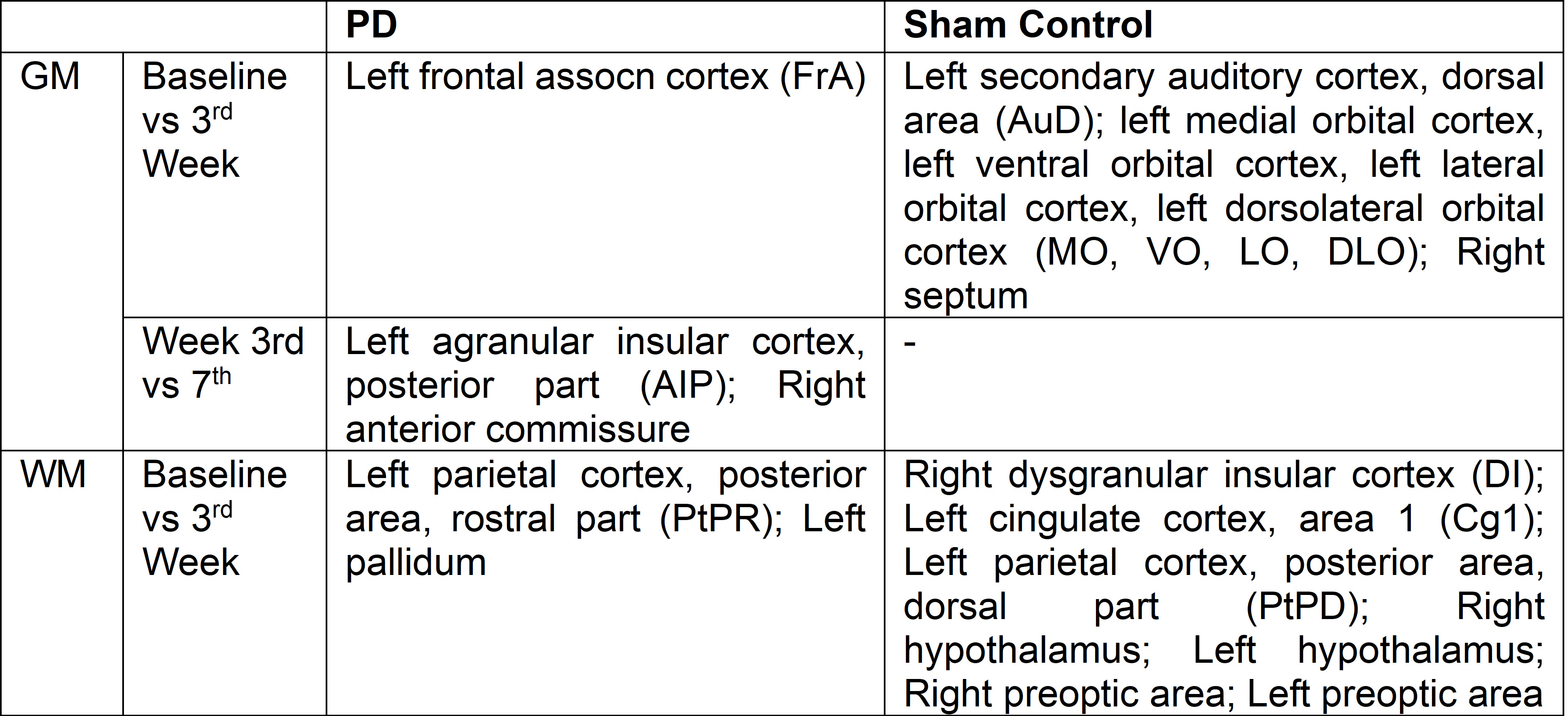
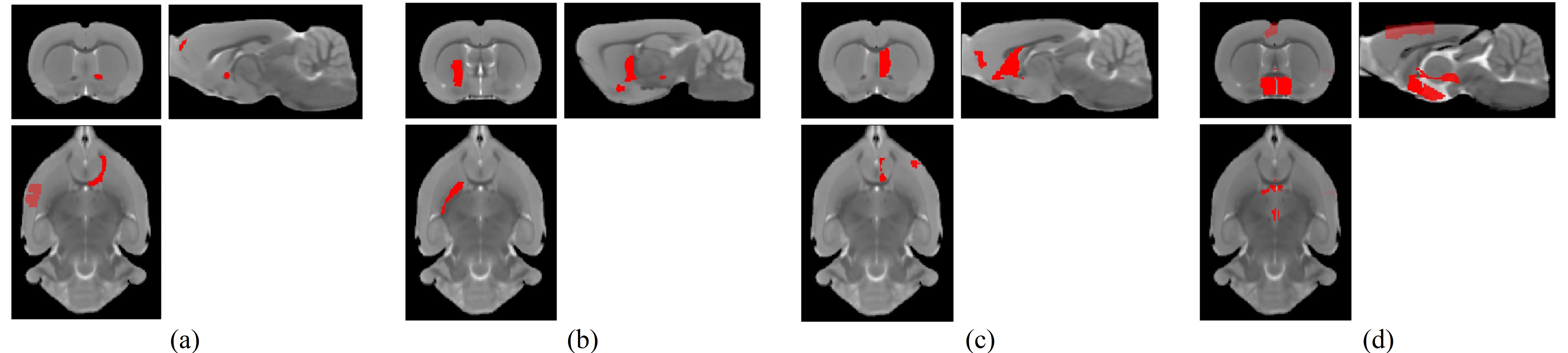
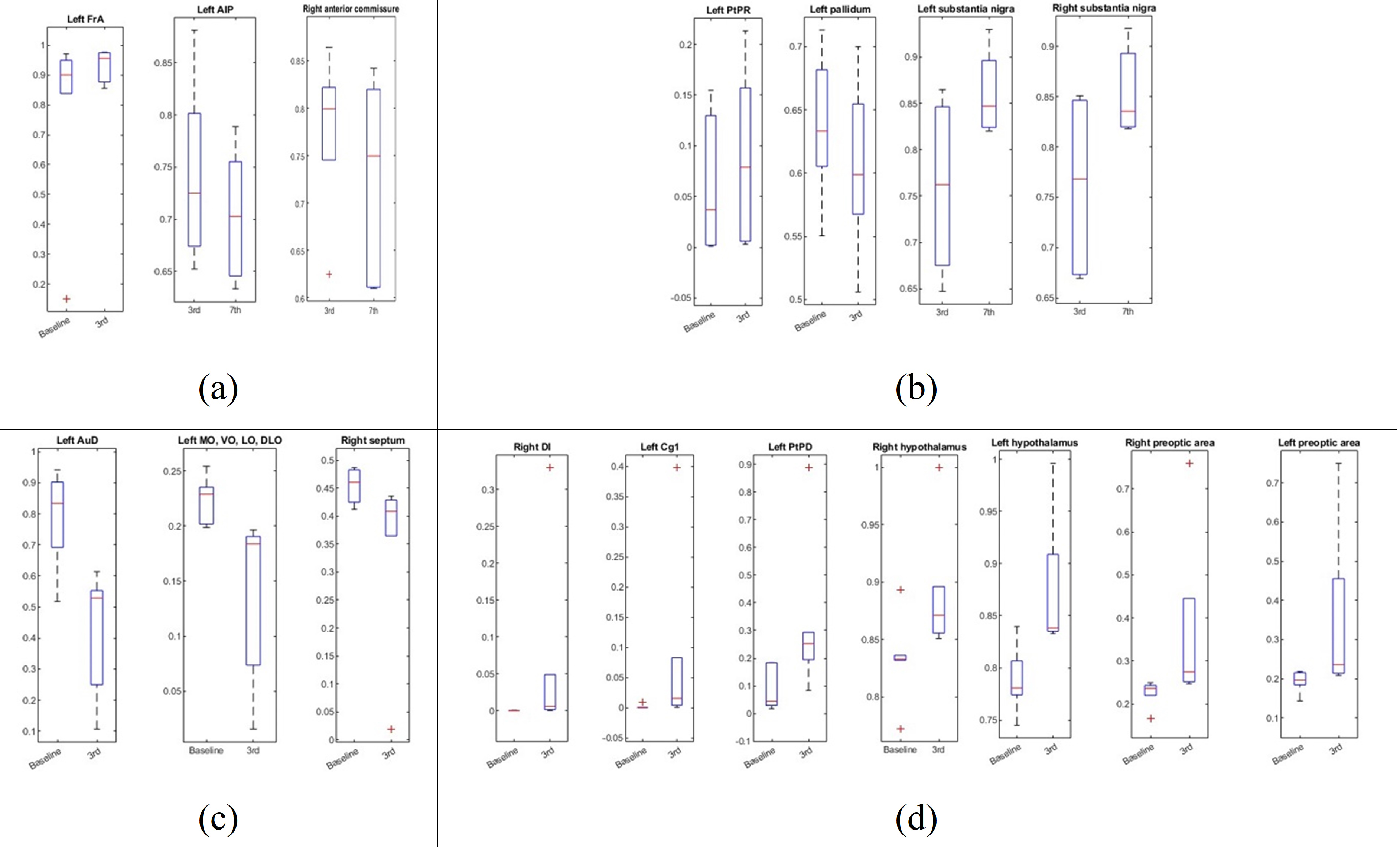
Figure 2. boxplots showing volume changes in (a) GM and (b) WM in PD, (c) GM and (d) WM in Sham Control
(FrA: frontal assocn cortex; AuD: secondary auditory cortex, dorsal area; MO: medial orbital cortex, VO: ventral orbital cortex, LO: lateral orbital cortex, DLO: dorsolateral orbital cortex; AIP: agranular insular cortex, posterior part; PtPR: parietal cortex, posterior area, rostral part; DI, Cg1: cingulate cortex, area 1; PtPD: parietal cortex, posterior area, dorsal part;)