4117
Predicting Pain caused by the Intervertebral Disc Degeneration in Large Animal Model Using CEST MRI Data1Bioengineering, University of California, Los Angeles (UCLA), Los Angeles, CA, United States, 2Biomedical Imaging Research Institute, Cedars Sinai Medical Center, Los Angeles, CA, United States, 3Emergency Medicine, Emory University, Atlanta, GA, United States, 4Biomedical Sciences, Cedars Sinai Medical Center, Los Angeles, CA, United States
Synopsis
Keywords: Biomarkers, CEST & MT
Motivation: Use MRI CEST data to predict lower back pain scores in porcine model.
Goal(s): To use a porcine model to correlate MRI-based biomarkers with backpain associated with IVD degeneration.
Approach: IVD injury was induced in the lower three lumbar discs while keeping the upper two intact. MRI scans were performed every 4 weeks upto 16 weeks post injury. Pearson Correlations were used for data analysis.
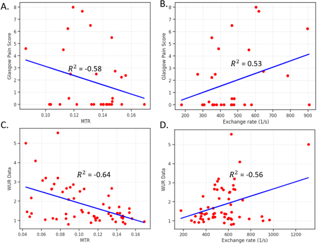
Results: MTR and Exchange rate signal clearly separate the injured and the healthy disks. There is a positive correlation between higher exchange rate signal and higher back pain while a negative correlation between MTR and pain scores.
Impact: The study bridges the gap between small animal models and human clinical studies by employing a clinically relevant large animal model. The development of MRI-based pain assessment biomarkers is a critical step toward advancing our understanding of lower back pain.
Introduction
Lower back pain is a pervasive health issue, with intervertebral disc (IVD) degeneration accounting for 26%-42% of the cases [1]. While small animal models have been valuable in studying back pain, their translational relevance to human disease remains limited. This study seeks to address this gap by employing a porcine model to correlate MRI-based biomarkers with backpain associated with IVD degeneration.Methods
IVD degeneration was induced in Yucatan minipigs (n=6) through controlled injury to the L3-L4, L4-L5, and L5-L6 intervertebral discs using a 16G needle stick injury (Fig 1). Biobehavioral assessments, including the Wind-up ratio (WUR) [2] and Glasgow pain scale [3] were conducted bi-weekly, starting at baseline and continuing until the pigs' sacrifice. Additionally, MRI imaging was performed every four weeks to monitor the development of IVD degeneration. For MRI data acquisition, conventional T1, T2, and Apparent Diffusion Coefficient (ADC) sequences were used, alongside Chemical Exchange Saturation Transfer (CEST) sequences to obtain Magnetization Transfer Ratio (MTR) and exchange rate maps. Multipool fitting analysis was used to extract the glycosaminoglycan (GAG) content (offset: +1.0 ppm). Exchange rate maps were generated using omega plot analysis for each pixel in the region of interest. Detailed scan parameters and analysis references can be found in our prior work [4].Results
Injured discs at week 4 exhibited a significant decrease in MTR (Fig 2A) and exchange rate values (Fig 2B), suggesting pronounced IVD degeneration. These trends continued at weeks 8, 12, and 16, with CEST signals (MTR and exchange rate) significantly differing in the injured region versus healthy control discs. Concurrently, both T1 (Fig 3A) and T2 (Fig 3B) demonstrated significant decreases starting at week 4. No significant changes occurred in the ADC values (Fig 3C).Linear regression analysis showed that MTR values (Fig 4A) exhibited a negative correlation, while the exchange rate demonstrated a positive correlation with Glasgow pain scores (Fig 4B). These trends were consistently observed with the Wind-up ratio, reinforcing the association between CEST values and pain assessment. (Fig 4C, 4D)
Discussion
This study underscores the potential of MRI-based biomarkers in quantifying pain within a porcine model of IVD degeneration. The observed reductions in MTR and exchange rate, indicative of decreased glycosaminoglycan content and increased tissue acidity, respectively, are consistent with pathological changes seen in back pain. Furthermore, the correlations between exchange rate, MTR and pain metrics suggests a novel avenue for understanding the pathophysiology of pain in IVD degeneration.The study bridges the gap between small animal models and human clinical studies by employing a more clinically relevant large animal model. The development of MRI-based pain assessment biomarkers in this model is a critical step toward advancing our understanding and treatment of lower back pain. An integrated regression model integrating all the imaging biomarkers may further improve the correlations with pain scores.
Conclusion
Establishing a pain assessment system in a large animal model may accelerate the translation of promising pre-clinical treatments to the clinic. The potential of MRI-based biomarkers, particularly MTR and exchange rate, to assess pain stemming from IVD degeneration holds great promise for improving the understanding and management of lower back pain.Acknowledgements
No acknowledgement found.References
[1] Peng B.-G. Pathophysiology, Diagnosis, and Treatment of Discogenic Low Back Pain. World J. Orthop. 2013;4:42–52.
[2] Zhu, G.C., Böttger, K., Slater, H., Cook, C., Farrell, S.F., Hailey, L., Tampin, B., Schmid, A.B. (2019). Concurrent validity of a low-cost and time-efficient clinical sensory test battery to evaluate somatosensory dysfunction. European Journal of Pain, 23(1), 135-144.
[3] Reid, J., Nolan, A.M., Hughes, J.M.L., Lascelles, D., Pawson, P., Scott, E.M. (2007). Development of the short-form Glasgow Composite Measure Pain Scale (CMPS-SF) and derivation of an analgesic intervention score. Animal Welfare, 16(S), 97-104. ISSN 0962-7286. Published by the Universities Federation for Animal Welfare.
[4] Zhou, Z., Bez, M., Tawackoli, W., Giaconi, J., Sheyn, D., de Mel, S., Maya, M.M., Pressman, B.D., Gazit, Z., Pelled, G., Gazit, D., Li, D. (2016). Quantitative Chemical Exchange Saturation Transfer MRI of Intervertebral Disc in a Porcine Model. Published in "Magnetic Resonance in Medicine," 76(6), 1677–1683.
Figures
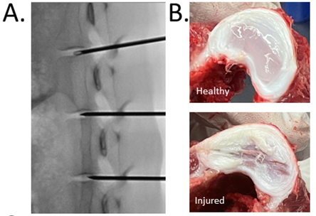
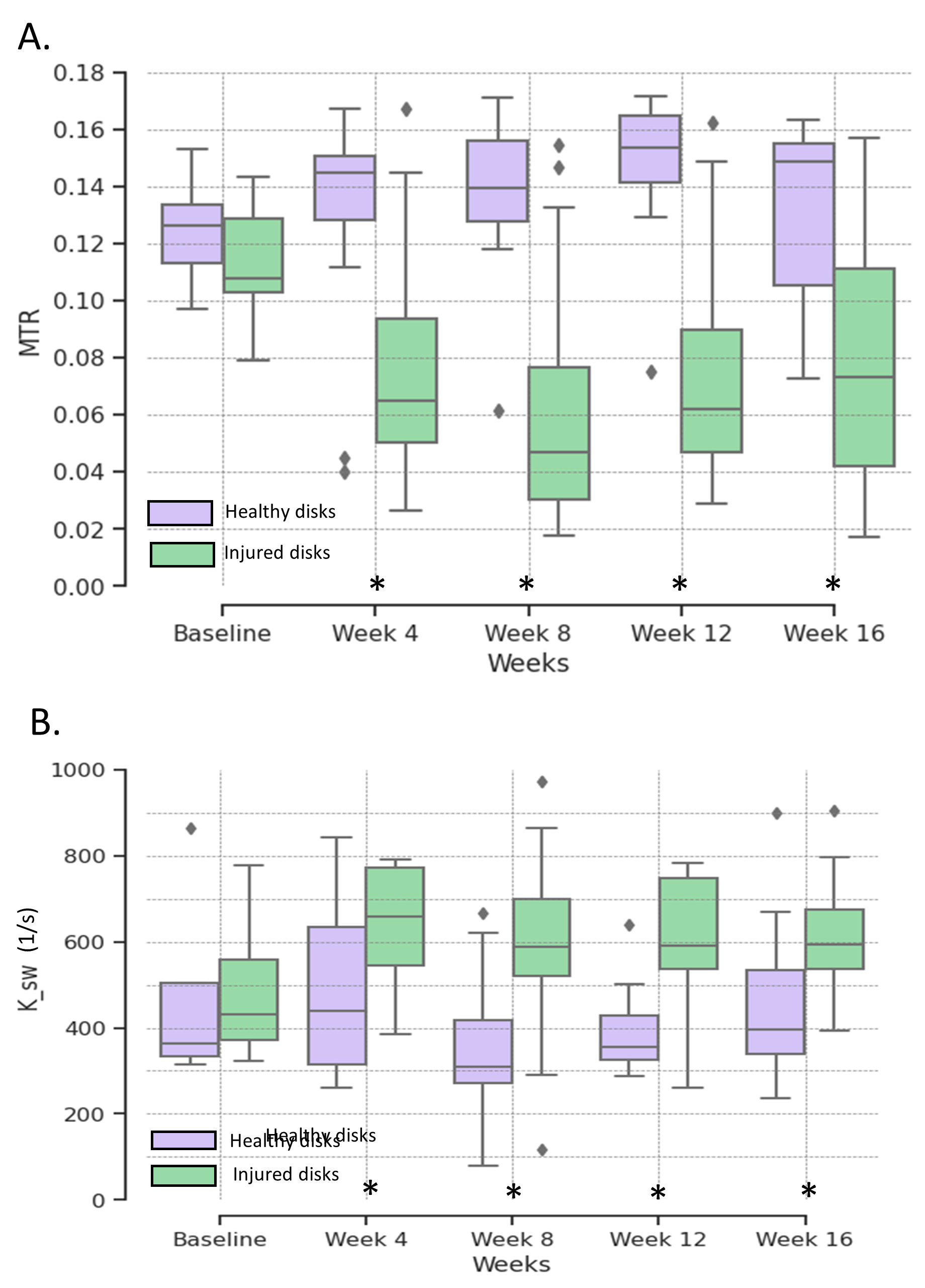
Figure 2: CEST Analysis. (A) Change in the Magnetization Transfer Ratio (MTR; proxy to GAG content) signal for injured and healthy disks. (B) Change in the Exchange rate (proxy to pH) signal for healthy and injured disks from baseline (before injury) to Week 16 after injury.
* Statistically significant difference between injured and healthy disks’ signal
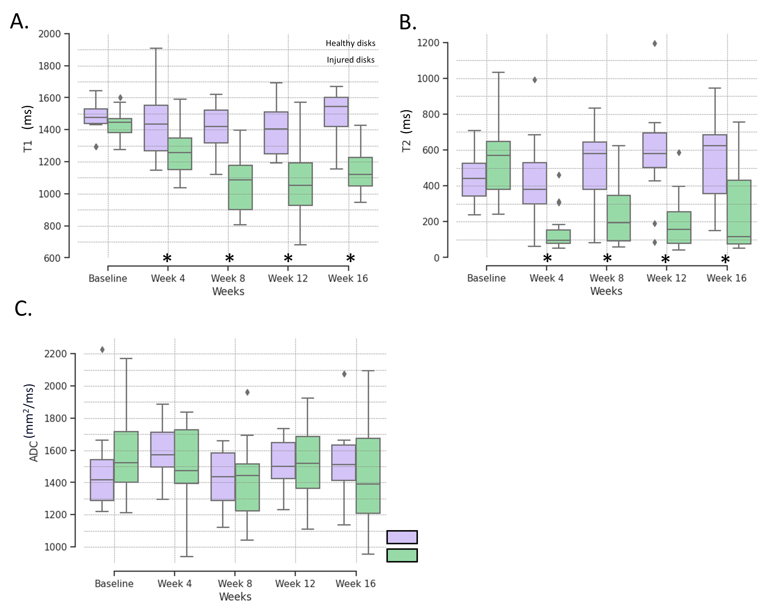
Figure 3: MRI Analysis. (A) Change in the T1 signal (B) T2 signal (C) Apparent Diffusion Coefficient (ADC) for injured and healthy disks from baseline (before injury) to Week 16. T1 and T2 signals separate the injured from the healthy disks after the injury.
* Statistically significant difference between injured and healthy disks’ signal