4115
Assessment of disease progression in the zQ175 Heterozygous Huntington’s mouse model1Calico Life Sciences, South San Francisco, CA, United States
Synopsis
Keywords: Biology, Models, Methods, Translational Studies, Huntington's
Motivation: Suitable mouse models of Huntington's disease are crucial for the evaluation and translation of potential new treatments.
Goal(s): We studied the zQ175 heterozygous model for Huntington's disease in comparison to wildtype controls.
Approach: We compared brain MRI and behavioral readouts over a large part of the lifespan to characterize and quantify phenotypes.
Results: The model only developed a mild brain atrophy phenotype, part of the size difference was shown to be due to reduced growth rather than atrophy.
Impact: This study can help researchers to make better informed decisions on mouse model selection for evaluation of potential treatment efficacy in translational studies of Huntington's disease.
Introduction
Huntington’s disease (HD) is an incurable genetic neurodegenerative disorder. It is caused by an expansion of CAG repeats in the Huntingtin gene, resulting in a mutated Huntingtin protein, which over time leads to aggregation, formation of inclusion bodies in the brain and consequently to neuronal damage. So far treatments options are limited, and there is an unmet need to develop drugs to treat HD. For testing of potential drug candidates, suitable animal models for HD are required. In the last years, a range of animal models of HD have been developed1. Brain MRI and behavioral readouts are key diagnostic markers to assess disease progression. Clinically, MRI can detect loss of striatal volume and white matter atrophy during the prodromal phase, years before disease onset2.Here, we studied the zQ175 heterozygous (HET) mouse model as it offers slow disease progression3. We compared MRI and behavioral readouts between zQ175 HET and wildtype control mice (WT) over a large part of their lifespan.Methods
All animal procedures were approved by the IACUC and followed AAALAC guidelines. Mice were anesthetized with isoflurane (2%) and kept at 37±0.5°C. Imaging was performed with a 9.4T Bruker BioSpec equipped with a 4 channel receive only mouse brain cryocoil. Anatomical brain images were acquired using a MSME sequence (FOV 14x14 mm2, resolution 0.055×0.55×0.5 mm3, 30 axial slices, 10 echoes with TEs from 12.5 to 192.5 ms, TR=2s, 2 averages, acquisition time 9 minutes). 14 WT control and 14 zQ175 HET mice were longitudinally studied from 3 to 18 months of age and underwent MRI and open field recordings.Images were processed in MATLAB using the antx2 toolbox4 and custom scripts. Image data were converted to Nifti format, images with different TEs were averaged, and brains were automatically skull-stripped and segmented. Brain images were registered to a customized in-vivo mouse brain atlas that was generated by averaging registered T2-weighted in-vivo brain images. Whole brain and brain area masks were transformed back into image space to measure brain structures.
Open field recordings were performed by placing mice individually into custom made acrylic boxes and recording their behavior with a video camera placed over the box for 10 minutes. Video data were analyzed using an ML-based mouse tracking model that was developed in house. Movement parameters like position, speed, distance covered, and rotations were measured, as well as time spent away from the walls.
Results
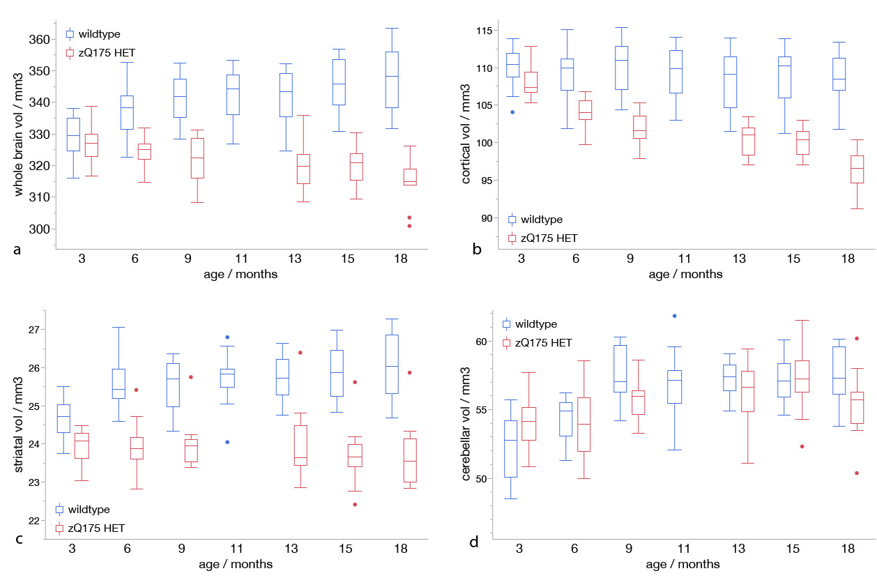
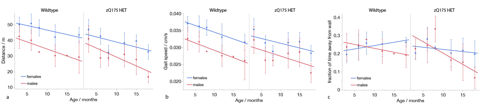
Longitudinal development of volume measurements for whole brain, cortex, striatum, and cerebellum from 3 to 18 months of age are shown in Fig. 1. Mean whole brain volume increased by 6 % in the WT mice and declined by 3.6% in the zQ175 HET mice. Mean striatal volume increased by 4% in the WT mice and was stable in the zQ175 HET mice. Cortical volume declined by 9.3% in the zQ175 HET mice and by 1.8% in the WT controls.Analysis of open field recordings showed a linearly declining covered distance during the recording as well as a declining gait speed (Fig. 2). Differences between male and female mice were observed, but not between WT and zQ175 HET mice. Male mice showed a gradual reduction in time spent in the center of the open field box, away from the walls, where the females increased time spent in the center over the duration of the study.
Discussion and Conclusion
Our results show significant differences in the development of whole brain, cortical and striatal volumes of the zQ175 HET compared to wildtype mice. Cerebellar volumes did not show a measurable difference between groups. Cortical volume in the zQ175 HET mice showed a reduction in size, but the difference of whole brain and striatal volumes between disease model and wildtype seems to be mostly reduced growth of the brain compared to WT rather than atrophy.Open field recordings did show a decline of measured distances and walking speed with increasing age of the mice from 3 to 18 months. In both groups, female mice walked further and faster, and spent slightly more time exploring the center with increasing age. We did observe some changes, but no strong reduction in exploratory behavior, indicating that repetition frequency was sufficiently low to avoid training effects. However, there was no detectable onset of a behavioral phenotype in the Huntington’s model.
In contrast to previously published results3, this indicates that the zQ175 HET model might be too mild to develop a sufficient phenotype that would allow for clear detection of potential treatment effects of drugs targeting HD.
Acknowledgements
We thank Stefan Koch and Philipp Boehm-Sturm for helpful support with the installation of ANTx2.References
1 Mahmoud A. Pouladi, A. Jennifer Morton & Michael R. Hayden. Nature Reviews Neuroscience 14, p. 708–721 (2013)
2 Scahill R. et. al., Lancet Neurol. 2020 Jun;19(6):502-512
3 Heikkinen et. al., PLOS One, Volume 7 (12), e50717 (2012)
4 Koch et al., J Cereb Blood Flow Metab, 2019 Feb; 39(2):313-323.
Figures