4113
Progressive brain volume alterations in the zQ175DN mouse model of Huntington’s Disease using in vivo MRI1Bio-Imaging Lab, University of Antwerp, Wilrijk, Belgium, 2µNeuro Center for Excellence, University of Antwerp, Wilrijk, Belgium, 3CHDI Foundation Inc, Princeton, NJ, United States
Synopsis
Keywords: Small Animals, Genetic Diseases, Volumetric
Motivation: Robust translational imaging biomarkers are needed to facilitate the development of disease-modifying treatments for Huntington’s disease (HD), a rare inherited neurodegenerative disease.
Goal(s): To determine the use of structural atrophy determined with MRI as a robust translational biomarker for testing therapeutics in mouse models prior to people with HD.
Approach: In this work, we used semi-automatic delineations and tensor-based morphometry (TBM) of 3D high-resolution anatomical MR images to assess structural anomalies in the knock-in zQ175DN model of HD at different ages.
Results: We detected progressive volumetric decrease in the zQ175DN mouse model, offering a powerful translational biomarker for the assessment of disease-modifying therapies.
Impact: We report the first brain-wide structural study of zQ175DN mice using in vivo MRI. Our work supports structural atrophy as a robust translational biomarker for testing therapeutics in mouse models prior to people with HD.
Introduction
Huntington’s disease (HD) is an inherited neurodegenerative disease caused by a CAG repeat expansion of the Huntingtin gene (Htt)1. Towards the development of disease-modifying treatments, robust translational imaging biomarkers are needed. The main pathological hallmark of HD, basal ganglia atrophy, was recently incorporated as first disease stage in the new HD staging system2. Thus, in this study, we assessed structural anomalies at different ages in a knock-in model of HD using semi-automatic delineations and tensor-based morphometry (TBM) of 3D high-resolution anatomical MR images.Methods
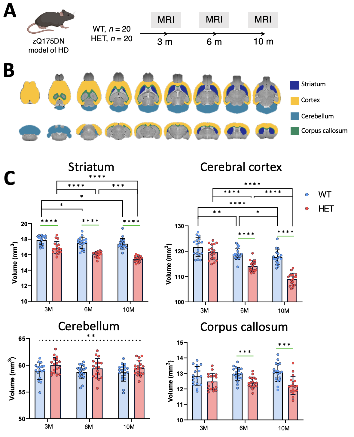
MRI data from 20 heterozygous (HET) zQ175DN and 20 WT littermates were acquired at 3, 6 and 10 months of age (M) (Fig. 1A) under 1-2% isoflurane with a 9.4T BioSpec scanner and CryoProbe (Bruker, Germany). 3D-TurboRARE images (TE/TR 51/1800ms) with a resolution of (78x78x78)µm3 were acquired. Whole brain and regions-of-interest (ROI; striatum, cortex, cerebellum and corpus callosum) volumes were extracted using manual delineations (Fig. 1B). Debiased 3D data were affine and non-linearly transformed to a 3M WT template to create a minimal deformation target (MDT). Next, the Jacobian determinants of the non-linear transformation to the MDT were log-transformed and statistically compared in a two-way ANOVA (genotype, age, and genotype x age) with FDR (ROI-analysis) or TFCE-FWE (voxel-analysis) correction.Results
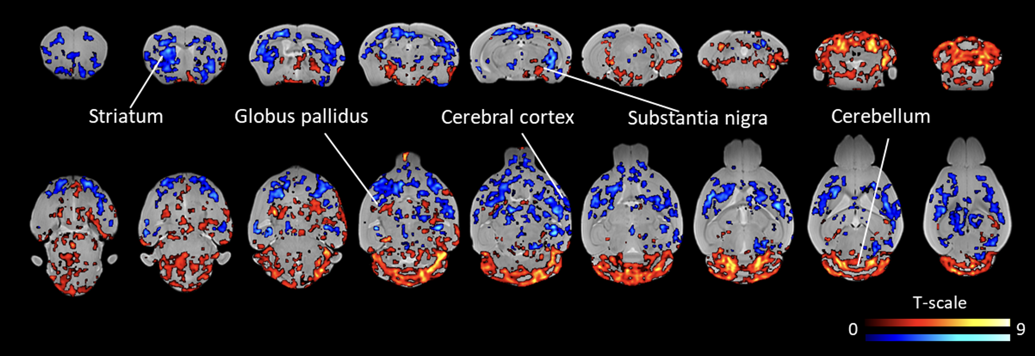
For the manually delineated volumes, we found that HET mice displayed a significant decrease in brain volume as of 6M (p<0.0001). The striatum was the first structure to display volumetric deficits already at 3M (p<0.0001), whereas progressive volume changes in cerebral cortex and corpus callosum were observed as of 6M (p<0.0001) of age onward (Fig. 1C). Interestingly, the cerebellum, a structure known to be less affected in HD, did not show a volumetric deficit (Fig. 1C). TBM analysis revealed local deformations, relative to the MDT after initial affine scaling, within the striatum, cerebral cortex, substantia nigra, globus pallidus and cerebellum of HET zQ175DN mice (Fig. 2A). ROI-based analysis of these areas identified progressive significant local structural susceptibility (p<0.05-p<0.0001).Conclusion
We demonstrated the sensitivity of MRI to detect subtle progression of structural anomalies in HET zQ175DN mice, even though this mouse model features neurodegeneration without overt neuronal loss3. These results indicate that volumetric decrease can be detected in the zQ175DN mouse model, offering a powerful translational biomarker for the assessment of disease-modifying therapies.Acknowledgements
This work was funded by CHDI Foundation, Inc., a nonprofit biomedical research organization. The computational resources and services were provided by the HPC core facility CalcUA, the VSC, funded by the Hercules Foundation, and the Flemish Government department EWI. The Bruker Biospec 9.4T system (Bruker, Ettlingen, Germany) was upgraded to AVANCE-NEO through Hercules foundation funding (Belgium, grant agreement I007120N) under the promotorship of AVDL.References
1. Bates, G. P. et al. Huntington disease. Nat Rev Dis Primers 1, 1–21 (2015).
2. Tabrizi, S. J. et al. A biological classification of Huntington’s disease: the Integrated Staging System. The Lancet. Neurology vol. 21 632–644 Preprint at https://doi.org/10.1016/S1474-4422(22)00120-X (2022).
3. Deng, Y., Wang, H., Joni, M., Sekhri, R. & Reiner, A. Progression of basal ganglia pathology in heterozygous Q175 knock-in Huntington’s disease mice. Journal of Comparative Neurology 529, 1327–1371 (2021).
Figures