4104
Discrepancy in the distribution of H217O and Gd-DTPA during and after intrathecal infusion in chronic unilateral hypoperfusion model mice.1National Institutes for Quantum Science and Technology, Chiba, Japan, 2Department of Diagnostic and Interventional Radiology, Hokkaido University, Sapporo, Japan
Synopsis
Keywords: Neurofluids, Neurofluids
Motivation: It is desirable to accurately evaluate water movement within the brain so that it can be compared with neurofluid flow observed with other tracers.
Goal(s): The aim of this study was to evaluate the motion of neurofluids in a mouse model of chronic unilateral hypoperfusion using H217O and Gd-DTPA as MRI tracers.
Approach: Dynamic T2WI and T1WI were performed during and after intrathecal infusion of H217O and Gd-DTPA, respectively.
Results: There was a clear difference in the distributions of H217O and Gd-DTPA after intrathecal injection. This suggests that different mechanisms are involved in the transport of water and other molecules in the brain.
Impact: The difference in distribution of the tracers in the UCCAO model suggests that this method may be useful for investigating the pathological mechanism of various brain diseases.
Intoroduction
Chronic cerebral hypoperfusion is a known cause of cognitive decline, but the pathomechanisms remain unclear. In recent years, the motion of neurofluids has attracted attention. For example, the cerebral spinal fluid (CSF) circulatory system is involved in the excretion of waste products from the brain, and disturbances in the excretory pathways may be involved in the pathogenesis of chronic cerebral hypoperfusion 1. The aim of this study was to evaluate the motion of neurofluids in a mouse model of chronic hypoperfusion with H217O and Gd-DTPA as MRI tracers.Methods
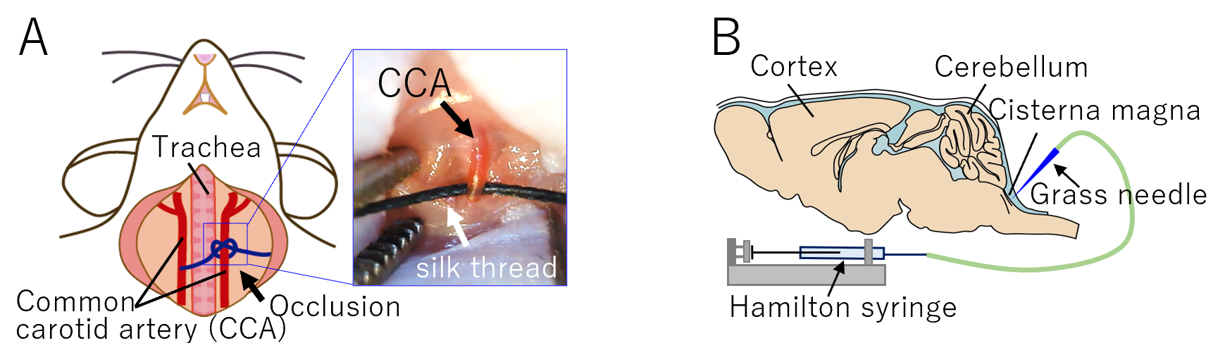
A mouse model of mild unilateral chronic cerebral ischemia was made by permanent unilateral common carotid artery occlusion (UCCAO, Fig. 1A) 2. One week after UCCAO surgery mice were anaesthetized with 2% isoflurane and positioned in a stereotaxic frame. A 50 µl glass Hamilton syringe loaded with tracer (90% H217O saline (n=6) or Gd-DTPA (50mmol/L, n=4)), was placed in a micro infusion pump and attached to a glass needle by PE10 tubing. After the dura mater overlaying the cisterna magna was exposed, the glass needle was advanced 1 mm into the cisternal space and anchored using superglue and fast setting resin (Fig. 1B). MRI was performed using a 7.0-T MRI scanner (Bruker Biospin, Germany) with a cryoprobe designed for the murine brain. The scanning protocol for all studies consisted of 5 min of baseline scans, followed by intrathecal infusion of the tracer (30 μl at 0.6 μl / min) over a period of 50 min. MRI data were continually acquired throughout and after intrathecal infusion for a total time of 150 min (112 min for one mouse due to machine trouble). The T2-shortening effect of H217O was exploited to image the distribution of H217O 3. T2-weighted images were acquired with a fast spin-echo sequence in the axial plane (13 slices, slice thickness = 0.9 mm, slice gap = 0.1 mm, TR/TE = 4,000 ms/120 ms, FA = 90 degree, echo train = 12, matrix size = 192 × 128, FOV = 19.2 × 12.8 mm2, repetitions = 388, 1 frame = 24 sec). To visualize the distribution of Gd-DTPA, 2D T1-weighted Gradient Echo images were acquired in the same slice location as above (TR/TE = 180 ms/3.539 ms, flip angle = 45 degree, matrix size = 192 × 128, FOV = 19.2 × 12.8 mm, repetitions = 404, 1 frame = 23 sec 40 msec). The change in transverse relaxation (ΔR2) and %change in the signal were used to evaluate the distribution of H217O and Gd-DTPA, respectively. Both quantities were calculated using the mean of the baseline images acquired before infusion and the images subsequently obtained after infusion began.Results
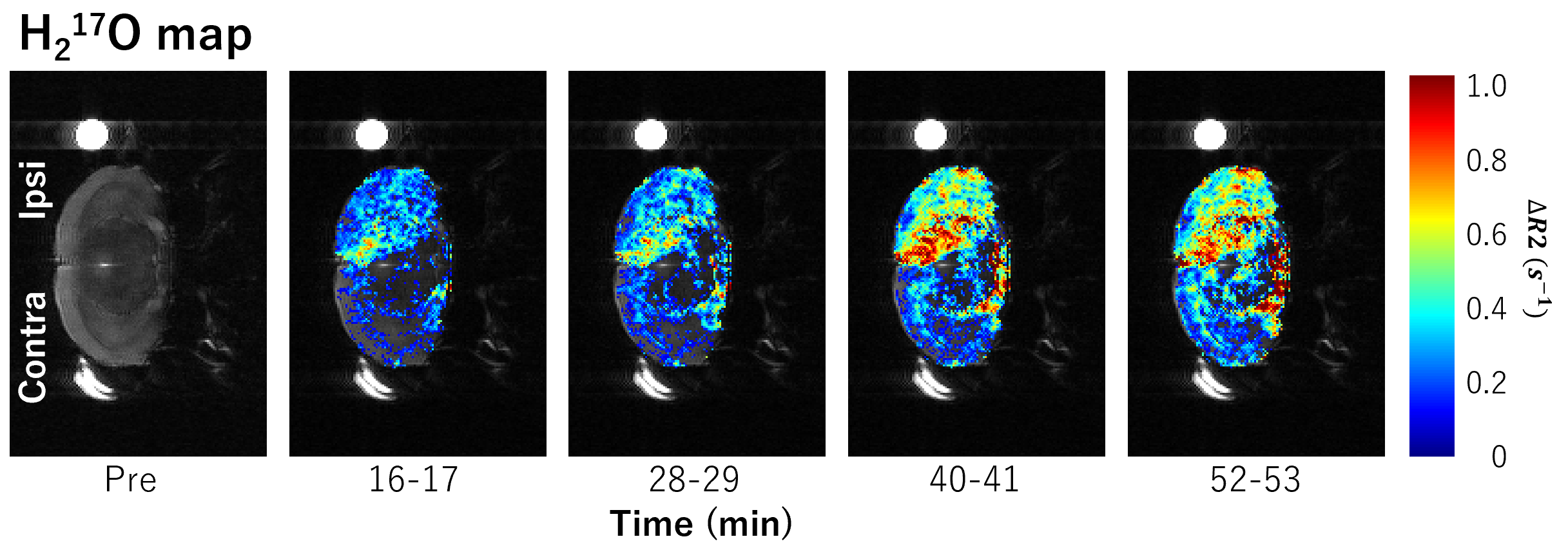
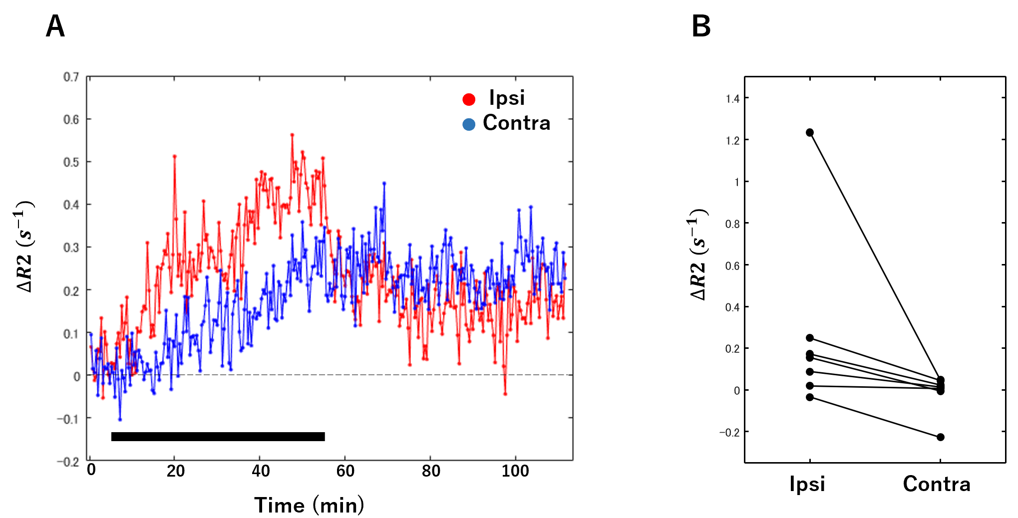
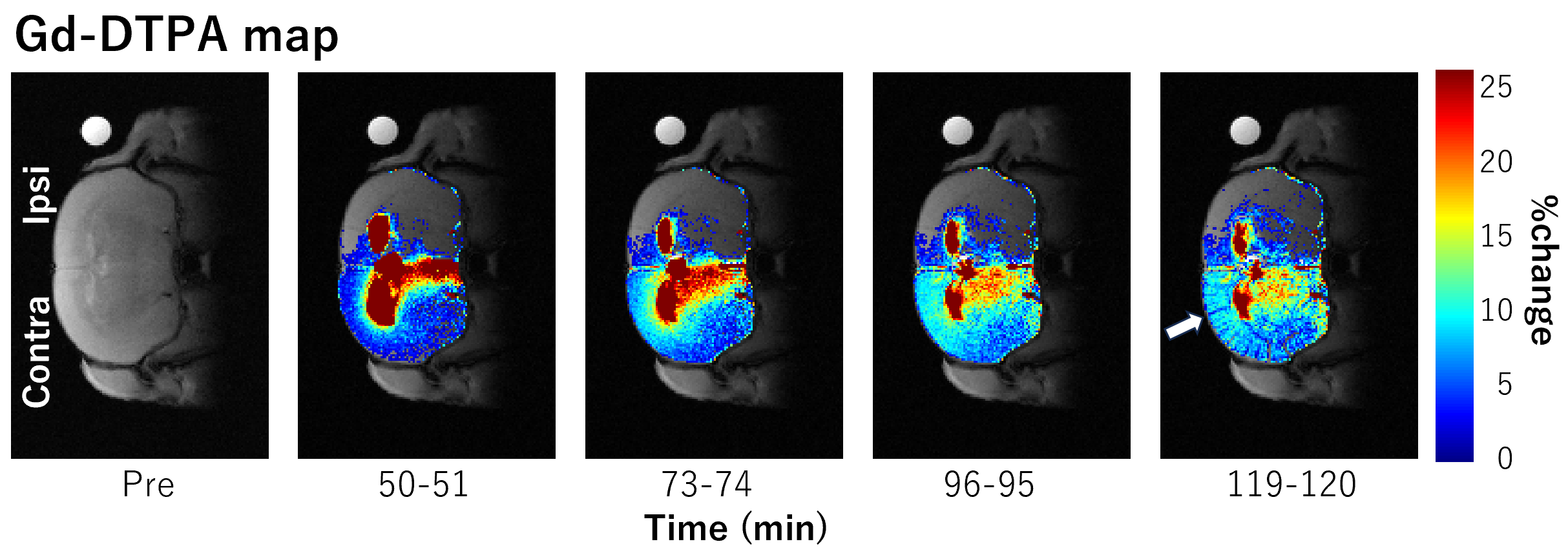
ΔR2 increased within the brain parenchyma more on the ipsilateral side during infusion of H217O into the cisterna magna (Fig. 2). The ΔR2 in the ipsilateral cortex was significantly larger than for the contralateral side (Fig. 3, p=0.16, Wilcoxon rank test). For three animals, the %change in the signal on the contralateral side during and after Gd-DTPA infusion was large compared to that on the ipsilateral side (Fig. 4). After the peak of the signal change, signal loss was noticeable in areas along the cortical arteries and veins in the brain cortex (arrow in Fig.4). For one other animal, the signal change was similar on both sides even though arterial spin-labeling showed that the UCCAO had been successful.Discussion
The significant increase in the ΔR2 during H217O infusion (Fig. 3A) on the ipsilateral side may be due to an increase of water exchange via AQP4 4. It is interesting that H217O and Gd-DTPA had different distributions in the brain parenchyma. Since arterial pulsation was weak on the ipsilateral side, the driving force in the perivascular space may have also been weak. It is possible that a decline in CBF is simply causing a decline in the neurofluid flow 5. Also, the arterial dilation induced by hypoperfusion and/or AQP4-related swelling of astrocyte endfeet may narrow the pathway for Gd-DTPA 4, 6.Conclusion
There was a clear difference in the distributions of H217O and Gd-DTPA after intrathecal injection in UCCAO model mice. This suggests that different mechanisms are involved in the transport of water and other molecules in the brain. Furthermore, the difference in distribution in the UCCAO model suggests that this method may be useful for investigating the pathological mechanism of various brain diseases.Acknowledgements
We thank Dr Sayaka Shibata for her support. This work was supported by a grant, Public/Private R&D Investment Strategic Expansion PrograM (PRISM) from Cabinet Office, and by a grant from the Ministry of Education, Culture, Sports, Science and Technology (MEXT), Japanese Government.References
1. Rajeev V, Chai YL, Poh L, et al. Chronic cerebral hypoperfusion: a critical feature in unravelling the etiology of vascular cognitive impairment. Acta Neuropathol Commun. 2023; 11:93.
2. Urushihata T, Takuwa H, Seki C, et al. Water Diffusion in the Brain of Chronic Hypoperfusion Model Mice: A Study Considering the Effect of Blood Flow. Magn Reson Med Sci. 2018; 17:318-324.
3. Kameda H, Kinota N, Kato D, et al. Magnetic Resonance Water Tracer Imaging Using 17O-Labeled Water. Invest Radiol. 2023.
4. Urushihata T, Takuwa H, Takahashi M, et al. Exploring cell membrane water exchange in aquaporin-4-deficient ischemic mouse brain using diffusion-weighted MRI. European Radiology Experimental. 2021; 5.
5. Taoka T, Naganawa S. Neurofluid Dynamics and the Glymphatic System: A Neuroimaging Perspective. Korean J Radiol. 2020; 21:1199-1209.
6. Stokum JA, Kurland DB, Gerzanich V, Simard JM. Mechanisms of astrocyte-mediated cerebral edema. Neurochem Res. 2015; 40:317-328.
Figures