4097
Scalable fast online-customized (FOCUS) pTx pulses for 3D TSE sequences at 7T1Siemens Healthcare GmbH, Erlangen, Germany, 2Department of Corporate Technology, Siemens AG, Munich, Germany
Synopsis
Keywords: Multiple Sclerosis, Parallel Transmit & Multiband
Motivation: 3D TSE sequences at 7T suffer from poor homogeneity, signal dropouts and local SAR limits.
Goal(s): Clinically acceptable image quality using scalable dynamic parallel transmit (pTx) pulses under SAR-constraints
Approach: Prior to the scan, a dictionary of preoptimized, symmetric pTx pulses is built. At the scanner, the best pulse for that subject is identified and serves as initialization for a then fast individual optimization constrained to maximum SAR, maximum voltage and temporal symmetry.
Results: Clinically acceptable image homogeneity is achieved with two different pulses at the expense of 0.4ms/0.9ms longer pulse duration and 220%/14% higher SAR than the commonly used 1Tx pulse.
Impact: A workflow to design customized and arbitrarily scalable pTx pulses is demonstrated, enabling homogeneous 3D TSE imaging of the head with variable flip angles at 7T. Various flip angle trains can be applied as flexibly as on 1Tx systems.
Introduction
Poor homogeneity of the B1+ fields in MRI at 7T causes regions of low signal1, which are particularly visible in TSE sequences. Dynamic parallel transmit (pTx) pulses have shown great potential to overcome these limitations and produce uniform flip angle (FA) distributions but are rarely used in a clinical setting due to their complex optimization2. For 3D TSE sequences, largely varying flip angle trains are often used while fulfilling the CPMG condition. Therefore, scalability of pTx pulses beyond the small tip angle domain is desirable. This can be achieved with universal pulses as demonstrated in 3,4. In this work, we design a non-parametrized, scalable and fast online-customized (FOCUS) pTx pulse5 for usage as excitation and refocusing pulse with arbitrary flip angle trains. Thereby we anticipate a clinically acceptable workflow regarding online calculation time, image homogeneity, T2-related blurring and SAR exposure.Methods
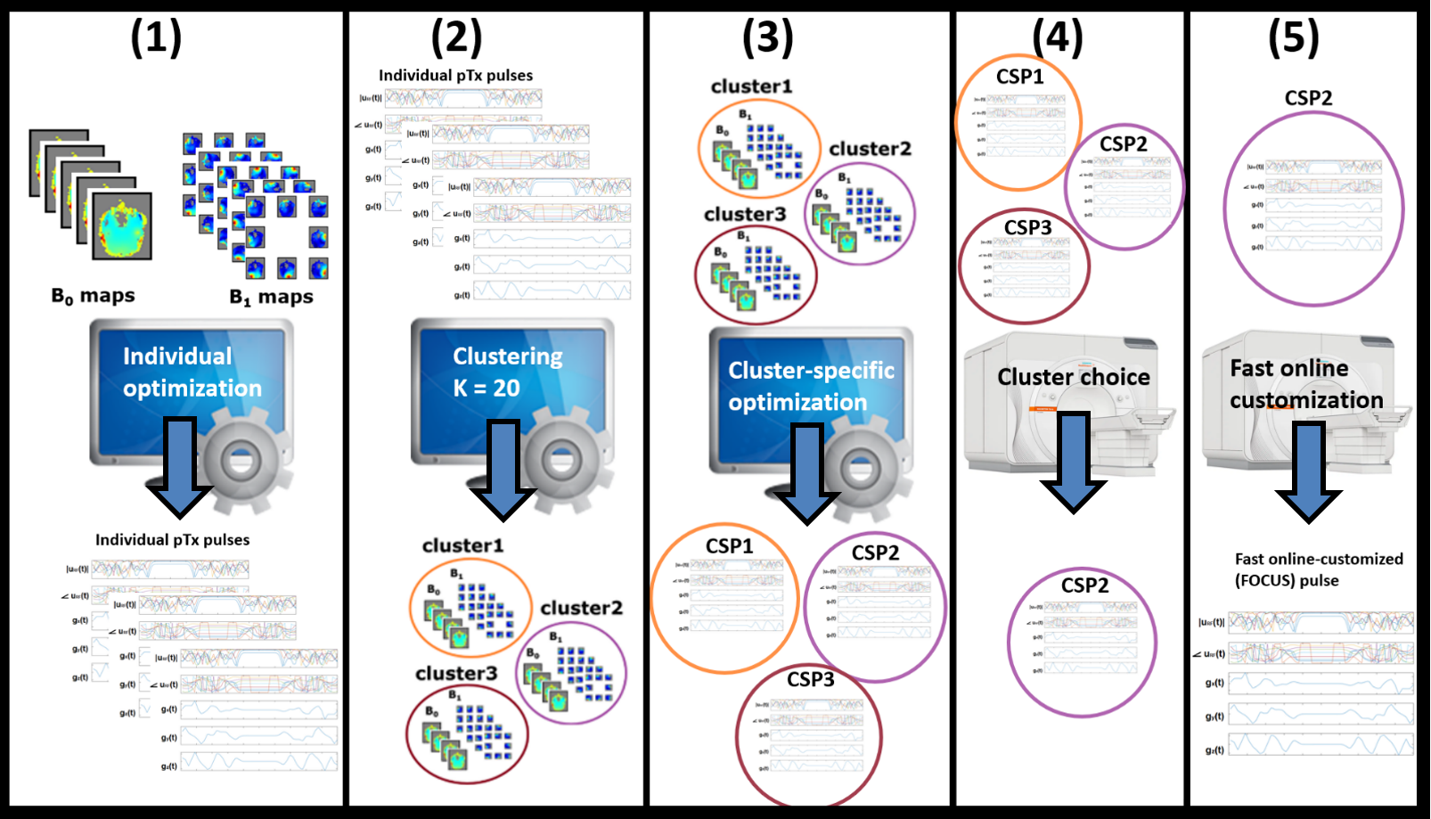
All measurements were performed on a Magnetom Terra.X (Siemens Healthcare GmbH, Erlangen) using an 8Tx/32Rx RF head coil (Nova Medical, Wilmington, USA). We designed two pTx pulse types called ‘Normal’/’LowSAR’ with 1ms/1.5ms pulse duration, maximum allowed FA-normalized specific absorption rate (SED)5 of 1.4 mJ/(kg °)/0.65 mJ/(kg °) and a sampling time of 20µs/30µs (i.e. time slots with new RF and gradient voltages). The pulses are compared to a routinely used circularly polarized (CP) pulse of 0.6ms duration. The optimization of the RF and gradient shapes was performed simultaneously with an interior-point method algorithm6. To ensure scalability up to 180°, we add linear constraints that ensure time-symmetric RF pulse shapes and antisymmetric gradient shapes as well as a maximum voltage limit of 1.05 V/° to the optimization. To reduce the calculation time, we use 8mm voxels and a rudimentary brain extraction. The local SAR supervision using 494 virtual observation points (VOPs) is incorporated into the pulse design as a SED constraint. As the high number of variables comes with many unfavorable local minima and a long calculation time for the optimization, we rely on good initialization values. Therefore, prior to the scan, we cluster 120 previously acquired B1/B0 maps based on the correlation of their respective excessively optimized pTx pulses (no time constraints, many initializations) and calculate 20 respective cluster-specific pulses (CSP)7. During the patient examination, individual B1 and B0 maps are acquired and Bloch simulations of all CSPs are performed. The individual optimization is then initialized with the CSP that yielded the lowest FA-RMSE enabling the optimizer to converge quickly in an acceptable local minimum (see Figure 1).Results
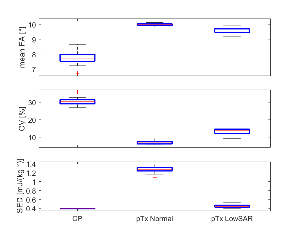
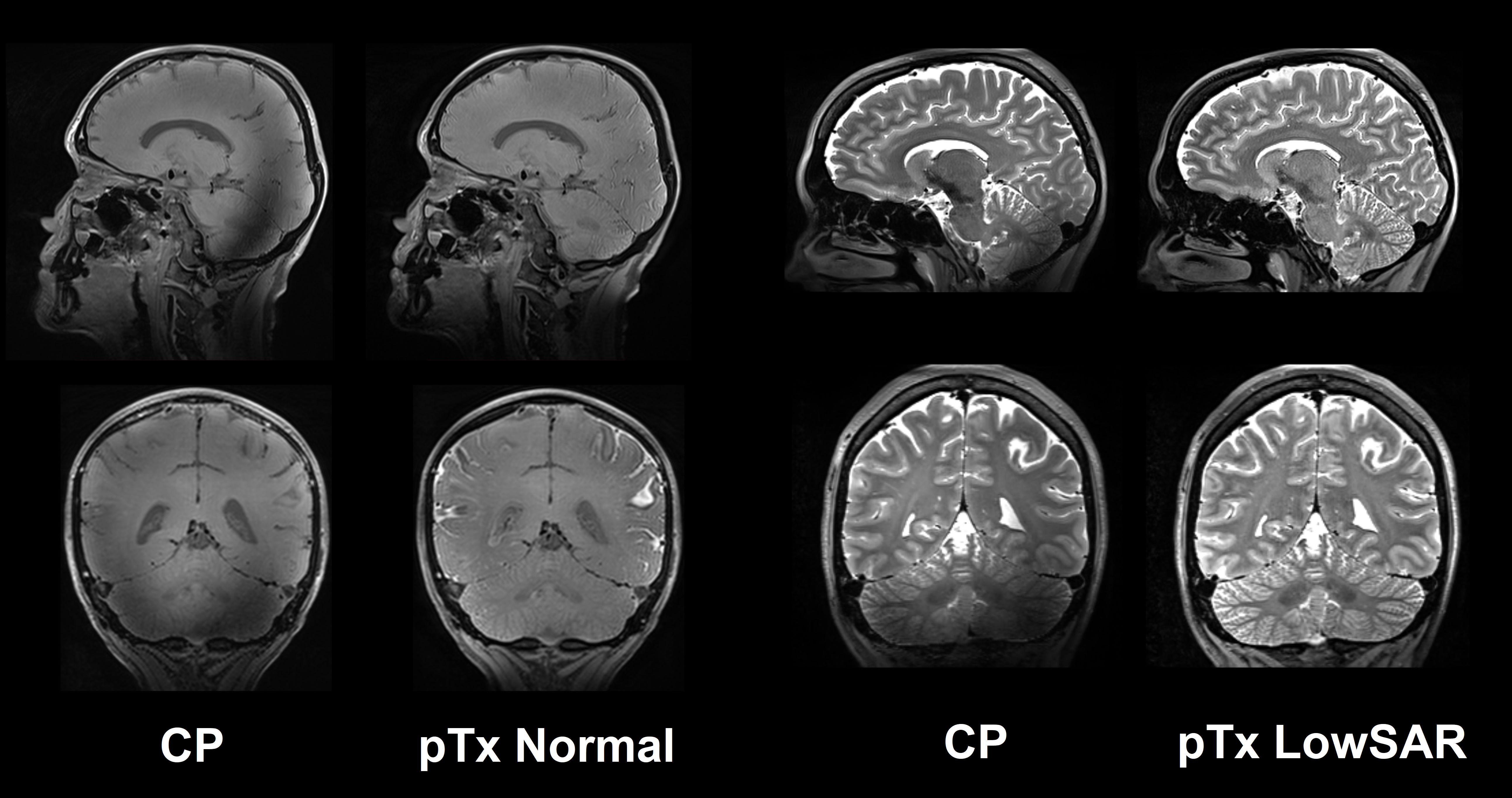
Figure 2 shows the mean FA within the brain region, its coefficient of variation (CV) and SED of the three mentioned pulses. The pTx pulses reach much better NRMSE values at the cost of longer pulse duration and higher SED values with different tradeoffs. If the pulses were played out with a k-factor based SAR supervision with 8 channel power limits for that same coil, the pTx pulse ‘Normal’ and ‘LowSAR’ would have 2.5 and 2.6 times higher SED values whereas the CP pulse would have only 1.5 times higher SED values. This difference indicates the benefits of inner-pulse SAR hopping for highly dynamic pTx pulses when using a full VOP model. Figure 3 shows two exemplary protocols with the pTx and CP pulses indicating the better homogeneity and proving that the CPMG condition was met with enforcing temporal symmetry. The online-customization time of both pTx pulses was 20 seconds.Discussion and Conclustions
The proposed pTx pulse design robustly generates homogeneous TSE images at 7T at the cost of online B1/ B0 mapping plus ~20s calculation time as well as higher SAR and longer echo spacing. Equipped with these pulses, SPACE sequences can flexibly apply arbitrary FA trains. CPMG condition was achieved through the scalability of the pulses and in combination with sufficiently conservative maximum voltage constraints per flip angle (1.05 V/°), the pulses are scalable up to 180°. Thereby we expect these pulses to be usable for magnetization preparation such as inversion, saturation and a T2-prep module8 where they may serve as a low-SAR alternative to their adiabatic counterparts. Accounting for the symmetry conditions to reduce the number of the optimization variables as well as machine learning techniques using a GPU might significantly accelerate the online-calculation. Furthermore, the pulses can be integrated to a signal-based optimization of the refocusing FA train such as the Discover method9. In such a setting, the signal of or contrast between specific tissues with their respective T1 and T2 values can be optimized while maxing out the available SAR budget.Acknowledgements
No acknowledgement found.References
1 Ladd, M. E. et al. Pros and cons of ultra-high-field MRI/MRS for human application. Prog Nucl Magn Reson Spectrosc 109, 1-50, doi:10.1016/j.pnmrs.2018.06.001 (2018).
2 Williams SN, McElhinney P, Gunamony S. Ultra-high field MRI: parallel-transmit arrays and RF pulse design. Phys Med Biol. 2023 Jan 18;68(2). doi: 10.1088/1361-6560/aca4b7. PMID: 36410046.
3 Van Damme L, Mauconduit F, Chambrion T, Boulant N, Gras V. Universal nonselective excitation and refocusing pulses with improved robustness to off-resonance for Magnetic Resonance Imaging at 7 Tesla with parallel transmission. Magn Reson Med. 2021 Feb;85(2):678-693. doi: 10.1002/mrm.28441. Epub 2020 Aug 4. PMID: 32755064.
4 Van Damme L, Mauconduit F, Chambrion T, Boulant N, Gras V. Universal nonselective excitation and refocusing pulses with improved robustness to off-resonance for Magnetic Resonance Imaging at 7 Tesla with parallel transmission. Magn Reson Med. 2021 Feb;85(2):678-693. doi: 10.1002/mrm.28441. Epub 2020 Aug 4. PMID: 32755064.
5 Herrler, J, Liebig, P, Gumbrecht, R, et al. Fast online-customized (FOCUS) parallel transmission pulses: A combination of universal pulses and individual optimization. Magn Reson Med. 2021; 85: 3140–3153. https://doi.org/10.1002/mrm.28643
6 Majewski, K. Simultaneous optimization of radio frequency and gradient waveforms with exact Hessians andslew rate constraints applied to kT-points excitation. J Magn Reson 326, 106941,doi:10.1016/j.jmr.2021.106941 (2021)
7 Herrler J, Liebig P, Majewski K, Gumbrecht R, Ritter D, Meixner C, Maier A, Doerfler A, Nagel A. Neural network-supported fast online-customized (focus) parallel transmit (ptx) pulses for slice-selective, large flip angle excitation. In 30th Annual Meeting of the International Society for Magnetic Resonance in Medicine, page 3312, London, 2022.
8 Vincent Gras, Eberhard D Pracht, Franck Mauconduit, Denis Le Bihan, Tony Stöcker, et al.. Robust non-adiabatic T 2 -preparation using universal parallel- transmit k T -point pulses for 3D FLAIR imaging at 7 Tesla. 2019. ffhal-02103512
9 Malik SJ, Beqiri A, Padormo F, Hajnal JV. Direct signal control of the steady-state response of 3D-FSE sequences. Magn Reson Med. 2015 Mar;73(3):951-63. doi: 10.1002/mrm.25192. Epub 2014 Mar 17. PMID: 24639096; PMCID: PMC7614097.
Figures