4094
Calibration-free Multidimensional Universal Refocusing Pulse Design for 3D Reduced Field-of-View Prostate Imaging1Department of Electrical Engineering and Computer Science, University of Michigan, Ann Arbor, MI, United States, 2Department of Radiology, University of Michigan, Ann Arbor, MI, United States, 3Department of Biomedical Engineering, University of Michigan, Ann Arbor, MI, United States
Synopsis
Keywords: RF Pulse Design & Fields, RF Pulse Design & Fields, Reduced FOV; 3D EPI;
Motivation: Universal pulses have been shown to be robust to B1+ inhomogeneity for brain imaging at 7T, without time-consuming online design. A similar approach may be useful for designing multidimensional RF pulses for the 3D prostate imaging at 3T.
Goal(s): To design a universal multidimensional refocusing pulse and demonstrate its potential use for reduced field-of-view imaging in the prostate.
Approach: A three-dimensional universal refocusing pulse was designed using 6 subjects and validated in 12 subjects and in vivo prostate imaging at 3T.
Results: The proposed 3D universal refocusing pulse achieved similar performance on seen and unseen subjects. We successfully acquired reduced field-of-view prostate images.
Impact: Our simulation and in vivo results demonstrated the potential to design one universal 3D refocusing pulses for most subjects in prostate imaging.
Introduction
The goal of this study is to design calibration-free multidimensional radiofrequency (RF) pulses using the concept of universal pulses [1] for reduced field-of-view (rFOV) prostate imaging. The use of multidimensional RF pulses in rFOV imaging could shorten the scan time or improve the spatial resolution. Previously, multidimensional RF pulses have been designed using an auto-differentiation method and spin-domain based optimization [2,3]. However, the performance of multidimensional pulses can be affected by B0 and B1+ inhomogeneities, thus requiring a tailored design using individually measured B0 and B1+ for each subject [4,5]. These tailored approaches are time-consuming, which prevents the further adoption of rFOV imaging in a clinical setting. The universal pulse concept has shown its robustness to B1+ inhomogeneities for parallel transmission at 7T. Similar to the brain imaging at 7T, prostate imaging at 3T is also strongly affected by the inhomogeneous B0 and B1+. In this work, we designed three-dimensional (3D) spatially selective refocusing pulses by combining the universal pulse concept with our spin-domain RF optimization [2]. We validated the performance of newly designed 3D RF pulses in numerical simulations and in vivo prostate imaging at 3T.Methods
RF pulse design: The 3D spatially selective universal pulse design problem in the spin-domain was formulated as the following optimization problem$$\arg\min_{\boldsymbol{b},\boldsymbol{g}}L=\sum_{s=1}^{S}\left(\sum_{i=1}^{M}w_i |\beta_{i}^2(\boldsymbol{b},\boldsymbol{g};s)-\beta_{D,i}^2|\right)^p,\quad\text{subject to}\quad\text{hardware limits},$$where $$$\boldsymbol{b}$$$ is the RF waveform, $$$\boldsymbol{g}$$$ is gradient waveforms, $$$\beta^2_{i}$$$ and $$$\beta^2_{D,i}=-1$$$ are simulated and desired spin-domain parameters representing the refocusing efficiency, $$$w_i$$$ is the weight for each spin, $$$M$$$ is the total number of spins, and $$$S$$$ is the total number of subjects used. Instead of directly minimizing the maximum error for each subject [1], we used $$$p$$$-norm ($$$p=4$$$) to approximate the infinity norm and performed the minimization by alternatingly updating $$$\boldsymbol{b}$$$ and $$$\boldsymbol{g}$$$. We constrained the optimization within the hardware limitations (i.e., peak B1 of 0.25 mT, the maximum gradient amplitude of 50 mT/m and the maximum slew rate of 120 mT/m/ms).We implemented this optimization in PyTorch using auto-differentiation with B0 and B1+ measured from the pelvic region of 6 subjects. A refocusing pulse for a target region-of-interest (ROI) of $$$10\times10\times6cm^3$$$ with a matrix size of $$$40\times40\times25$$$ was designed to cover the prostate gland. The design was initialized by the small-tip-angle approximation [6].
Numerical Simulations: Together with the universal pulse, RF pulses without considering B0 and B1+ and tailored for specific B0 and B1+ were also designed. The performance of these RF pulses was evaluated in simulations using B0 and B1+ acquired from additional 12 subjects at 3T.
In vivo validation: The 3D spatially selective universal pulse was inserted into a 3D spin-echo stack-of-EPIs sequence by replacing the conventional refocusing pulse using Pulseq [7]. The method covered an FOV of $$$152\times152\times96mm^3$$$ with a matrix size of $$$152\times152\times32$$$ in 60 seconds. 7 subjects were scanned on a 3T scanner (Magnetom Vida, Siemens) to validate the performance of our designed RF pulse.
Results
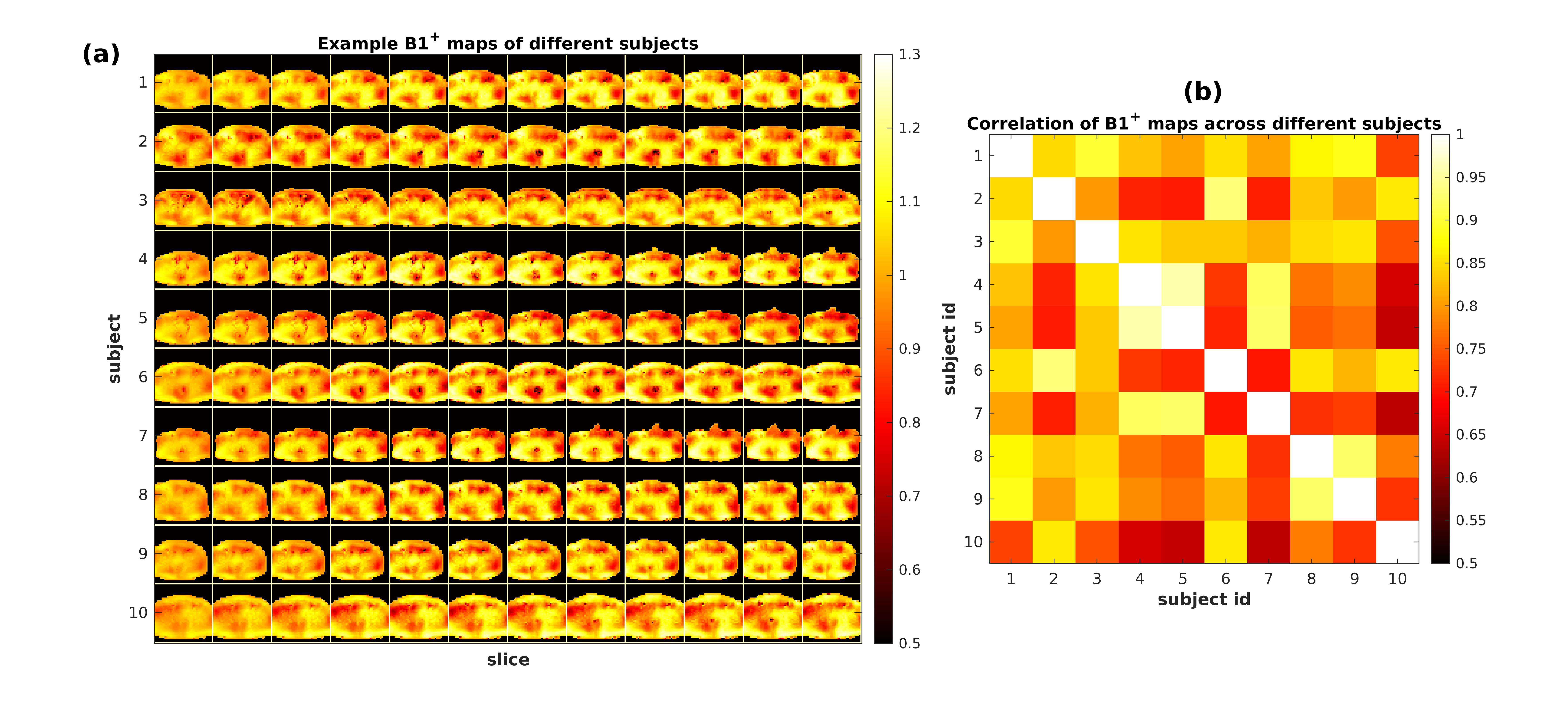
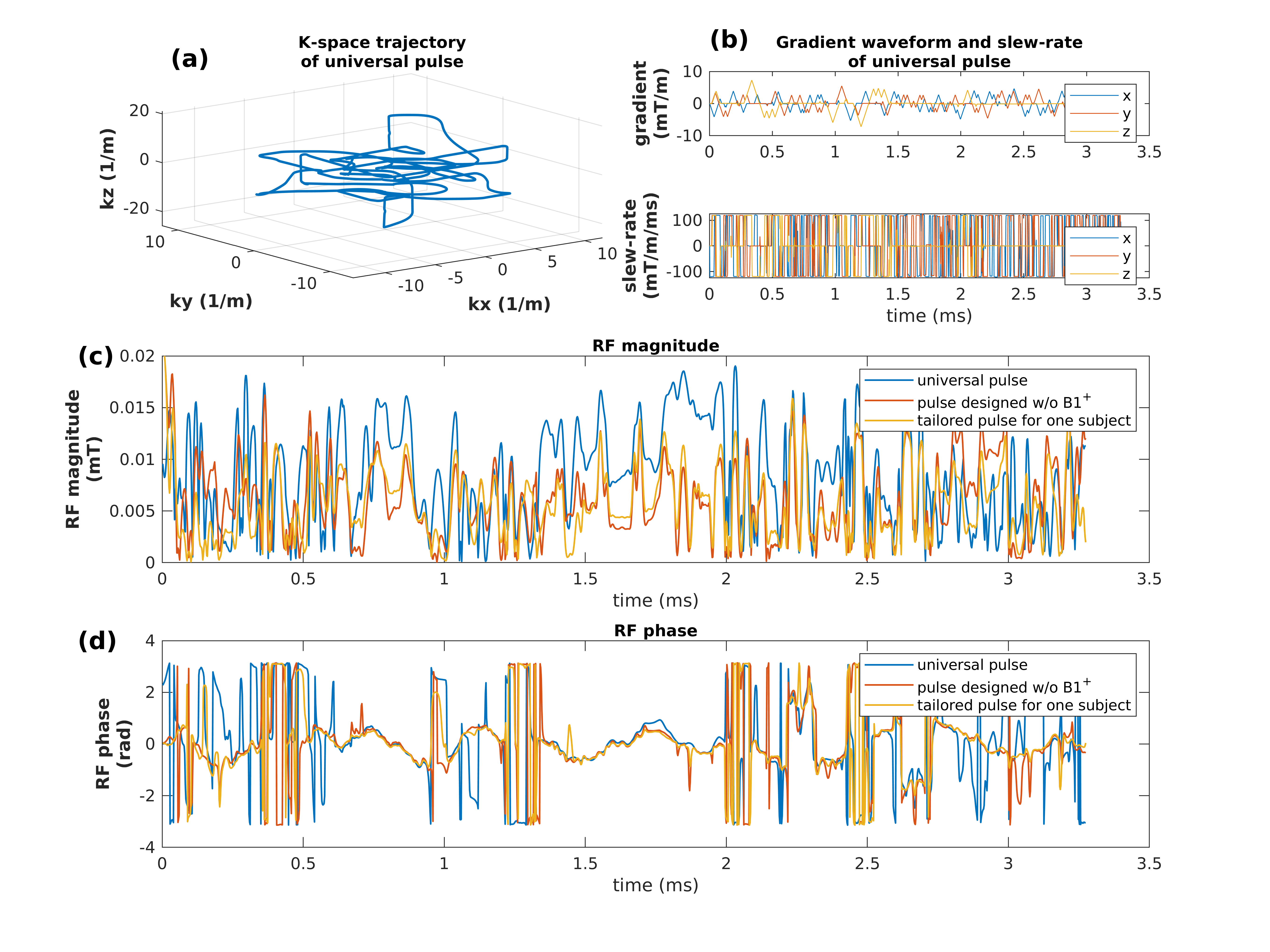
Figure 1a shows prostate B1+ of 10 subjects measured at 3T. Figure 1b shows the correlation of B1+ among these subjects, with coefficients ranging from 0.65 to 1. It demonstrates that B1+ values of the pelvic region at 3T exhibit a high level of correlation, suggesting that a group of subjects may potentially be used to represent others.Figure 2 shows the gradient waveform, its slew rate and k-space trajectory, and the optimized RF waveforms. The universal pulse was optimized over 12 hours using 6 subjects, the pulse designed without B0 and B1+ and the tailored pulse were optimized over 2 hours.
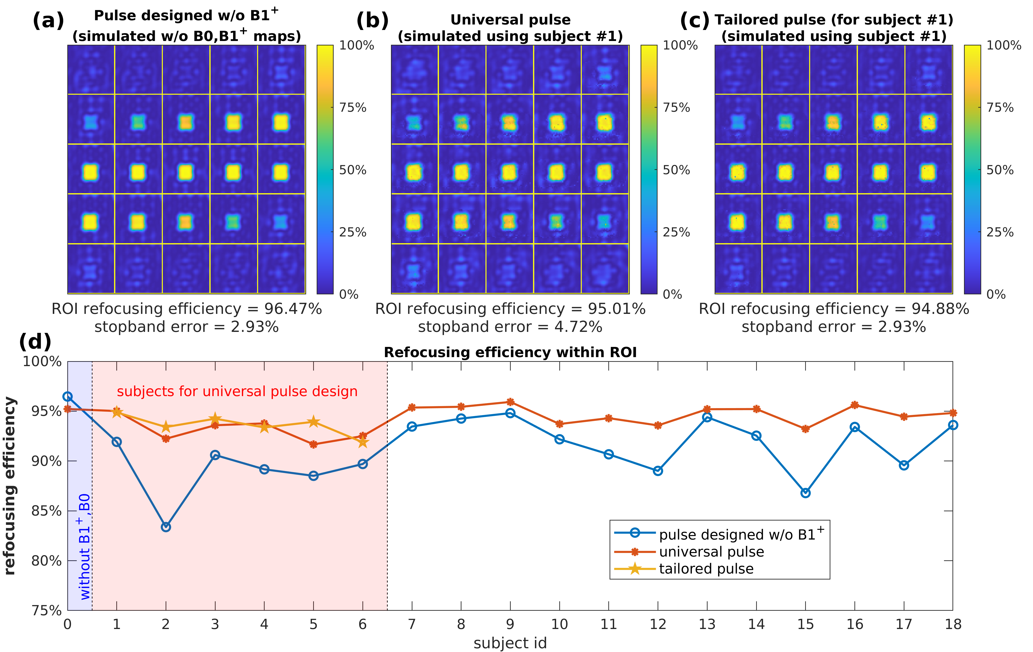
Figure 3 shows the comparison of different RF pulses in simulations. Compared to the RF pulse designed without B0 and B1+, the universal pulse achieves better performance in term of the refocusing efficiency in both seen and unseen subjects.
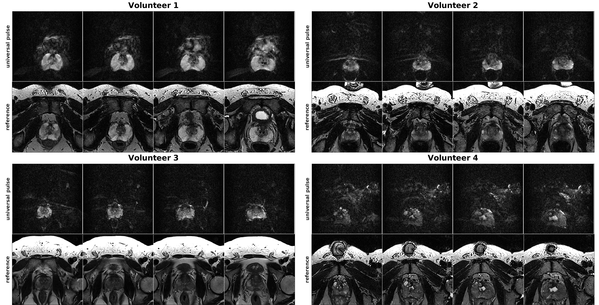
Figure 4 shows representative images from four subjects acquired using 3D EPI with the universal pulse, as well as corresponding T2 reference images. Our results demonstrate our designed universal pulse is effective in achieving rFOV refocusing for prostate imaging at 3T.
Conclusions and Discussion
In this study, we developed an algorithm that designs a multidimensional spatially selective universal refocusing pulse. We evaluated the performance of the RF pulse through simulations and in vivo prostate imaging. Our results show that a 3D universal pulse designed using 6 subjects could achieve similar performance on unseen subjects, indicating the potential to move the pulse design offline. Furthermore, our in vivo results validated that rFOV prostate imaging could be achieved for most subjects using universal pulses, which could lead to the possibility for achieving high-resolution diffusion imaging with 3D rFOV EPI imaging.Acknowledgements
This study was supported by NIH grants R37CA263583 and R01CA208236, and Siemens Healthcare.References
[1] Gras, Vincent, et al. "Universal pulses: a new concept for calibration‐free parallel transmission." Magnetic resonance in medicine 77.2 (2017): 635-643.
[2] Luo, Tianrui, et al. "Joint Design of RF and gradient waveforms via auto-differentiation for 3D tailored excitation in MRI." IEEE Transactions on Medical Imaging 40.12 (2021): 3305-3314.
[3] Yang, Jiayao, et al. "Multidimensional RF pulse design in spin-domain using auto-differentiation for 3D refocusing pulse", 2023 ISMRM Annual Meeting, Toronto, ON, Canada
[4] Hao, Sun, et al. "Joint design of excitation k-space trajectory and RF pulse for small-tip 3D tailored excitation in MRI." IEEE transactions on medical imaging 35.2 (2015): 468-479.
[5] Setsompop, Kawin, et al. "Magnitude least squares optimization for parallel radio frequency excitation design demonstrated at 7 Tesla with eight channels." Magnetic Resonance in Medicine: An Official Journal of the International Society for Magnetic Resonance in Medicine 59.4 (2008): 908-915.
[6] Pauly, John, et al. "A k-space analysis of small-tip-angle excitation." Journal of Magnetic Resonance (1969)81.1 (1989): 43-56.
[7] Layton, Kelvin J., et al. "Pulseq: a rapid and hardware-independent pulse sequence prototyping framework." Magnetic resonance in medicine 77.4 (2017): 1544-1552.
Figures