4092
Design of dynamic kT-point pTx pulses for renal imaging at 7T1Institute of Radiology, University Hospital Erlangen, Friedrich-Alexander-Universität Erlangen-Nürnberg (FAU), Erlangen, Germany, Erlangen, Germany, 2Siemens Healthcare GmbH, Erlangen, Germany, Erlangen, Germany, 3Department of Nephrology and Hypertension, Friedrich-Alexander-Universität Erlangen-Nürnberg (FAU), Erlangen, Germany, Erlangen, Germany, 4Division of Medical Physics in Radiology, German Cancer Research Centre (DKFZ), Heidelberg Germany, Heidelberg, Germany
Synopsis
Keywords: RF Pulse Design & Fields, High-Field MRI
Motivation: In ultra-high field MRI, transmit magnetic field (B1+) inhomogeneities are affecting kidney imaging.
Goal(s): Our goal was evaluate dynamic kT-point pulses for homogenous excitation in bilateral renal imaging.
Approach: Channel-wise B1+ maps of the kidneys were acquired for 15 subjects. Universal and individual kT-point pulses with different number of subpulses were calculated and evaluated. For one subject anatomical images were acquired with phase shims and dynamic kT-point pulses.
Results: While fixed-phase shims still suffer from flip-angle inhomogeneity, universal and subject-specific dynamic pulses with non-selective 5kT-point trajectories improves flip-angle homogeneity in the kidney and enable imaging with homogeneous excitation.
Impact: Our study evaluated universal and subject-specific kT-point pulses for kidney MRI at 7T. kT-point pulses substantially improve flip-angle homogeneity and universal pulses enable calibration-free imaging at ultra-high field, promising advances in high-resolution renal imaging.
Introduction
One of the major challenges in ultra-high field MRI is the inhomogeneous transmit magnetic field (B1+), which leads to a spatially non-uniform flip-angle (FA) influencing the image contrast. Due to the larger imaging volume, these artifacts are more prevalent in body imaging and affect kidney imaging at 7T.1Parallel transmission (pTx) aims to improve FA homogeneity by designing dedicated RF pulses.² In the human torso universal (UP) and subject-specific kT-point pulses have been successfully applied for cardiac imaging.³ In the following, the concept of dynamic pTx using nonselective kT-point trajectories for imaging of both human kidneys is evaluated.
Methods
Fifteen subjects (7w, 8m, 21.4+/-2.7kg/m², 26.3+/-2.4years) were scanned on a 7T MR-system (MAGNETOM Terra.X, Siemens Healthcare GmbH, Erlangen, Germany) with informed consent and approval of the local ethics committee, using an 8Tx/16Rx coil (Rapid Biomedical GmbH, Rimpar, Germany). All measurements were performed under free breathing.Channel-wise, relative B1+ maps were acquired with a density-adapted 3D radial sequence (DA-3D-RAD)4 in the small FA regime (FOV (320mm)³, spatial resolution (4mm)³; FA=8°; TE/TR=2.02ms/4.5ms, TA=6min)5. Channel-wise, absolute B1+ estimations6 were obtained by a calibration scan (AFI7, TR1/TR2=90ms/20ms, FA1/FA2=70°/8°).
kT-point pulses were calculated with different numbers of subpulses and a target FA of 5°. The length of each subpulse was set to 0.15ms and the length of the gradient blips was set to 0.09ms.3 The SED limit was set to 0.1256J/kg. Pulses were calculated for 1-7 kT-points to investigate the effect of multiple subpulses.
Initially, for each number of subpulses 100 universal pulses8 (UP) were calculated for twelve subjects (UP12) with random initial values (IV). The performance of the different pulses was compared using the coefficient of variation (CV=std(FA)/mean(FA)) within a region of interest (ROI) that included both kidneys. For each number of subpulse the UP12 with the lowest median CV was selected and tested on three unseen subjects. Additionally, the corresponding UP12 was set as IV for the calculation of individual pulses for all subjects.9
The performance of the kT-pulses was also compared to the null shim and a vendor provided default phase shim, optimized for cardiac imaging.
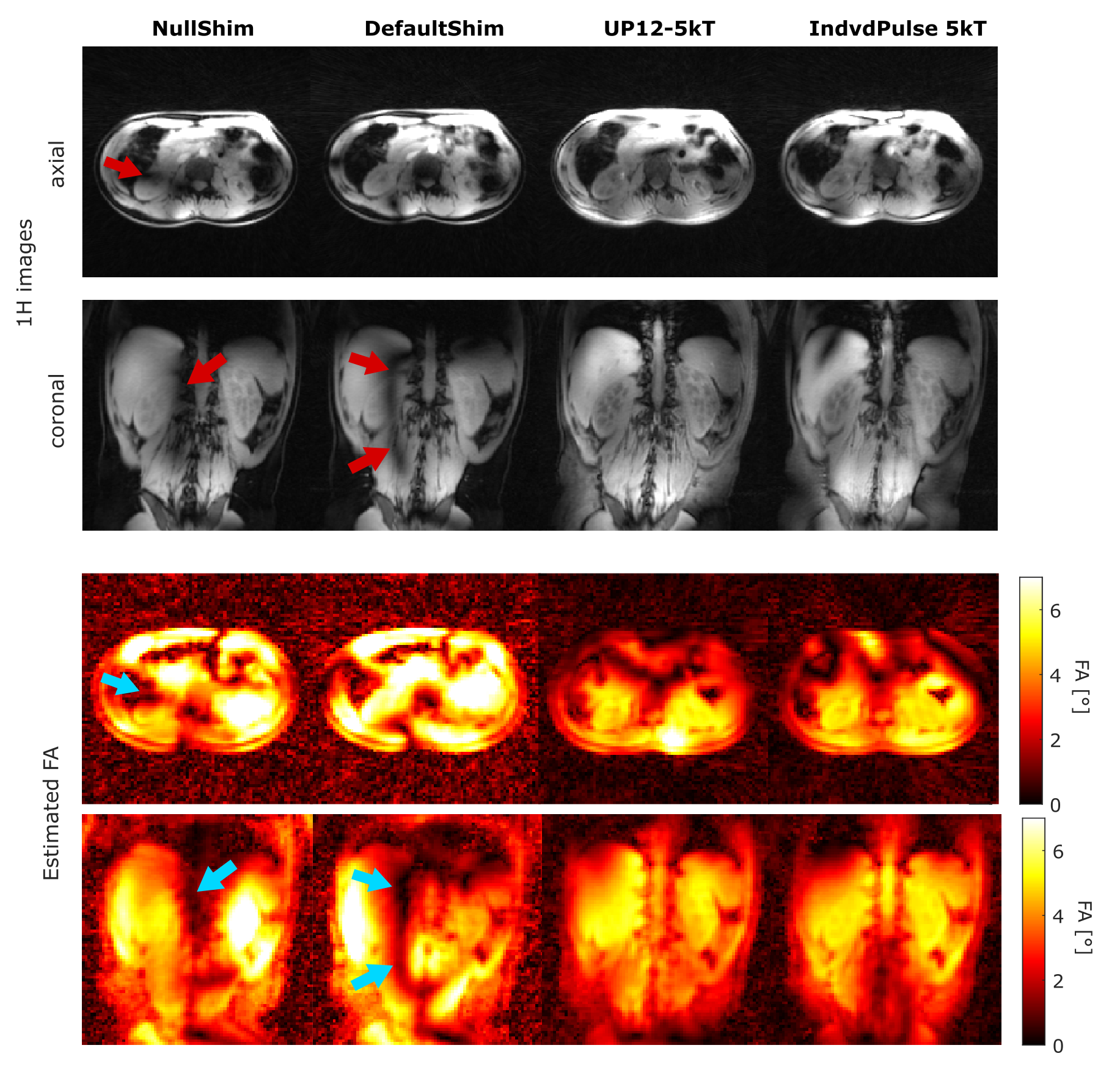
For subject 15, anatomical images were acquired with the DA-3D-RAD sequence with a spatial resolution of (1.4mm)³, FOV=(320mm)³, FA=5° and TA=6min with NullShim, DefaultShim, the UP12-5kT, and an individual 5kT-point RF pulse.
Results
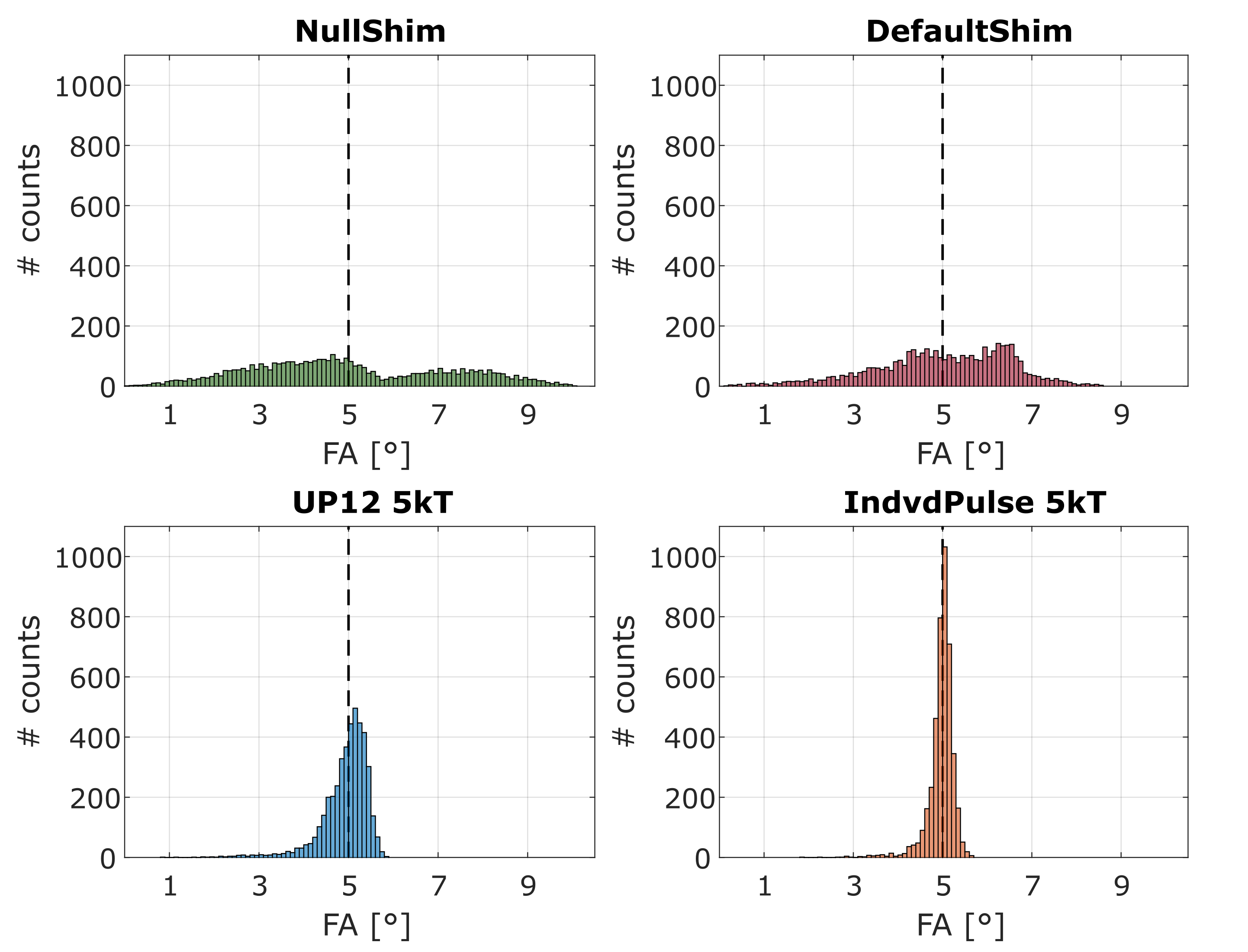
For individual and universal pulses, the median CV decreases with a rising number of subpulses although the improvement is less considerable after 5kT-points (see Figure 1). The individually optimized pulses have a systematically lower CV compared to the UPs. The CVs of the UP12s applied to the three subjects not included in the UP12 library lie within the range of the subjects included in the optimization. NullShim and DefaultShim result in large CV values across the subjects with median values of 41.3% and 32.1% respectively (see Figure 2), while the UP12-5kT and individual-5kT pulses reduce the CV to 10.0% and 6.1%. The estimated FA distributions for subject 15 for NullShim and DefaultShim are very broad around the target FA (see Figure 5). In contrast, the FA distributions of the UP12-5kT and individual-5kT pulse are very narrow and centered around the target FA with (4.9+/-0.6)° for UP12-5kT and (5.0+/-0.3)° for the individual-5kT pulses.The estimated FAs fit the measured signal intensities and both show signal dropouts for the phase shims and homogeneous excitation for the kT pulses (see Figure 4).
Discussion and Conclusion
Our results show that dynamic pTx with (universal and subject-specific) kT-point pulses substantially enhances FA homogeneity in UHF MRI kidney imaging. The CVs of the UP12s for the unseen subjects are within the range of the CVs of the subjects included in the library, indicating that the UP12s are robust for unseen subjects. Additionally, the CVs for individual and universal kT-point pulses are comparable to CV values from literature for cardiac imaging at 7T.³ Therefore, UPs enable scan-time reduction by allowing calibration free imaging of the kidneys. As long pulses durations are more prone to B0 and T2* related artifacts, a trade-off has to be made between the number of kT-points and the homogeneity of the excitation.10For our measurement setup the 8Tx/16Rx coil can be combined with a 23Na birdcage and used for interleaved imaging11 of 1H and 23Na at the heart. Universal pTx pulses could be applied to interleaved measurements of the human kidney improving 1H imaging, without the need of additional calibration time.
Acknowledgements
This project was funded by the Deutsche Forschungsgemeinschaft (DFG) under 509149993 (TR 374).References
1. Erturk MA, Li X, Van de Moortele PF, Ugurbil K, Metzger GJ. Evolution of UHF Body Imaging in the Human Torso at 7T: Technology, Applications, and Future Directions. Top Magn Reson Imaging. 2019;28(3):101-124.
2. Ibrahim TS, Lee R, Baertlein BA, Kangarlu A, Robitaille PL. Application of finite difference time domain method for the design of birdcage RF head coils using multi-port excitations. Magn Reson Imaging. 2000;18(6):733-42.
3. Aigner CS, Dietrich S, Schaeffter T, Schmitter S. Calibration-free pTx of the human heart at 7T via 3D universal pulses. Magn Reson Med. 2022;87(1):70-84.
4. Nagel, A.M., Laun, F.B., Weber, M.-A., Matthies, C., Semmler, W. and Schad, L.R. 2009, Sodium MRI using a density-adapted 3D radial acquisition technique. Magn. Reson. Med., 62: 1565-1573.
5. Pierre-Francois Van de Moortele and K. Ugurbil. “Very Fast Multi Channel B1 Calibration at High Field in the Small Flip Angle Regime”. In: Proc Intl Soc Mag Reson Med 17 2009, p. 367.
6. Dietrich, S, Aigner, CS, Kolbitsch, C, et al. 3D Free-breathing multichannel absolute Mapping in the human body at 7T. Magn Reson Med. 2021; 85: 2552–2567.
7. Yarnykh, V.L. Actual flip-angle imaging in the pulsed steady state: A method for rapid three-dimensional mapping of the transmitted radiofrequency field. Magn. Reson. Med. 2007, 57: 192-200.
8. Gras V, Vignaud A, Amadon A, Le Bihan D, Boulant N. Universal pulses: A new concept for calibration-free parallel transmission. Magn Reson Med. 2017 Feb;77(2):635-643.
9. Herrler, J, Liebig, P, Gumbrecht, R, et al. Fast online-customized (FOCUS) parallel transmission pulses: A combination of universal pulses and individual optimization. Magn Reson Med. 2021; 85: 3140–3153.
10. Cloos MA, Boulant N, Luong M, Ferrand G, Giacomini E, Le Bihan D, Amadon A. kT -points: short three-dimensional tailored RF pulses for flip-angle homogenization over an extended volume. Magn Reson Med. 2012;67(1):72-80.
11. Ruck L, Wilferth T, Gast LV, et al. Interleaved 23Na/1H MRI of the human heart at 7 Tesla. In Proceedings of the 30th Annual Meeting of ISMRM, London, UK, 2022. Abstract 276.
Figures
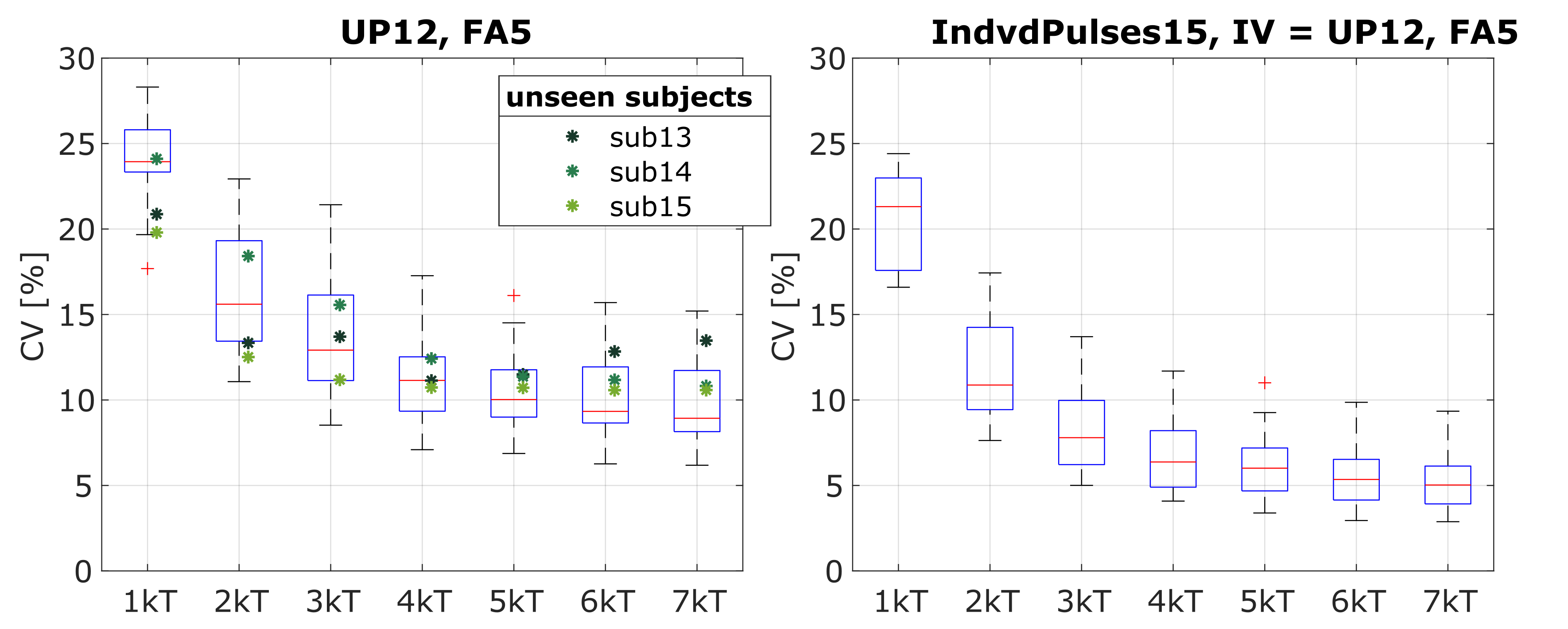
left: for UP12 and its performance for subjects within (boxplot) and separate from (green stars) the UP12 library. The CVs of unseen subjects lie in the range of the subjects included in the UP12.
right: for individual pulses for 15 subjects, with UP12 as IV.
More subpulses lead to a decrease in the median CV of the FA. The median CV of the individual pulses is systematically lower than the median CV of UP12.
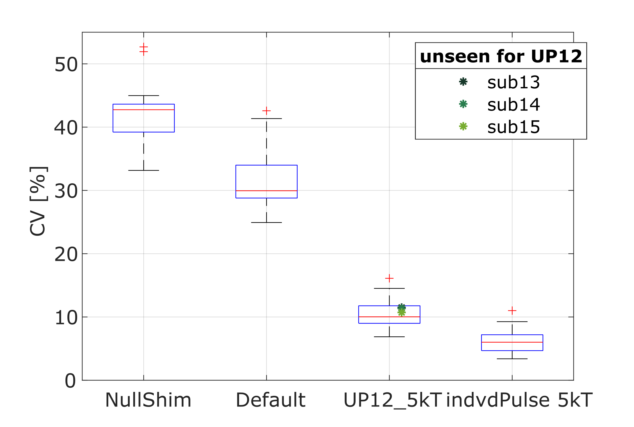
Figure 2: CV values for the B1+ datasets from subject 1 – 15, for NullShim, DefaultS, UP12-5kT and the individual 5kT point pulse. As subjects 13-15 are not included in the UP12 library they are marked in green. The dynamic pTx pulses yield a considerably higher FA homogeneity than the fixed phase shims.
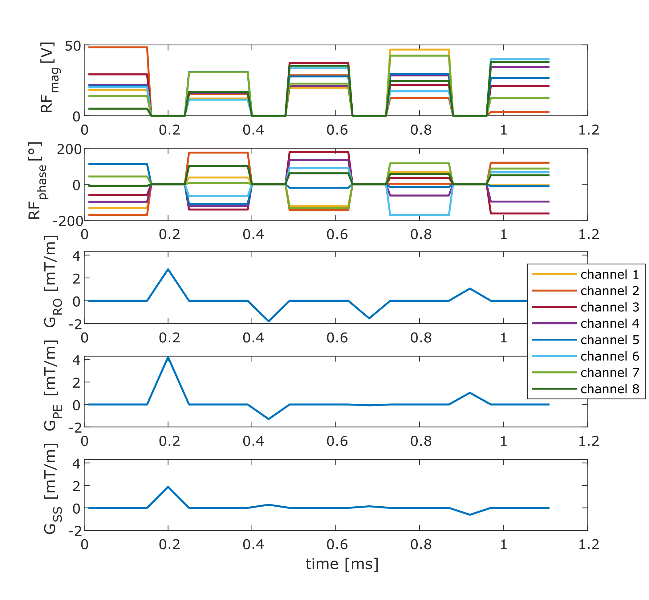
Figure 3: Pulse scheme for UP12-5kT. The magnitude and phase of the five rectangular subpulses and the intervening 3D gradient blips of the UP12-5kT are shown.