4089
Parallel transmit spatial-spectral pulse design with specific absorption rate control for water excitation: validation in humans at 7 Tesla1Center for Biomedical Imaging Research, Department of Biomedical Engineering, School of Medicine, Tsinghua University, Beijing, China, 2Tiantan Neuroimaging Center of Excellence, Beijing Tiantan Hospital, Capital Medical University, Beijing, China, 3Department of Radiology and Imaging Sciences, University of Utah, Salt Lake City, UT, United States, 4Center for Magnetic Resonance Research, Radiology, Medical School, University of Minnesota, Minneapolis, MN, United States
Synopsis
Keywords: Parallel Transmit & Multiband, Parallel Transmit & Multiband
Motivation: Last year we demonstrated the utility of our new parallel transmit spatial-spectral pulse design for robust water excitation, but only using Bloch simulations without experimental proof.
Goal(s): Our goal here was to validate our method via phantom and human scans at 7 Tesla.
Approach: We validated our design method by collecting 3D GRE data in a water-fat phantom and in the human brain. All data were obtained on a Siemens Terra using the commercial Nova 8-channel transmit coil.
Results: Our method outperformed existing approaches, producing uniform water excitation across the whole brain with nearly complete fat suppression even in the challenging areas.
Impact: Validated in humans at 7 Tesla, our design method provides an effective solution for volumetric uniform water excitation, eliminating the need for additional fat saturation and holding a promise to many applications including high-resolution functional MRI at ultrahigh field.
Introduction
Previously, we introduced a new parallel transmit (pTx) spatial-spectral (spsp) pulse design with explicit SAR management for robust uniform water excitation and demonstrated its utility for whole-brain water excitation at 7 Tesla (7T) using Bloch simulation1. Here, we validated our design in humans, and demonstrated its usability for designing universal pulses2, comparing to existing approaches.Methods
Experiments were conducted on a Siemens Terra (Siemens, Erlangen, Germany) equipped with a body gradient (200 T/m/s slew-rate, 80 mT/m Gmax).Data were acquired using the commercial Nova 8-channel transmit 32-channel receive coil (Nova Medical Inc.). Calibration including $$$\Delta$$$B0 and B1+ mapping was obtained using vendor-provided pulse sequences with 3-mm resolution, 100×108 matrix size, and 80 axial slices. A pTx-enabled sequence was developed to acquire 3D GRE images.
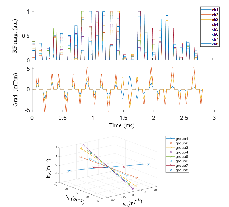
Our pulses (Fig. 1) were designed using a 2-step approach as described previously1. Local SAR control was informed by SAR calculation based on 1669 virtual observation points3,4.
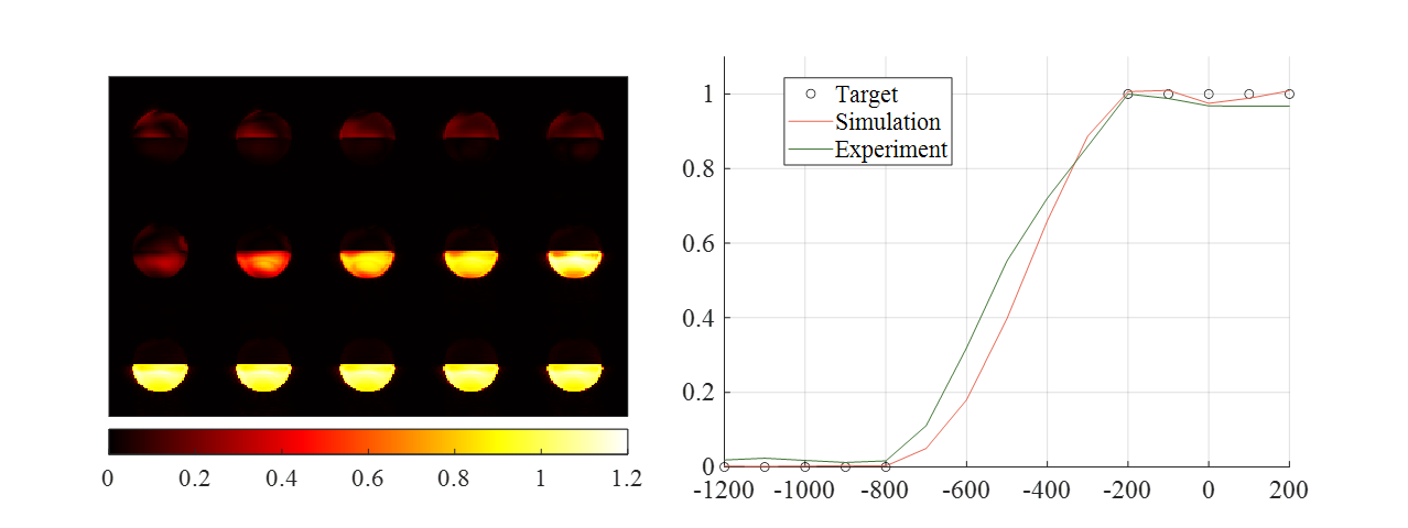
We started by validating our method in a phantom constructed by filling a cylinder (diameter: 10 cm; length: 13 cm) with 500 ml of oil and 500 ml of physiological saline. Our pulses’ frequency responses over the frequencies of interest were measured by incrementing RF frequency and acquiring corresponding 3D GRE images with 3-mm resolution, 2-fold acceleration, 8-ms TE and 50-ms TR.
We then validated our method in humans. Brain masking was achieved by brain extraction based on a 3D whole-brain GRE. To demonstrate the utility of our pulses, high-resolution whole-brain 3D GRE images were acquired with 1-mm resolution, 220×220×220 matrix size, 2-fold acceleration, 8-ms TE, and 50-ms TR.
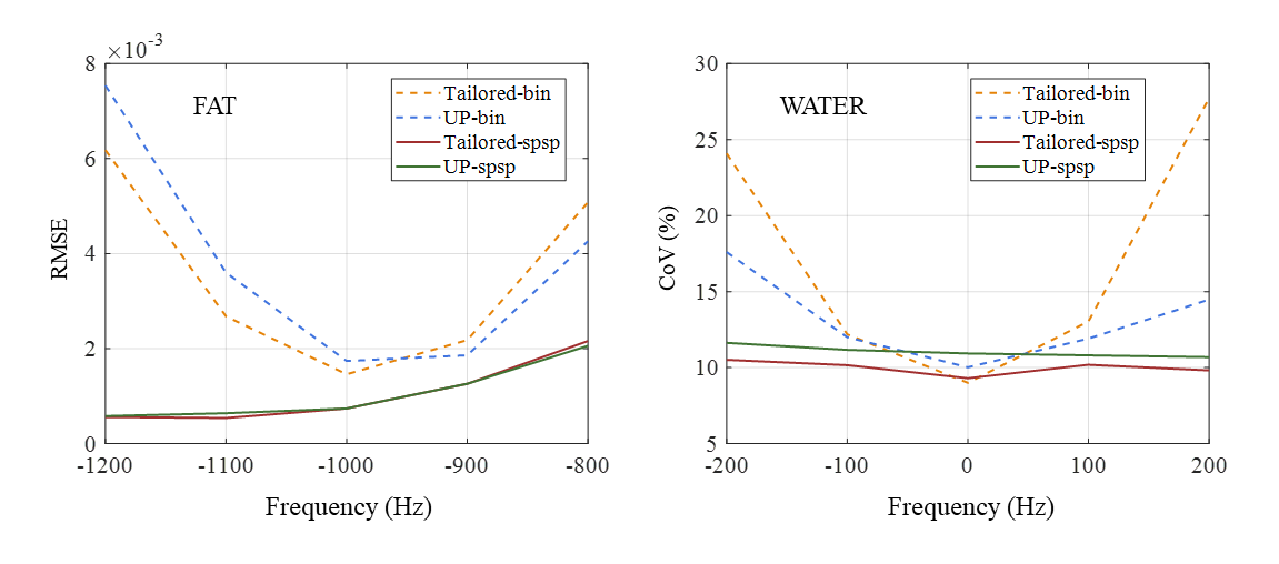
We also investigated how our method would perform when used to design universal pulses (UP). A calibration dataset comprising brain masking, $$$\Delta$$$B0 and B1+ mapping obtained in 10 healthy volunteers (five men) were used. Simulations were conducted to evaluate the performances of our UP based on a 10-fold cross-validation. Root mean square error (RMSE) and coefficient of variation (CoV), averaged across folds, were calculated to evaluate performances for fat suppression and water excitation, respectively. Results were compared to tailored, subject-specific design.
To demonstrate the utility of our method, we designed UP using entire calibration and use it to scan a new volunteer using the same 3D GRE protocol as in the above validation.
For comparison, imaging in same volunteers with matched parameters was also performed using 1) the Nova coil operated in its circularly polarized (CP) mode mimicking single-transmit, and 2) pTx pulses designed with the interleaved binomial approach5.
Results
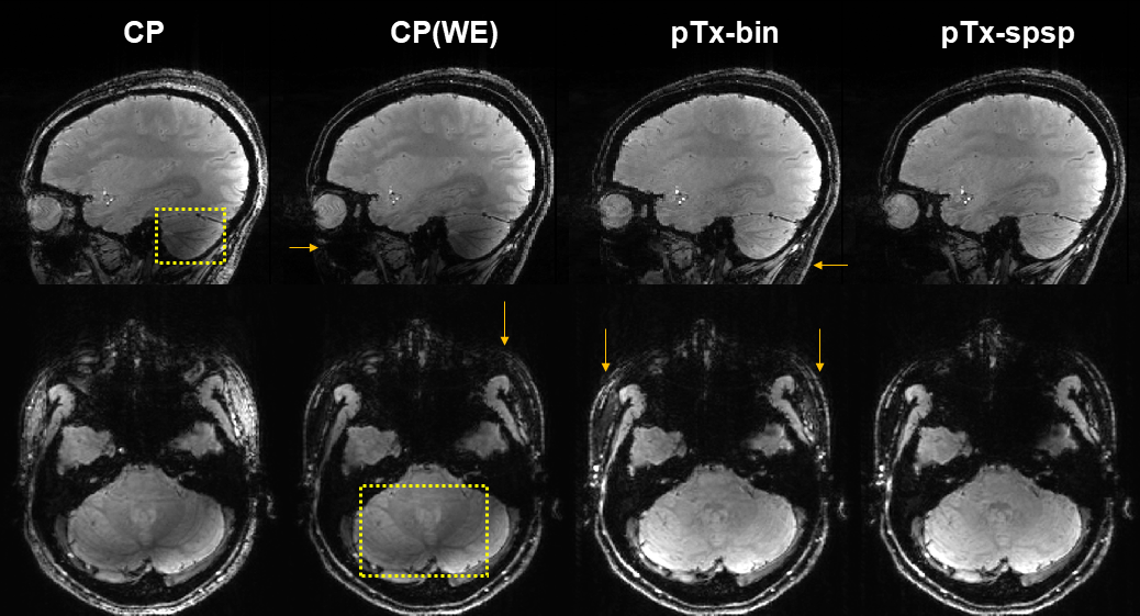
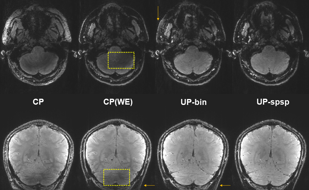
Our design was validated in the phantom (Fig. 2), achieving water-selective excitation at the water resonance frequency while producing the desired spectral selectivity across the entire frequency range of interest.In humans, our method outperformed existing approaches (Fig. 3), improving not only water excitation (relative to excitation in the CP mode) but also fat suppression (compared to the interleaved binominal approach) across the entire brain even in challenging areas.
When used to design UP, our method was found robust against inter-subject variabilities (Fig. 4), inheriting most of water-excitation performances from the tailored design while outperforming both UP and tailored designs with the interleaved binomial approach in achieving robust simultaneous water excitation and fat suppression over a 400-Hz bandwidth.
As shown in Fig. 5, our UP when used to scan an unseen volunteer, improved water-selective excitation across the entire brain, effectively restoring signal loss in the cerebellum (compared to CP mode approaches) and robustly suppressing fat even in challenging regions (relative to UP designed with the interleaved binomial approach).
Discussion
We have validated our previously proposed pTx spsp pulse design capable of robust uniform water excitation across an extensive image volume even in the presence of strong RF inhomogeneity and large off-resonances. Our 7T human results show that our design can produce high-quality uniform water excitation across the entire brain, outperforming existing approaches. Our results also show that our method can be used to design universal pulses without sacrificing its performances, suited to be used for plug-and-play pTx.Further SAR calculation suggests that our method can reduce local SAR by as much as ~31% relative to the interleaved binomial approach for comparable pulse lengths, thanks to explicit local SAR control incorporated in pulse design.
Part of our future work is to integrate our pulses into GRE-EPI to promote whole-brain functional MRI at ultrahigh field.
Conclusion
In humans at 7T, we have validated that our new pTx spatial-spectral pulse design can achieve quality uniform water excitation across the entire brain. We believe our new design will have many applications including high-resolution functional MRI at ultrahigh field.Acknowledgements
The authors would like to acknowledge Patrick Liebig from Siemens Healthineers for his assistance with calculation of multichannel B1+ mapping using the vendor sequence. KU and XW and all work carried out at the University of Minnesota were supported in part by USA NIH grants (NIBIB P41 EB027061 and U01 EB025144).References
1. Xin S, Xiaodong M, Hua G, Kamil U, Xiaoping W. Parallel transmit spatial spectral pulse design with specific absorption rate control: demonstration for robust water excitation at 7 Tesla. ISMRM. Canada, 2023. p. 6123.
2. Gras V, Vignaud A, Amadon A, Le Bihan D, Boulant N. Universal pulses: A new concept for calibration-free parallel transmission. Magnetic Resonance in Medicine 2017;77(2):635-643.
3. Eichfelder G, Gebhardt M. Local Specific Absorption Rate Control for Parallel Transmission by Virtual Observation Points. Magnetic Resonance in Medicine 2011;66(5):1468-1476.
4. Guérin B, Gebhardt M, Cauley S, Adalsteinsson E, Wald LL. Local Specific Absorption Rate (SAR), Global SAR, Transmitter Power, and Excitation Accuracy Trade-Offs in Low Flip-Angle Parallel Transmit Pulse Design. Magnetic Resonance in Medicine 2014;71(4):1446-1457.
5. Löwen D, Pracht ED, Stirnberg R, Liebig P, Stöcker T. Interleaved binomial kT-Points for water-selective imaging at 7T. Magnetic Resonance in Medicine 2022;88(6):2564-2572.
Figures