4088
Design of pTx pulses with free RF and gradient waveforms for cardiac MRI at 7T: Initial results and comparison to kT-point pulses1Institute of Radiology, University Hospital Erlangen, Friedrich-Alexander-Universität Erlangen-Nürnberg (FAU), Erlangen, Germany, 2Siemens Healthcare GmbH, Erlangen, Germany, 3Division of Medical Physics in Radiology, German Cancer Research Centre (DKFZ), Heidelberg, Germany
Synopsis
Keywords: RF Pulse Design & Fields, Heart, parallel transmission, 7T, ultra-high field MRI
Motivation: Parallel transmission (pTx) pulses with fixed gradient trajectory such as kT-points have successfully demonstrated to benefit cardiac imaging at 7T. However, it is possible to optimize pTx pulses without restrictions regarding RF and gradient waveforms.
Goal(s): Our aim was to optimize first pTx pulses with free RF and gradient trajectory for cardiac imaging.
Approach: We designed tailored free pulses for 35 heart subjects and compared them in terms of flip angle (FA) homogeneity and applied energy dose to kT-point pulses.
Results: Comparable performance in FA homogeneity was obtained for both pulse types, but the free pulses additionally achieved a reduction of SED.
Impact: Generating homogeneous excitation while complying with RF power deposition regulations is a challenge for cardiac imaging at 7T. Designing pTx pulses with free RF and gradient waveforms allows for reduced SED and similar homogeneity compared to kT-point pulses.
Introduction
To mitigate B1+ inhomogeneities that occur at higher magnetic field strengths (B0≥7T) in particular in human body applications, dynamic parallel transmission (pTx) pulses are used. Tailored and pre-computed universal pulses (UPs)1 with a kT-point trajectory2 have already proven to benefit the flip angle (FA) homogenization in cardiac MRI at 7T3. However, the use of a fixed k-space trajectory reduces the number of degrees of freedom for the pulse optimization and with more efficient optimization algorithms available, the free optimization of both RF waveform and excitation k-space trajectory becomes feasible. Therefore, the aim of this work was to design first pTx pulses with free RF amplitudes and phases, as well as gradient trajectory (“free pulses”) for the human heart and compare these pulses to kT-point pulses.Methods
3D channel-wise, relative B1+ maps4 of the human heart of 35 subjects (12 female, 23 male, 22-62 years, 16.4-32.2 kg/m2) were acquired with a density-adapted 3D radial GRE sequence5 (nominal FA=8°, TE/TR=2.02/4.5ms, special resolution Δx3=(4mm)3, FOV=(320mm)3, TA=6:00min) and with an 8Tx/16Rx transceiver array (Rapid Biomedical GmbH, Rimpar, Germany) at 7T (Magnetom Terra, Siemens Healthcare GmbH, Erlangen, Germany). Absolute B1+ estimations were obtained by multiplying the relative B1+ maps with a factor6 determined from an actual flip angle imaging7 scan. SAR safety was ensured using channel-power limits.kT-point and free pulses were designed using an interior-point method-based optimization algorithm8. 1000 4kT-point UPs with an individual subpulse length of 0.15ms and a gradient blip length of 0.09ms and 50 free UPs with a total RF pulse length of 0.87ms were calculated for a target FA of 5°, with the full library of 35 subjects and randomized starting values. The UP35 with the lowest coefficient of variation (CV) was used as initial value (IV) for calculating the corresponding tailored pulses. The calculation time for a tailored pulse was 17s (kT) and 29s (free) (Intel Xeon W-2245 CPU; 3.9 GHZ; 64GB RAM). The tailored pulses were evaluated according to their CV and specific energy dose (SED) values. Subsequently, 3D GRE measurements were acquired with the vendor provided cardiac shim and both tailored pulses (nominal FA=5°, TE/TR=2.02/7.0ms, Δx3=(1.4mm)3, FOV=(320mm)3, TA=7:00min).
Results
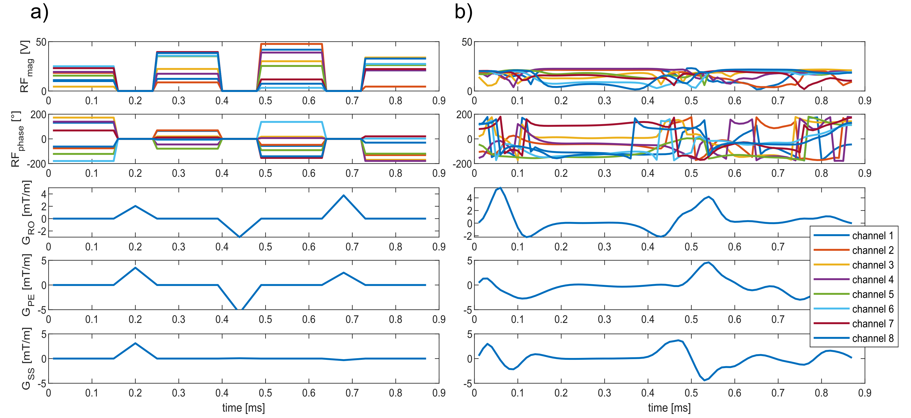
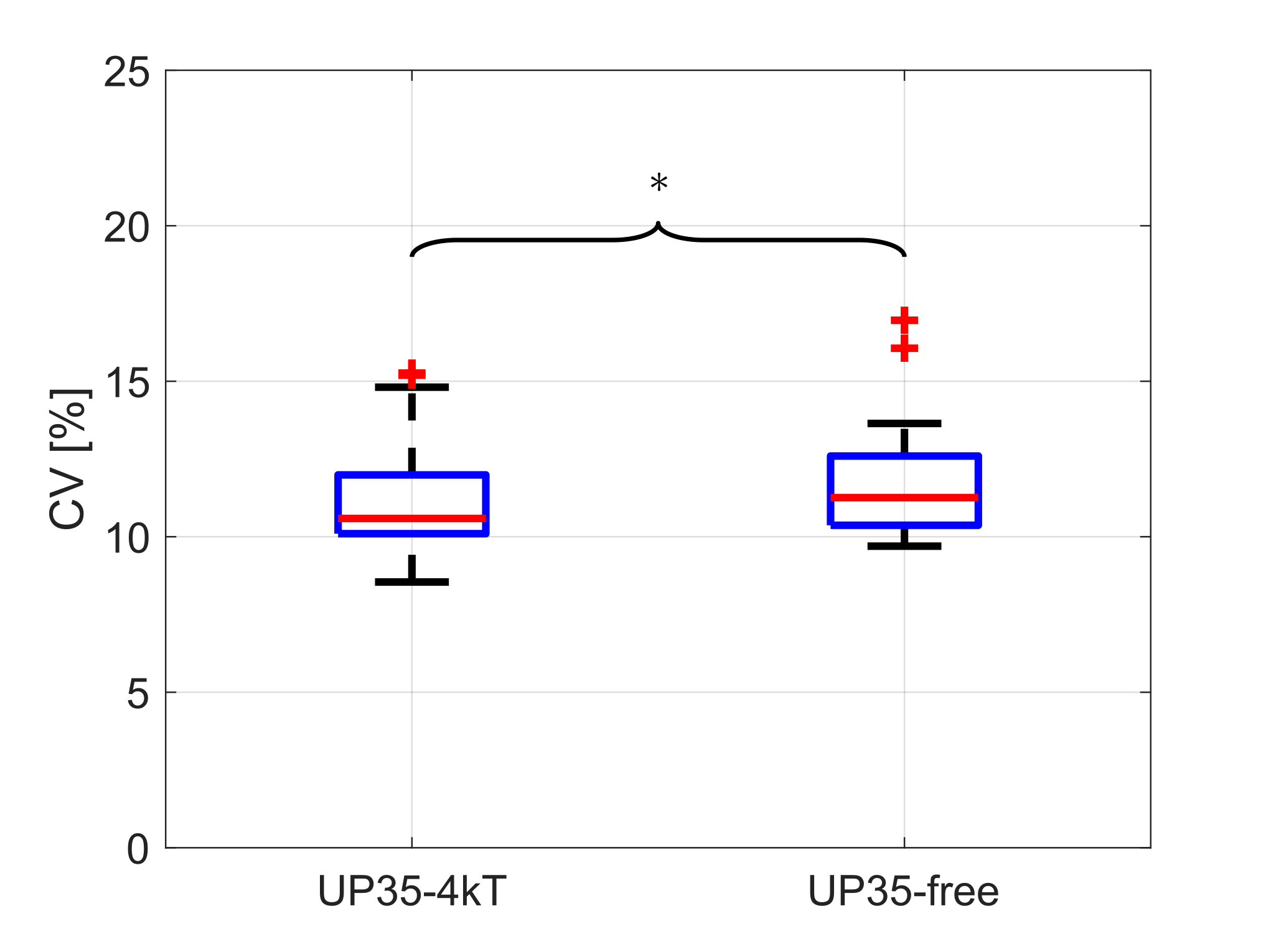
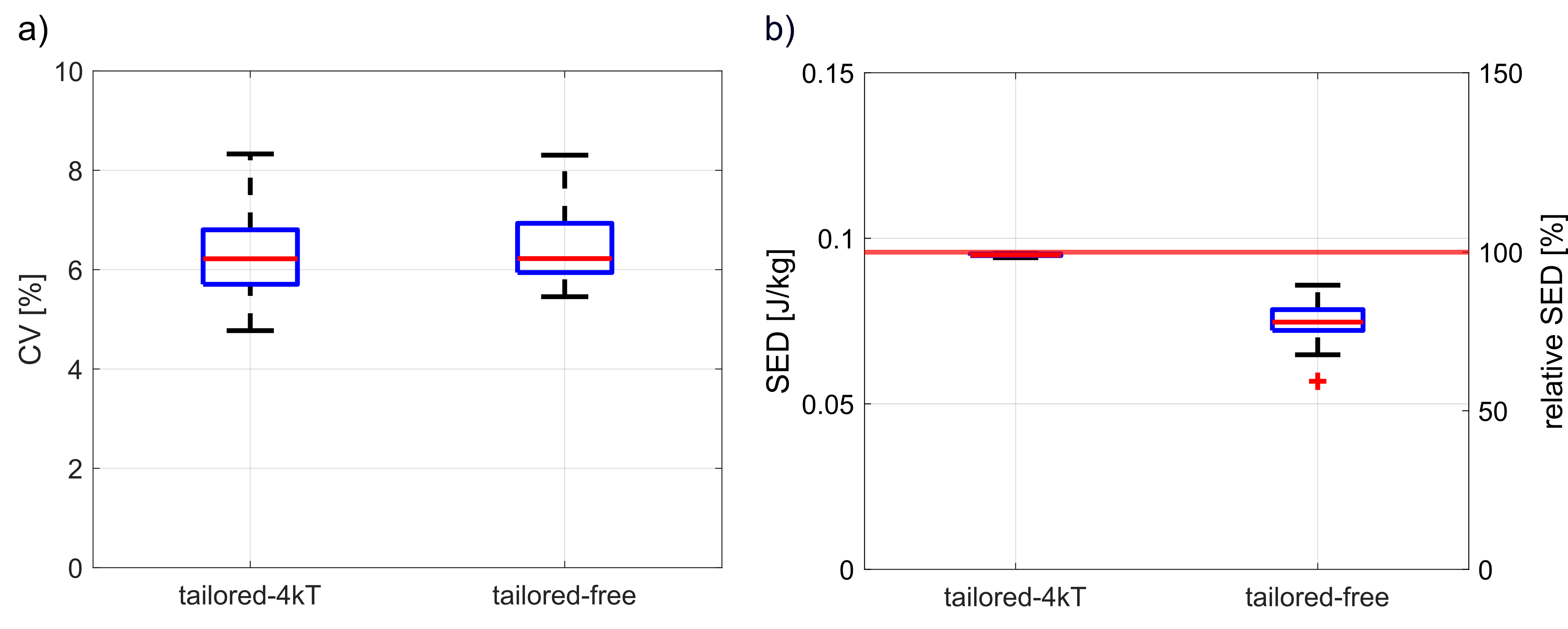
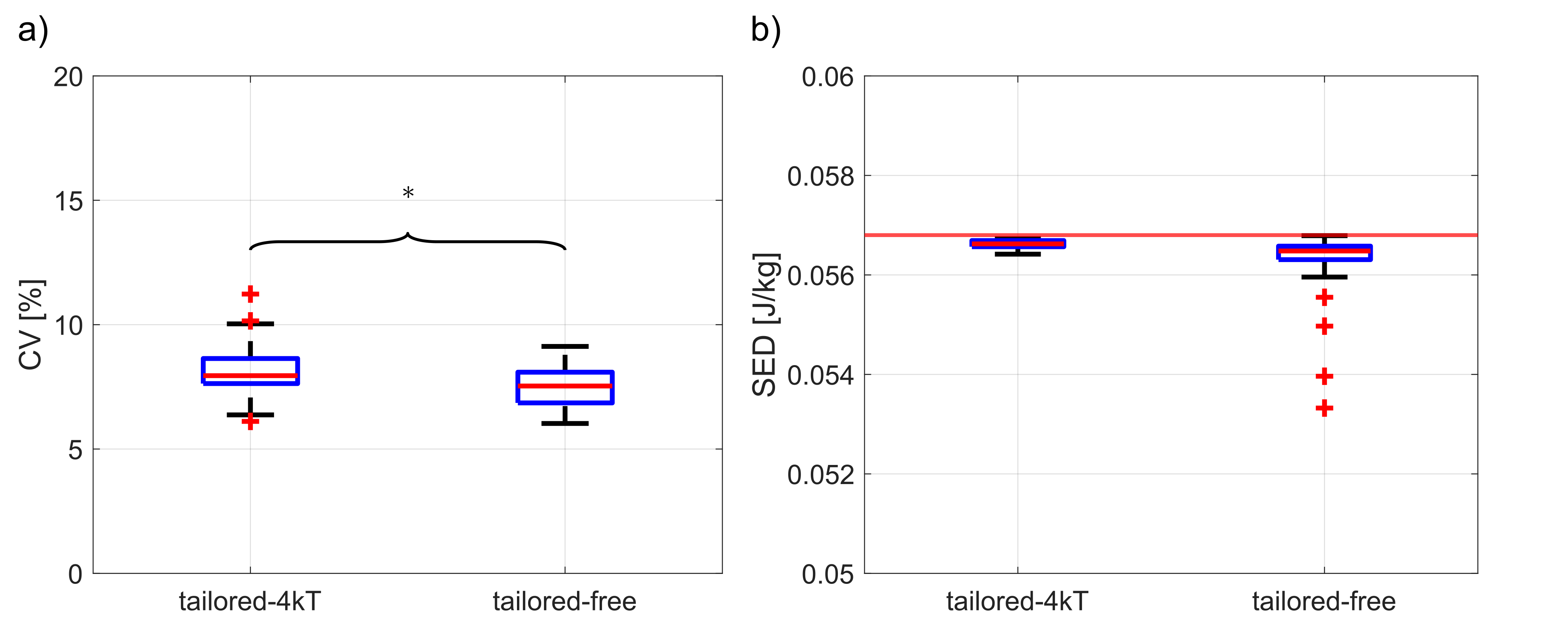
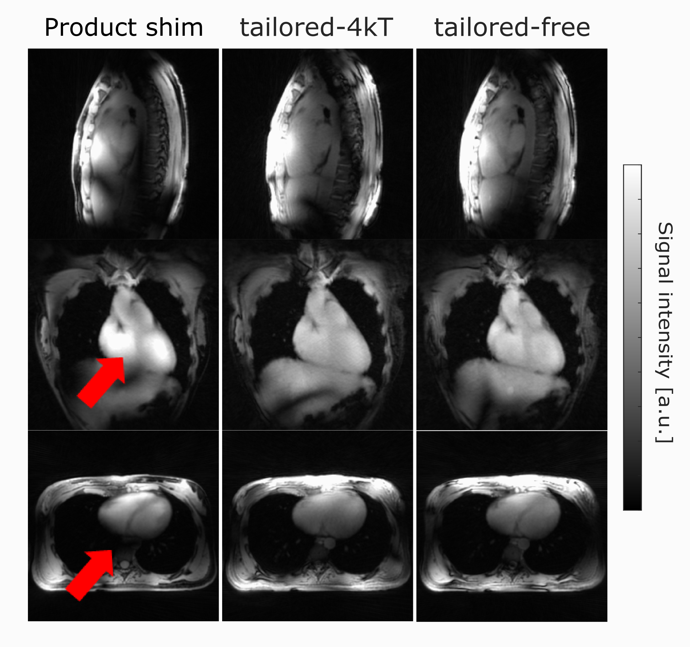
Figure 1 illustrates an RF pulse diagram for a 4kT-point and free pulse for one subject. Figure 2 shows the performance of the UP35-4kT (median CV: 10.6%) and UP35-free (median CV: 11.3%) with the minimum CV of all optimized UPs evaluated on 35 subjects. Figure 3 depicts the CV and SED values of the tailored-4kT and tailored-free pulses using the corresponding UP35 as initial value for the calculation. For an initial comparison of both pulse types, the SED limit for the optimization was chosen such that an equal median CV (6.22%) for both was obtained. For a comparable homogeneity, the tailored-4kT pulses result in a median SED of 0.0949 J/kg, whereas the median SED of the tailored-free pulses is 0.0747 J/kg. To investigate the performance when both pulse types reach the SED limit, the SED constraint was subsequently set to the minimum SED value of the tailored-free pulses (see Fig.4). The tailored-4kT pulses yield a median CV of 7.95% (6.11-11.23%) and the tailored-free pulses lead to a slightly lower median CV of 7.53% with smaller variations (6.02-9.13%). Figure 5 shows the GRE images acquired with the product shim and the tailored pulses with the SED limits from Fig.3 for one subject. For the default shim, signal dropouts within the heart can be observed. The tailored-4kT and tailored-free pulse show a similar performance with improved homogeneity.Discussion
By using a UP as initial value for calculating the tailored free pulses, online pulse optimization is feasible within 20-30s. However, it is possible that the optimization does not manage to optimize all degrees of freedom perfectly. Different approaches, such as e.g. other IV need to be investigated, since the choice of IV seems to be crucial for the success of the optimization. Additionally, further evaluations with different SED limits need to be performed. However, to achieve similar homogeneity, the tailored-free pulses require only about 75% of the SED of 4kT-point pulses. This advantage can also be further explored to push SED savings to the limit. Furthermore, the use of a virtual observation points model9 might favor the use of free pulses.Conclusion
We successfully designed kT-point and first free pulses for 35 B1+ data sets of the human heart. Both pulses improved the FA homogeneity at 7T compared to the default shim, which was confirmed by in-vivo measurements. For a comparable homogeneity to the 4kT-point pulses, the free pulses require less SED.Acknowledgements
No acknowledgement found.References
1. Gras V, Vignaud A, Amadon A, Le Bihan D, Boulant N. Universal pulses: A new concept for calibration-free parallel transmission. Magn Reson Med. 2017;77(2):635-643.
2. Cloos MA, Boulant N, Luong M, et al. kT -points: short three-dimensional tailored RF pulses for flip angle homogenization over an extended volume. Magn Reson Med. 2012; 67(1):72-80.
3. Aigner CS, Dietrich S, Schaeffter T, Schmitter S. Calibration-free pTx of the human heart at 7T via 3D universal pulses. Magn Reson Med. 2021; 87: 70–84.
4. Van de Moortele, PF. Very Fast Multi Channel B1 Calibration at High Field in the Small Flip Angle Regime. Proc Intl Soc Mag Reson Med 17 (2009): 367.
5. Nagel AM, Laun FB, Weber MA, Matthies C, Semmler W, Schad LR. Sodium MRI using a density-adapted 3D radial acquisition technique. Magn Reson Med. 2009; 62(6):1565-1573.
6. Dietrich S, Aigner CS, Kolbitsch C, et al. 3D Free-breathing multichannel absolute B1+ Mapping in the human body at 7T. Magn Reson Med. 2021; 85(5): 2552-2567.
7. Yarnykh VL. Actual flip-angle imaging in the pulsed steady state: A method for rapid three-dimensional mapping of the transmitted radiofrequency field. Magn. Reson. Med. 2007; 57: 192-200.
8. Majewski K. Simultaneous optimization of radio frequency and gradient waveforms with exact Hessians and slew rate constraints applied to kT-points excitation. J Magn Reson. 2021; 326: 106941.
9. Eichfelder G, Gebhardt M. Local specific absorption rate control for parallel transmission by virtual observation points. Magn Reson Med. 2011; 66(5):1468-1476.Figures