4084
Design and application of a 16-channel field-probe insert for a 16Tx/32 Rx coil at 9.4 Tesla1High Field Magnetic Resonance, Max-Planck Institute for Biological Cybernetics, Tübingen, Germany, 2Department of Biomedical Magnetic Resonance, Eberhard Karls University Tübingen, Tübingen, Germany, 3German Center for Neurodegenerative Diseases (DZNE), Bonn, Germany
Synopsis
Keywords: System Imperfections, System Imperfections: Measurement & Correction, field camera, gradients, spiral, rf coils, physiological, fluctuations
Motivation: Gradient imperfections and significant field fluctuations due to physiology at higher field can degrade the image quality and thus limit the use of certain readout trajectories.
Goal(s): To perform concurrent monitoring of the encoding gradients and other spatio-temporal field variations during imaging at 9.4 Tesla.
Approach: We optimized, designed and manufactured a highly customizable field-probe insert for a 16Tx/32Rx RF array and performed an initial in-vivo experiment.
Results: The locations of the probes yield good conditioning for measuring field up to second order spherical harmonics and showed an acceptable reduction in decay time. We demonstrated concurrent field monitoring with a 2D spiral experiment.
Impact: Correction of trajectory and field deviations due to system imperfections and physiological effects is important for measurements with long readouts and echo times at ultra-high fields. We present a coil insert that allows simultaneous field monitoring at 9.4 T.
Introduction
NMR field probes enable the measurement of fluctuations and imperfections of the static magnetic field as well as gradients with very high precision and high temporal resolution1,2. Therefore, they are of great importance for trajectory corrections during image reconstruction particularly for non-Cartesian sequences, but also to correct for physiological field fluctuations, for example due to breathing. In order to capture the mentioned effects reliably, the probes need to be placed in a fixed position with respect to the gradient system. Additionally, at least a few probes need to be placed close to the sample if a correction of physiological correction is desirable. The usage of transmit only / receive only coils at ultra-high field thus requires the positioning between the transmit and the receive layer of the coil3,4. Here, we describe the optimization of a field-probe insert for a 16Tx/ 32Rx coil and show initial results of in-vivo experiments at 9.4 Tesla.Methods
The position of 16 19F field probes (‘clip-on-camera’, Scope, Switzerland) was numerically optimized by performing a grid search in Matlab on an elliptic cylinder (214 mm x 254mm, height 320mm) which was designed such that it fits between the transmit and the receive array of the coil5. The position search was carried out using the following constraints: Each field probe should be placed at least 20mm from the nearest conductor of the TX array to minimize RF interference and the distance between the individual probes should be at least 80mm. For each possible location, the system matrix was calculated with the fields up to the 2nd-order spherical harmonics. During the optimization, the condition number of this matrix was minimized4. The resulting positions of the field probes were then translated to FreeCAD in which the design of the holder was carried out before it was printed on an Ultimaker S5 from PETG. The employed lattice structure allowed routing the cables such that the interaction with the TX array was minimal (Fig. 1). However, even in case of careful routing, there may be changes in the transmit field and SAR distribution. Thus, the influence of the whole setup on the coil performance was simulated in CST and validated in phantom experiments. This work is presented elsewhere6 .The performance of the combined assembly was evaluated during an initial experiment with one healthy volunteer after obtaining written consent. The participant was asked to perform normal breathing, deep breathing and a breath hold while the field probe signals were recorded. In addition, a 2D spiral experiment (32 shots, 7 ms readout, matrix 320 x320, resolution 0.75x0.75x1.5mm, TR 400ms) was acquired to demonstrate the concurrent monitoring.
Results
Renderings of the created holder with optimized positions are depicted in Fig. 1A and all components of the coil assembly are shown in Fig. 1B. Although the measured covariance matrix of the actual assembly differs slightly from the simulations, the positioning of the probes still allows for better capturing of some 2nd order components than the holder provided by the vendor (Fig.2). When the probe-insert was inside the RF coil, a decrease in T2* of the probes compared to placing them in the provided plastic holder can be observed (Fig. 3). The in-vivo measurements showed that some of the NMR field probes were able to capture B0 fluctuations due to breathing nicely (Fig.4). Fields measured by the probes placed towards the foot end (2&12) were phase shifted compared to those placed towards the head end (6&5). Taking the measured trajectory into account significantly improved the quality of the images acquired with spiral trajectory7 (Fig.5). Taking an additionally acquired B0 field map into account improved the quality even further.Discussion and Conclusion
Although other field probe holders have been presented in the past3,4, this represents the first implementation and validation for 9.4 T applications. The observed moderate reduction in T2* indicates that the probes on the holder do not experience excessive off-resonances in the presence of a volunteer. The lifetime is therefore sufficiently long to monitor even long trajectories relevant for in-vivo experiments at 9.4 T. The discrepancy between the predicted gradient pulse response and the measured trajectories is minimal due to the short readout times and the relaxed gradient duty cycle during the first test. Nevertheless, an improvement in image quality could be demonstrated. The in-vivo experiment also showed that only a few probes were affected by field fluctuations due to respiration. Future work will therefore focus on investigating whether this is sufficient to correct for dynamic B0 changes in time series and how the field-probe signals relate to FID navigators8.Acknowledgements
The financial support of Max-Planck society is gratefully acknowledged.References
1. De Zanche N, Barmet C, Nordmeyer-Massner JA, Pruessmann KP. NMR probes for measuring magnetic fields and field dynamics in MR systems. Magnetic Resonance in Medicine. 2008;60(1):176-186. doi:10.1002/mrm.21624
2. Vannesjo SJ, Haeberlin M, Kasper L, et al. Gradient system characterization by impulse response measurements with a dynamic field camera: Gradient System Characterization with a Dynamic Field Camera. Magn Reson Med. 2013;69(2):583-593. doi:10.1002/mrm.24263
3. Chu S, Gras V, Boulant N, Gunamony S. 16-Channel 11.7T Transmit Array with Integrated Field Probes. 32nd Annual Meeting and Exhibition of the ISMRM 2023: Toronto, ON, Canada; #4589.
4. Brunheim S, Mirkes C, Dietrich BE, et al. Replaceable field probe holder for the Nova coil on a 7 Tesla Siemens scanner. ISMRM and SMRT Virtual Conference & Exhibition 2020; #3389.
5. Nikulin A, Scheffler K, Avdievich N. Optimization of the Double-Row 16-channel Transmit-only RF Array Coil for Human Brain Imaging at 9.4T. 31st Joint Annual Meeting ISMRM-ESMRMB 2022: London, UK; #3315.
6. Berezko E, Solomokha, Georgiy, Avdievich N, Bause J, Lindig T, Scheffler K. Numerical Evaluation of the Specific Absorption Rate Change of Transmit Arrays at 9.4T due to Presence of B0 Field Probes, Submitted to ISMRM 2024.
7. https://github.com/praveenivp/SpiralReco.git
8. Vaculčiaková L, Podranski K, Edwards LJ, et al. Combining navigator and optical prospective motion correction for high‐quality 500 μm resolution quantitative multi‐parameter mapping at 7T. Magnetic Resonance in Med. 2022;88(2):787-801.
Figures
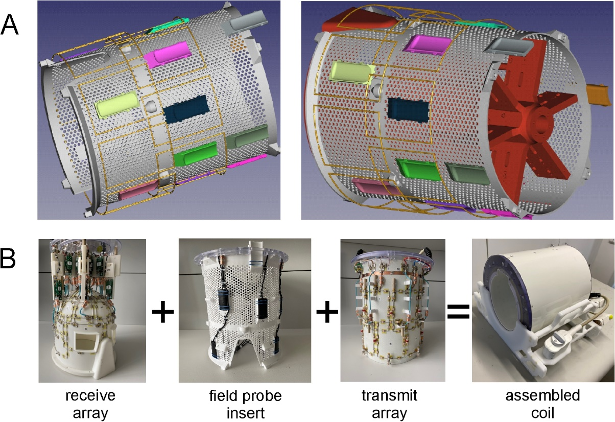
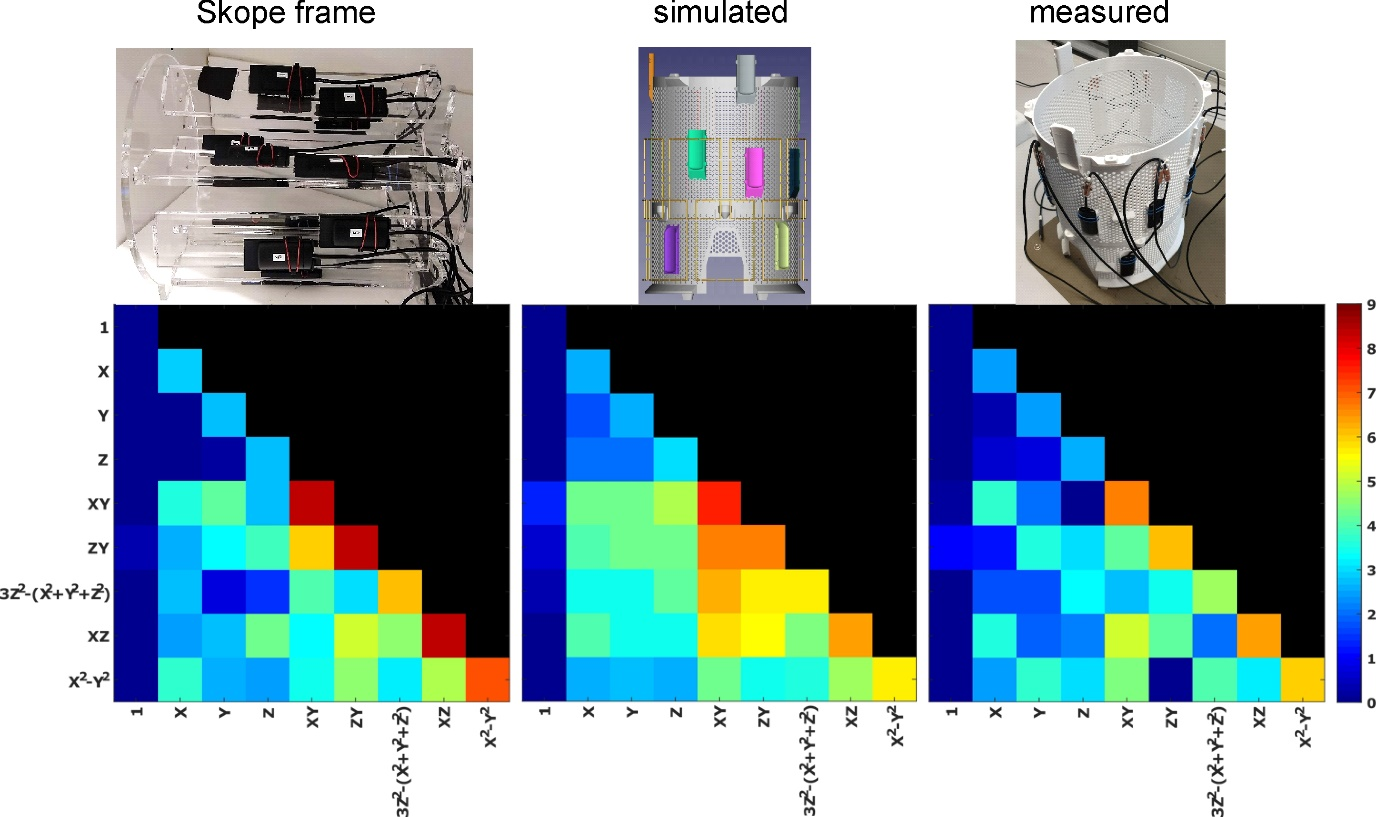
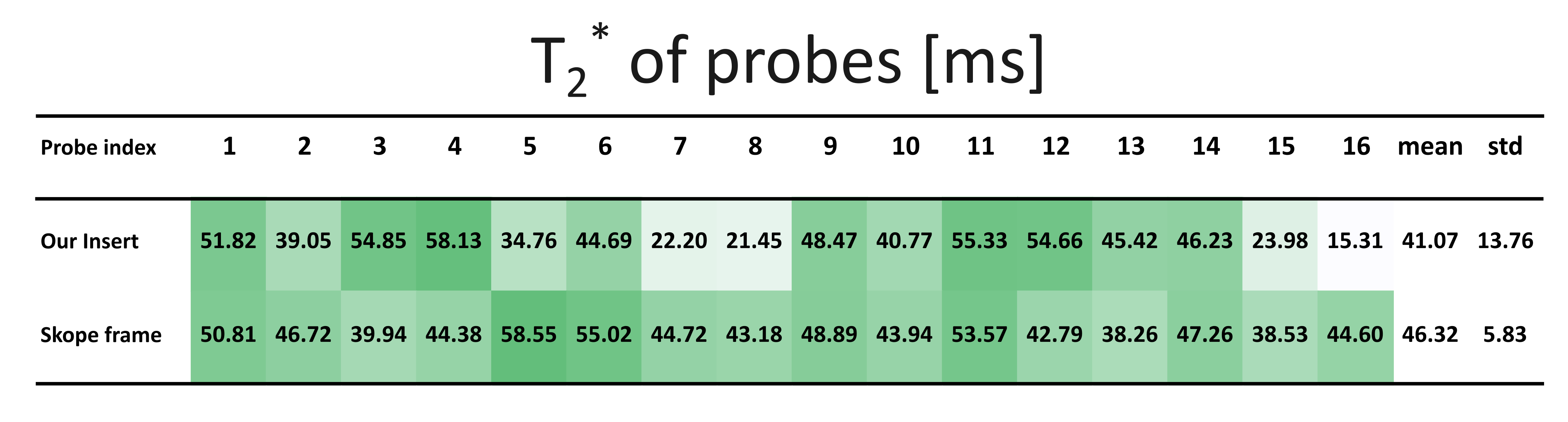
Figure 3: T2* in ms of the individual probes when placed on our probe-insert and in the default provided frame. The data from for the insert were obtained during the breath hold experiment with a volunteer inside the coil.
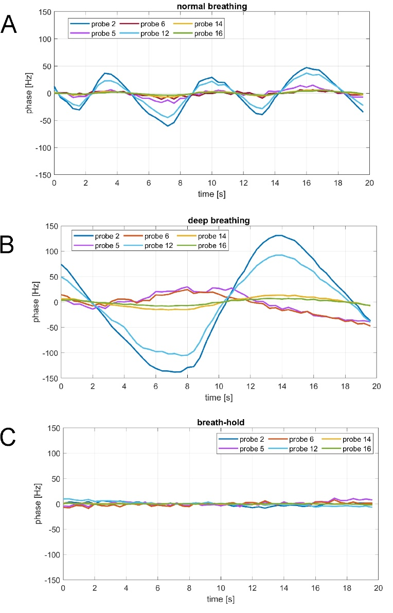
Figure 4: Phase variations measured on some sample probes during different breathing tasks. Some probes experienced strong field fluctuations, while others appeared to be unaffected by physiological B0 changes during normal breathing (A). During deep breathing (B), other probes also show phase variations. In these cases, however, movement cannot be completely excluded as an additional source of field changes. During a breath hold, the measured phase variations are only in the range of a few Hertz, indicating a high stability of the experiment itself.
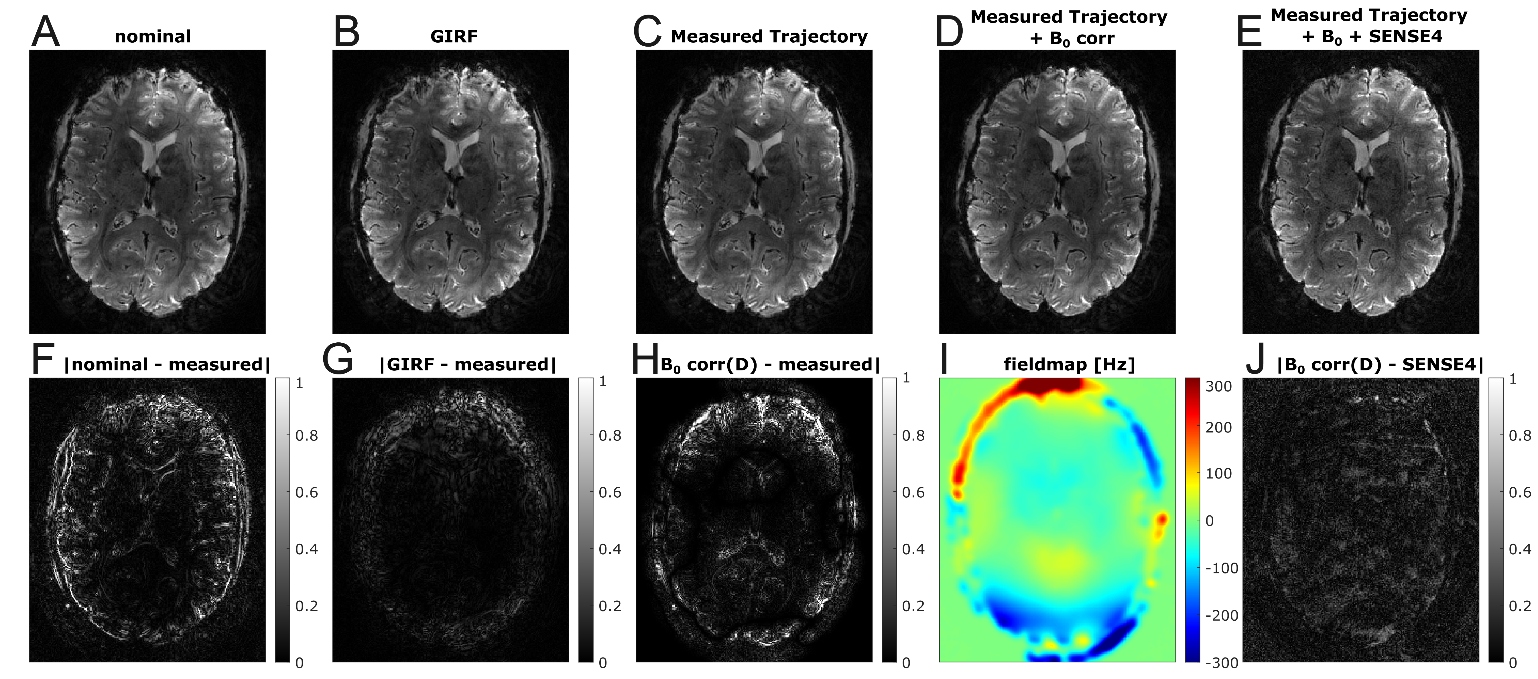
Figure 5: Reconstruction results with concurrent trajectory measurement. A 2D 0.75 mm multi-shot spiral (7 ms readout) is reconstructed with nominal trajectory (A), trajectory from gradient impulse response function (GIRF) predictions (B) , measured trajectory (C), measured trajectory with B0 correction (D) and parallel imaging (E). The second row shows the image differences to highlight the improvements (F-H,J) and field map (I) used for B0 correction.