4081
A 64-channel ex-vivo brain coil array for temperature-controlled diffusion imaging with the Connectome 2.0 MRI scanner1Institute of Medical Physics and Radiation Protection, Mittelhessen University of Applied Sciences, Giessen, Germany, 2Athinoula A. Martinos Center for Biomedical Imaging, Massachusetts General Hospital and Harvard Medical School, Charlstown, MA, United States
Synopsis
Keywords: RF Arrays & Systems, RF Arrays & Systems
Motivation: Ex-vivo brain DWI with long scan times poses the problem of temperature-related drift of diffusion measurement results.
Goal(s): The construction of a 64-channel ex-vivo brain coil with time-course temperature stabilization for obtaining accurate DWI measurements.
Approach: Combining a newly developed high-density ex-vivo brain coil array with a forced-air cooling system and a multi-channel temperature recording.
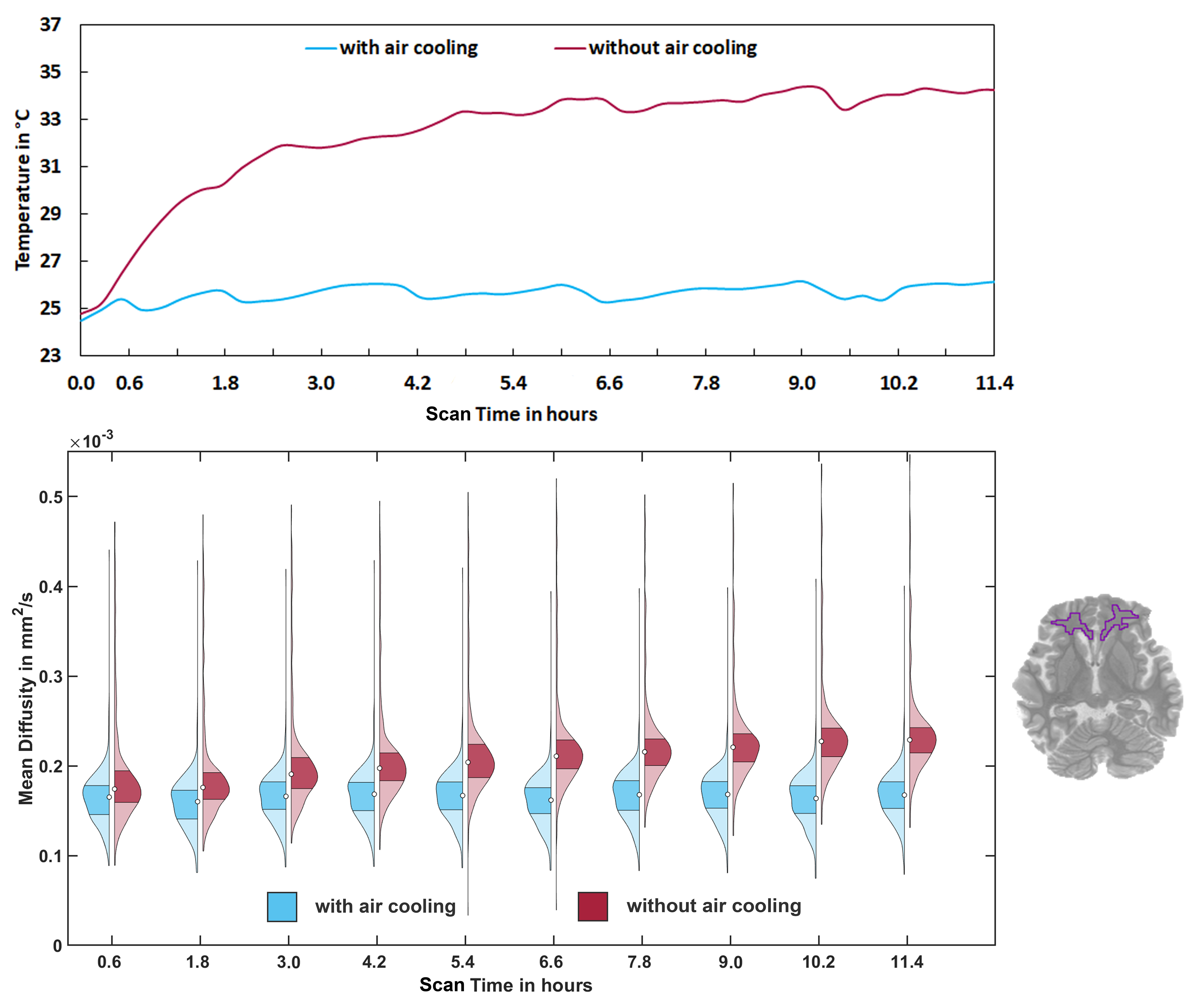
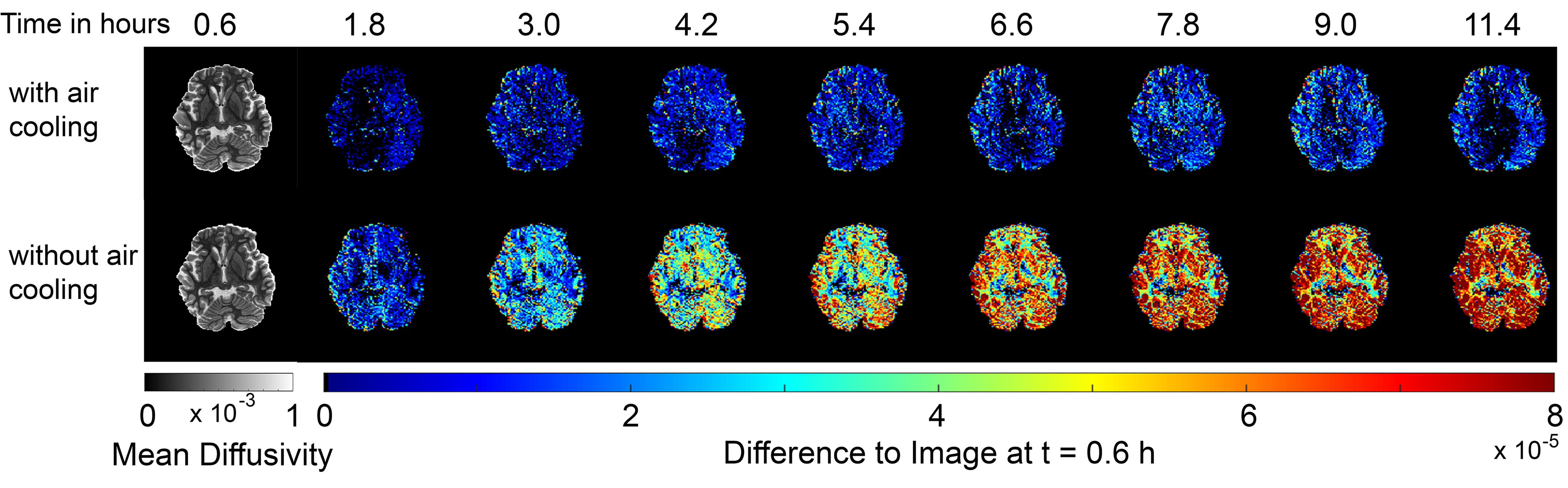
Results: The air circulation system was able to maintain the ambient temperature of the coil and, thus, stabilizing the mean diffusivity values over repeated lengthy scans. Without cooling, a drift of the mean diffusivity was overserved, peaking at a 35%-offset at approximately 11 hours.
Impact: Temperature-stabilized post-mortem brain samples for diffusion MRI in combination with a dedicated large channel count ex-vivo brain coil improves image quality in terms of achievable SNR and greatly reduced temperature-induced diffusivity shifts.
Introduction
Ex-vivo MRI offers several advantages over in-vivo imaging, such as unlimited scan time, absence of motion artifacts, and reduction of susceptibility artifacts [1]. In particular for connectome brain studies, ex-vivo imaging can provide detailed connectivity pathways in high resolution, when imaging is performed over long periods of time. However, there is no thermal body regulation in ex-vivo specimens. Therefore, the temperature rise of the ex-vivo sample, due to RF excitation and dissipated heat of the surrounded coil detector electronics, cannot be compensated. This is particularly critical as many ex-vivo scans are carried out, where the intended in-vivo SAR limit is deliberately far exceeded. Since the diffusivity of molecules depends on temperature, the DWI results are being distorted by the rising tissue temperature. To mitigate these limitations in ex-vivo high-resolution diffusion-weighted images, we developed a dedicated 64-channel ex-vivo brain receive coil, outfitted with temperature probes and a forced-air circulation system. The coil was designed for the new Connectome 2.0 scanner with a Gmax of 500 mT/m and a SRmax of 600 T/m/s.Methods
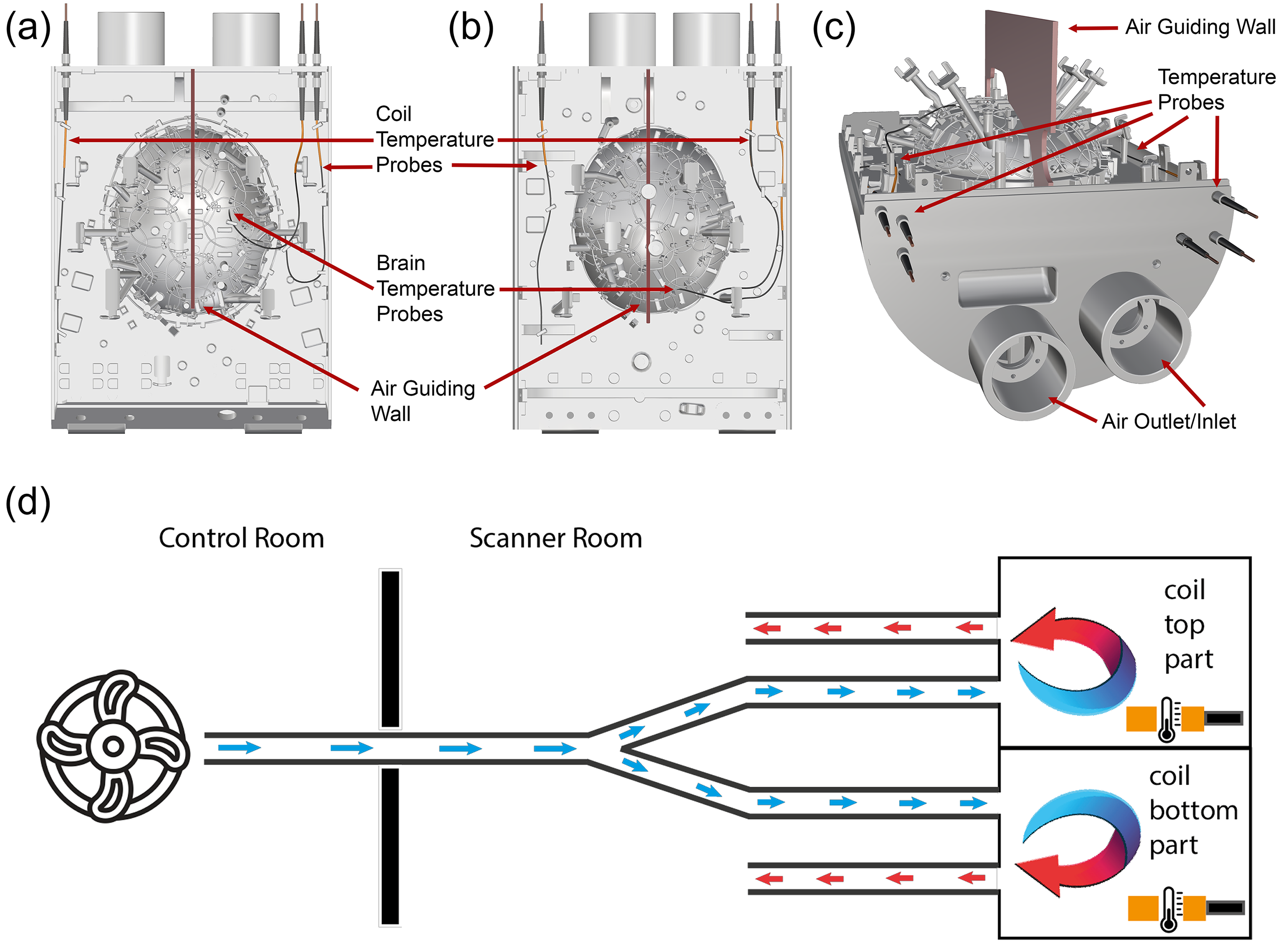
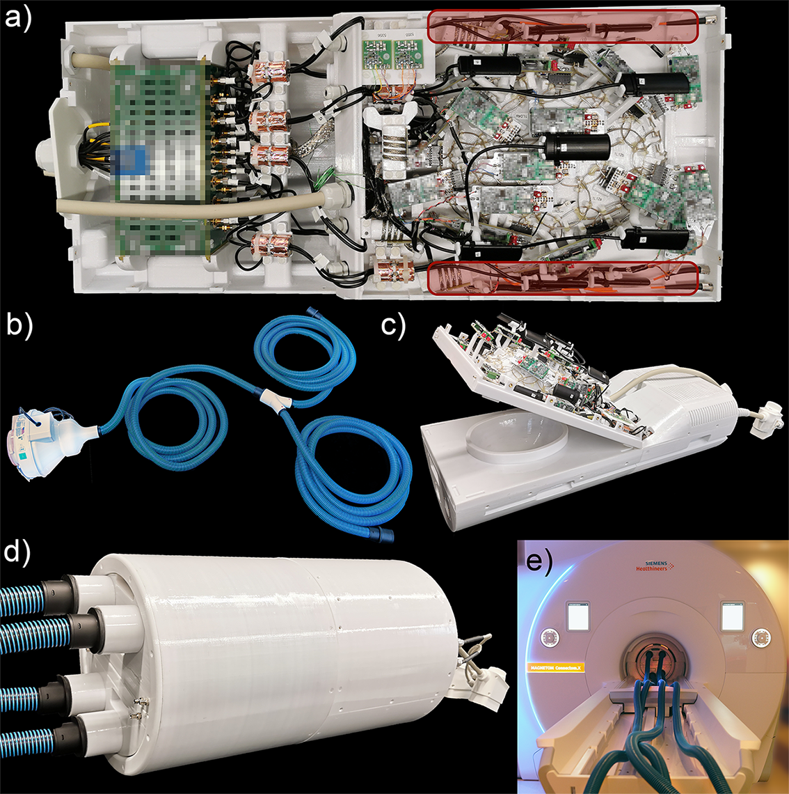
Radiofrequency Coil: The anatomically conormal ex-vivo receive coil comprises 64 loops arranged to completely enclose a whole brain specimen [2]. It is equipped with an integrated field monitoring system (Skope, Zurich, Switzerland), that enhances the coil's suitability for diffusion imaging with ultra-high performance gradient coils.Temperature stabilization system: The 64-channel ex-vivo array coil is outfitted with six fiber optic temperature probes (PRB-100-STM-MRI, OSENSA Innovations Corp, Burnaby, BC, Canada) and a circulating forced-air cooling system (Figure 1). Two temperature probes are located directly at the brain sample compartment of the array coil to measure the brain’s ambient temperature. The other four probes are placed inside the coil housing (upper and lower segment) to monitor the airflow temperature, which is supplied via to the coil via connected air hoses (Figure 1 a-c). A duct fan outside the scanner room blows (250 cfm @ 3/8 in. of H₂O) room temperature air into the air hose, which passes through a waveguide into the scanner room and to the coil. Inside the coil housing, built-in walls guide the forced air throughout the coil segments to the designated air outlets. This ensures a constant exchange of the accumulated air inside the coil housing (Figure 1 and 2).
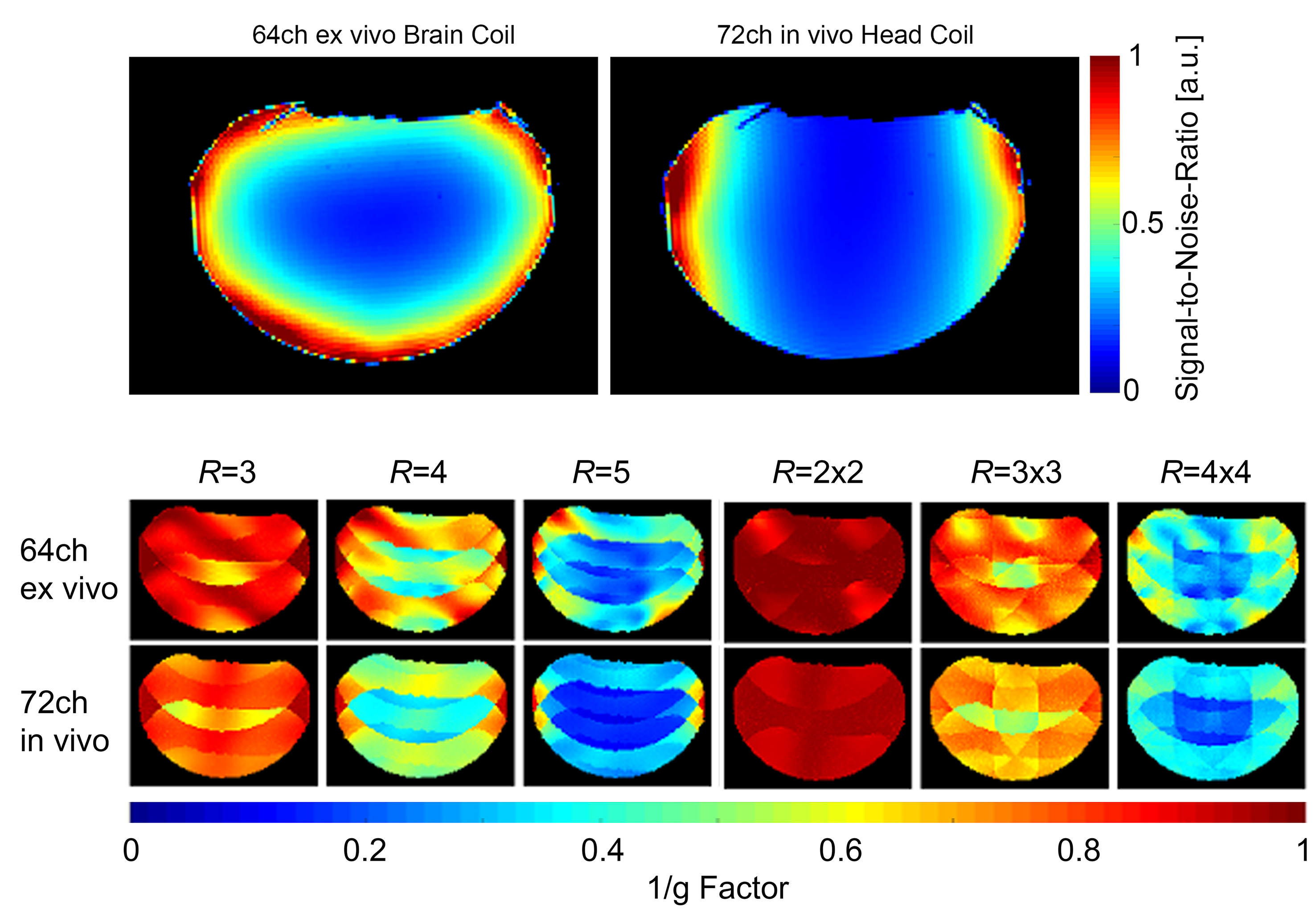
Measurements: SNR and g-maps were measured from GRE images obtained from an agar brain-shaped phantom using the 3T Connectome 2.0 scanner (MAGNETOM Connectom.X, Siemens Healthineers, Erlangen, Germany) [3] and compared to 72-channel in-vivo head coil. To assess the cooling system’s capability for stabilizing the temperature dependent diffusivity drifts during long scans, we carried out two measurement series of the brain sample’s mean diffusivity (MD) with and without forced air cooling, while monitoring the temperature (acquisition parameters in Figure 3 and 4).
Results
Rx coil: SNR and g-factor-maps measurements (Figure 3) showed an increased SNR and slightly lower g-factor performance of the developed 64-channel brain coil, when compared to the 72-channel in-vivo head coil.Temperature system: The forced air-cooling system was able to maintain the ambient temperature of the coil and, thus, stabilizing the mean diffusivity of the brain sample, when measured 29 repeated DWI scans with a total acquisition time of 11.6 hours. Without any cooling, the temperate of the coil gradually increased up to 10 °C (Figure 4a), leading in a severe increase of mean diffusivity values of white matter tissue by 35% (Figure 4b and 5).
Discussion
Long scan-time ex-vivo DWI with ultra-high performance gradient systems suffer from temperature-related diffusivity drifts [4,5]. In addition, high b-value diffusion images suffer from low SNR. The constructed ex-vivo brain coil addresses this limitation, by increasing the reception sensitivity utilizing a high-density coil array, populated onto an anatomical conormal ex-vivo brain coil former. The coil system is designed to enable accurate quantitative post-mortem MRI scans by keeping the temperature of the brain sample stable over long scan times. This is crucial, since T1, T2, and water diffusivity are temperature dependence, which can lead to a strong bias in parametric imaging [6]. We used forced air as a cooling medium, because it does not generate any signal in the image. Our approach benefits from excellent performance despite its simplicity and easy handling due to the direct coil integration.Conclusion
The constructed array coil is well-suited for imaging post-mortem brain samples for long scan times. The integrated temperature stabilization system can effectively minimize temperature drifts in DWI, thus improving the parametric accuracy of the DWI results in ex-vivo studies. The high SNR performance of the coil enables high-resolution ex-vivo diffusion images with high b-value.Acknowledgements
This work was supported by the National Institute of Biomedical Imaging and Bioengineering of the National Institutes of Health under award number U01EB026996.
Moreover, we would like to thank Andreas Potthast, René Gumbrecht, and Jasmine Fischer for the assistance in integrating the constructed coil into the Connectome 2.0 scanner. Special thanks also go to Susie, Chiara and Gabriel for your efforts preparing the long scan sessions last weekend and in putting together the air-cooling system.
References
[1] Roebroeck, Alard, Karla L. Miller, and Manisha Aggarwal. "Ex vivo diffusion MRI of the human brain: Technical challenges and recent advances." NMR in Biomedicine 32.4 (2019): e3941.
[2] Scholz, Alina, Mahmutovic, Mirsad, Alem, Mona, Müller, Roland, Schlumm, Torsten, Möller, Harald E., Yendiki, Anastasia, Ramos-Llorden, Gabriel, Wald, Lawrence L., Huang, Susie Y., Keil, Boris. “Design of a 64-Channel ex vivo Brain Rx Array Coil with field monitoring and temperature control for DWI at 3T” Annual Meeting of Intl Soc Magn Reson Med, Toronto, Canada (2023) #5548.
[3] Huang, Susie Y., et al. "Connectome 2.0: Developing the next-generation ultra-high gradient strength human MRI scanner for bridging studies of the micro-, meso-and macro-connectome." NeuroImage (2021): 118530.
[4] D’Arceuil, Helen E., Susan Westmoreland, and Alex J. de Crespigny. "An approach to high resolution diffusion tensor imaging in fixed primate brain." Neuroimage 35.2 (2007): 553-565.
[5] Rieger, Sebastian W., et al. "A temperature‐controlled cooling system for accurate quantitative post‐mortem MRI." Magnetic Resonance in Medicine 90.6 (2023): 2643-2652.
[6] D’Arceuil, Helen E., Susan Westmoreland, and Alex J. de Crespigny. "An approach to high resolution diffusion tensor imaging in fixed primate brain." Neuroimage 35.2 (2007): 553-565.
Figures