4070
MR-based glymphatic function: association between DTI-ALPS and perivascular space1Radiology, ASAN MEDICAL CENTER, SEOUL, Korea, Republic of
Synopsis
Keywords: Aging, Diffusion Tensor Imaging, Other diffusion models
Motivation: The glymphatic system is presumed to be associated with perivascular space (PVS).
Goal(s): To identify the relationship between the glymphatic system (ALPS index), PVS, and cerebral white matter hyperintensities (WMH) according to age.
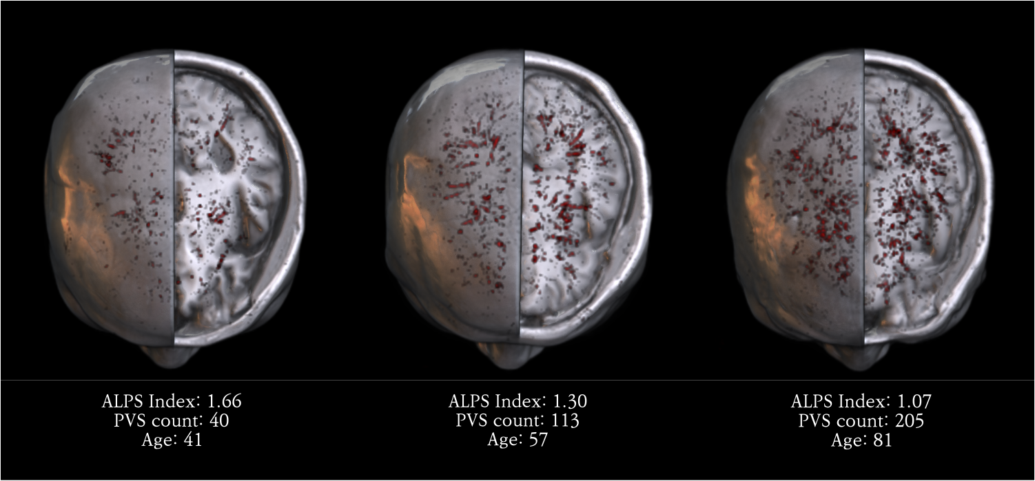
Approach: Participants indicated for brain MRI for cognitive decline (n=611) were retrospectively included. The glymphatic function was assessed by ALPS index calculated from diffusion tensor imaging along the perivascular space (DTI-ALPS). PVS volumes were automatically quantified via deep learning segmentation model. FLAIR WMH were also automatically quantified.
Results: ALPS index showed a significant negative correlation with WMH and PVS in the age group of 50–59.
Impact: WMH show a negative correlation with ALPS index, indicating poor glymphatic function. However, only age group of 50–59 shows such relationship. This suggests that aging and enlarged PVS might have diminished glymphatic function as reflected in decreased ALPS index.
Introduction
Recently, the glymphatic system has received much research attention with its role as a clearance pathway in the CNS. The gold standard of assessing the glymphatic system is the serial follow-up MRI after intrathecal gadolinium contrast agent, which is an invasive technique. To overcome this limitation, diffusion tensor imaging along the perivascular space (DTI-ALPS) has emerged as the alternative non-invasive method for assessing the glymphatic function in many previous studies. Briefly, DTI-ALPS is based on the water molecule diffusion along the ‘perivascular space’, which is often visible on high-resolution MRI. The ALPS index can be calculated from DTI with lower values (near 1) indicating poor glymphatic function. In the current study, we sought to find the potential association between DTI-ALPS (ALPS index) and MRI-visible perivascular space (PVS) and cerebral white matter hyperintensities (WMH) on FLAIR.Methods
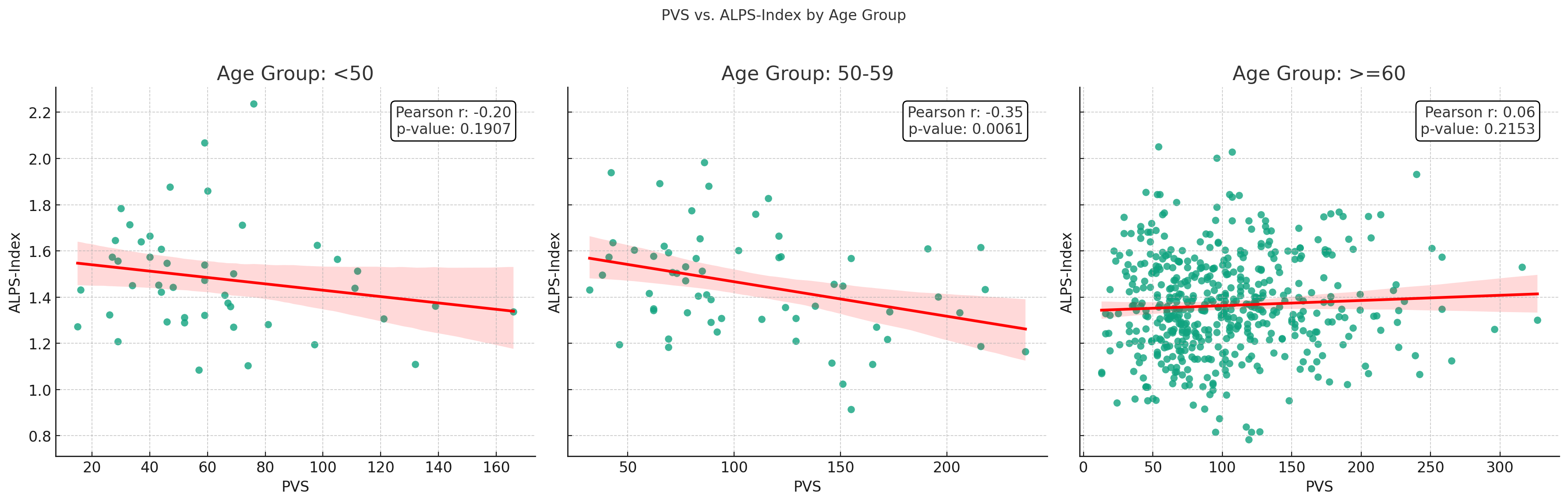
In a cohort of 611 subjects with MRI indications for cognitive decline, we calculated the ALPS index from three region-of-interests drawn onto projection, association, and subcortical directions on DTI. FLAIR WMH and perivascular spaces (PVS) were automatically quantified based on deep learning segmentation models. For PVS quantification, only PVS volumes above the threshold of 5 mm3 were selected. Correlation analyses between the ALPS index and WMH, PVS, and age were performed. Linear regression analyses were also performed with the ALPS index as the dependent variable. Furthermore, for subgroup correlation analysis, we stratified WMH and PVS according to age groups: <50, 50–60, and >60.Results
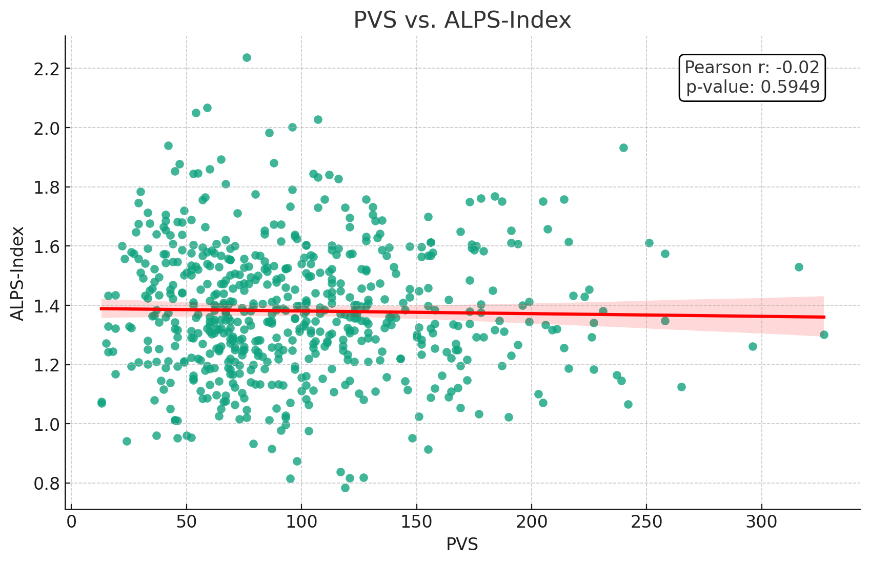
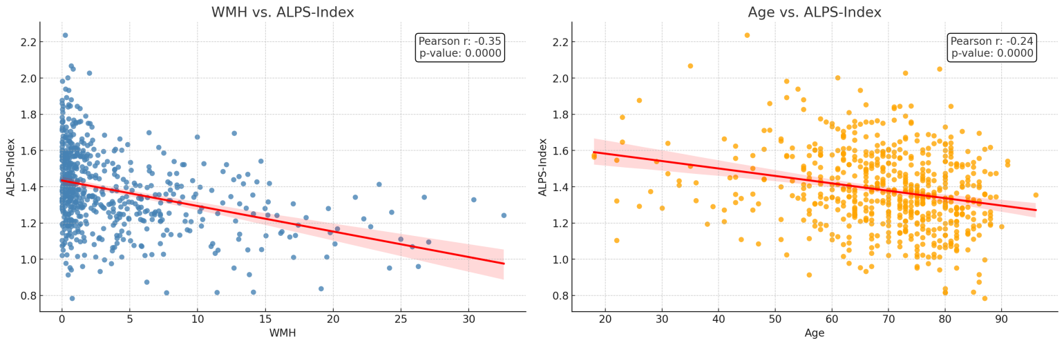
The ALPS index demonstrated significant negative correlations with age (ρ = -0.24, p < 0.001), WMH (ρ = -0.35, p < 0.001), but not with PVS (ρ = -0.02, p = 0.59). In subgroup analysis according to age groups, WMH demonstrated significant negative correlations with the ALPS index in all age groups (ρ =-0.29–-0.34, p < 0.05). However, only the age group of 50–60 demonstrated a significant negative correlation with the ALPS index (ρ =-0.35, p = 0.006). In univariate linear regression analysis, WMH (R2=0.12, p<0.001) and age (R2=0.06, p<0.001) were significantly associated with the DTI-ALPS while PVS was not (R2=0.0005, p=0.59). Similarly, in multivariate linear regression, WMH (p<0.001) and age (p=0.002), but not PVS (p=0.182), were significantly associated with the DTI-ALPS.Discussion
WMH on FLAIR is associated with aging and is regarded as an imaging biomarker for various neurodegenerative diseases. The significant negative correlation between WMH and ALPS index indicates that there might be a link between WMH and the glymphatic function. WMH is often attributable to chronic ischemic changes of the cerebral white matter due to small vessel diseases, which may impair the tiny perforating vessels along the perivascular spaces. Interestingly, although WMH is closely associated aging, the reduction in the ALPS index attributable to WMH is more pronounced (ρ = -0.35, R2=0.12) than that associated with aging alone (ρ = -0.24, R2=0.06), which suggests that WMH might have a more influence on the glymphatic function than aging.Unlike our assumption, a significant correlation was found between the ALPS index and PVS only in the age group of 50–60. In subjects older than 60 years of age, a substantial number of subjects had enlarged PVS volumes >5mm³, which might indicate the diminished function of PVS. This condition could compromise the molecular diffusion along the PVS, whereby an increase in the number of PVS has little impact on the ALPS index.
Conclusion
Aging and cerebral WMH had significant negative correlations with the ALPS index, suggesting they negatively affect the glymphatic function. In age group younger than 60, PVS volumes had a significant negative correlation with the ALPS index while older subjects had no significant association. Older subjects had more enlarged PVS, which subsequently would have little influence on the DTI-ALPS.Acknowledgements
No acknowledgement found.References
1. Cacciaguerra, Laura, et al. "Magnetic resonance imaging evaluation of perivascular space abnormalities in neuromyelitis optica." Annals of Neurology 92.2 (2022): 173-183.
2. Ke, Zhihong, et al. "Glymphatic dysfunction mediates the influence of white matter hyperintensities on episodic memory in cerebral small vessel disease." Brain Sciences 12.12 (2022): 1611.
3. Ma, Xinxin, et al. "Diffusion tensor imaging along the perivascular space index in different stages of Parkinson’s disease." Frontiers in Aging Neuroscience 13 (2021): 773951.
4. Liu, Hui, et al. "Associations among diffusion tensor image along the perivascular space (DTI-ALPS), enlarged perivascular space (ePVS), and cognitive functions in asymptomatic
5. Hablitz, Lauren M., and Maiken Nedergaard. "The glymphatic system." Current Biology 31.20 (2021): R1371-R1375.patients with carotid plaque." Frontiers in Neurology 12 (2022): 789918.
Figures