4065
Cerebral macro- and micro-structural impairment are neuroradiological mediators for serum uric acid variance-related cognitive decline1Department of Radiology, beijing friendship hospital, Capital Medical University, Beijing, China
Synopsis
Keywords: Dementia, Dementia, Serum uric acid; Magnetic resonance imaging; Brain tissue volume; White matter microstructural integrity; Cognitive function
Motivation: The longitudinal effects of changes in serum uric acid (SUA) levels on brain health are largely unknown.
Goal(s): This study aimed to evaluate the longitudinal associations of SUA variance with neuroimaging indices and cognitive function.
Approach: The multivariate-adjusted associations of SUA variance with brain MRI markers and cognitive function were examined using generalized linear models and logistic regression models. Mediation analyses were performed to assess whether brain MRI markers mediate the relationship between SUA variance and cognitive function.
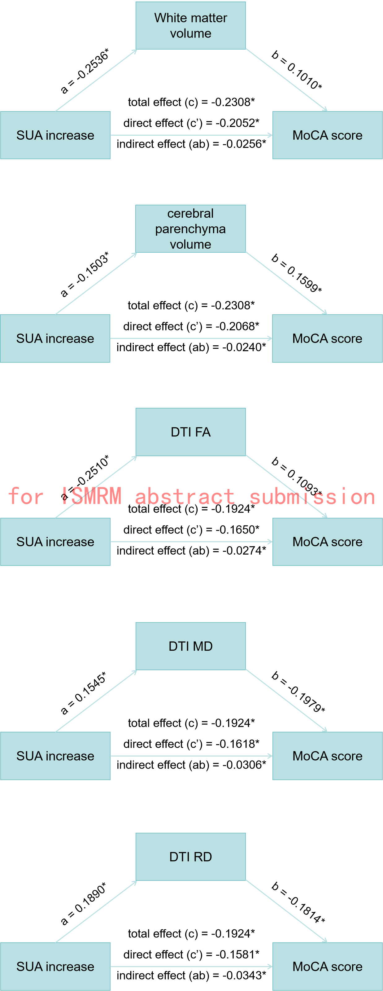
Results: Changes in SUA concentration, particularly elevated SUA levels damages brain health, manifested as smaller brain tissue volume, impaired microstructural integrity, and poorer cognitive performance.
Impact: In conclusion, our study findings deepen the comprehension of an integrated relationship between SUA variance and neuroimaging indices of brain health. Long-term prevention of SUA fluctuation is essential for protecting brain health and preventing early-stage dementia.
Methods: This cohort study recruited 1,111 participants aged 25-83 years from the Kailuan study. The SUA concentrations were measured every two years from 2006 to 2018. We primarily assessed SUA variance as the average slope incorporating seven measurements and further defined the direction of changes. The multivariate-adjusted associations of SUA variance with brain MRI markers [brain tissue volumes, microstructural integrity, white matter hyperintensity (WMH), and cerebral small vessel disease] and cognitive function were examined using generalized linear models and logistic regression models. Mediation analyses were performed to assess whether brain MRI markers mediate the relationship between SUA variance and cognitive function.
Results: Compared with the stable group, brain white matter volume decreased irrespective of the increase or decrease in SUA levels (beta=−0.26, 95% confidence interval [CI] −0.40 to 0.12 and beta=−0.15, 95% CI −0.28 to −0.02). Elevated SUA levels were also associated with a smaller cerebral parenchyma volume (beta=−0.14, 95% CI −0.25 to −0.04). Participants with progressively increased SUA exhibited lower global fractional anisotropy (beta=−0.27, 95% CI −0.41 to −0.13), as well as higher mean diffusivity (beta=0.20, 95% CI 0.07 to 0.33) and radial diffusivity (beta=0.23, 95% CI 0.10 to 0.36). Elevated SUA during follow-up was also associated with cognitive decline (beta=−0.20, 95% CI −0.33 to −0.06). Additionally, cerebral parenchyma and white matter atrophy, and impaired brain microstructural integrity mediated the impact of SUA increase on cognitive decline.
Conclusion: Changes in SUA concentration, particularly elevated SUA levels detrimentally affect brain health, manifested as smaller brain tissue volume, impaired microstructural integrity, and poorer cognitive performance. Long-term prevention of SUA fluctuation is essential for protecting brain health and preventing early-stage dementia.
Acknowledgements
The authors would like to thank all the involved study investigators, clinicians, nurses, and technicians for dedicating their time and skills to the completion of this study.References
Tang X, Song ZH, Cardoso MA, Zhou JB, Simo R. The relationship between uric acid and brain health from observational studies. Metab Brain Dis 2022;37:1989-2003.5.
Verhaaren BF, Vernooij MW, Dehghan A, et al. The relation of uric acid to brain atrophy and cognition: the Rotterdam Scan Study. Neuroepidemiology 2013;41:29-34.6.
Shih CY, Chen CY, Wen CJ, Liu HM, Kuo HK. Relationship between serum uric acid and cerebral white matter lesions in the elderly. Nutr Metab Cardiovasc Dis 2012;22:154-159.7.
Beydoun MA, Canas JA, Dore GA, et al. Serum Uric Acid and Its Association with Longitudinal Cognitive Change Among Urban Adults. J Alzheimers Dis 2016;52:1415-1430.10.
Wang T, Wu Y, Sun Y, Zhai L, Zhang D. A Prospective Study on the Association between Uric Acid and Cognitive Function among Middle-Aged and Older Chinese. J Alzheimers Dis 2017;58:79-86.
Figures