4064
Association of MRI-derived Glymphatic Function with Cognition and Cerebral Small Vessel Disease1Institute of Science and Technology for Brain-Inspired Intelligence, Fudan University, Shanghai, China, 2Key Laboratory of Computational Neuroscience and Brain-Inspired Intelligence (Fudan University), Ministry of Education, Shanghai, China, 3Department of Materials Science, Fudan University, Shanghai, China, 4Department of Neurology, Yueyang Hospital of Integrated Traditional Chinese and Western Medicine, Shanghai University of Traditional Chinese Medicine, Shanghai, China, 5USC Viterbi School of Engineering, University of Southern California, Shanghai, China, 6Laboratory of FMRI Technology, USC Mark & Mary Stevens Neuroimaging and Informatics Institute, Keck School of Medicine, University of Southern California, Shanghai, China, 7MR Collaboration, Siemens Healthcare Ltd., Shanghai, China, 8Department of Neurology, Zhongshan Hospital, Fudan University, Shanghai, China
Synopsis
Keywords: Dementia, Dementia, cerebral small vessel disease, glymphatic system
Motivation: Both cerebral small vessel disease (CSVD) and disfunction of glymphatic system contribute to cognition decline, yet the interactions between them remains unclear.
Goal(s): Identify associations of glymphatic function with cognitive function and CSVD.
Approach: 111 CSVD subjects were involved and underwent 7T MRI scans. CSVD makers, neuropsychological test and clinical characteristics were collected. Linear regression and mediation model were used to access the associations.
Results: Age and CSVD markers were associated with glymphatic function in the multivariable model. The relationship between glymphatic function and cognition was mediated by CSVD burden.
Impact: MRI-derived glymphatic features might be utilized to predict early CSVD-induced cognitive decline.
Introduction
The glymphatic system is a pivotal component in the central nervous system responsible for clearing metabolic waste, and its wellness is crucial for cognitive function and overall brain health1. Diffusion-weighted MRI has emerged as a minimally invasive method to assess glymphatic function by measuring diffusion Along the Perivascular Space (DTI-ALPS) index2. Previous research has established a correlation between this MRI feature and factors such as cognition, Alzheimer's disease, and cerebral small vessel disease (CSVD)3,4, however, the complex interactions involved remain unclear. This study aims to systematically evaluate the associations of glymphatic function with cerebrovascular risk factors, cognitive function and various imaging markers of CSVD. Moreover, mediation analysis models are used to evaluate potential pathophysiological linkages.Methods
This study was approved by the local ethics committee. Subjects in this study were part of an ongoing single-center prospective longitudinal 7T MRI study5. All individuals underwent multimodality MRI with one 7.0T scanner (MAGNETOM Terra, Siemens Healthcare, Erlangen, Germany) using a 32-channel brain phased array coil. The MRI protocol included: (1) T1-weighted 3D-MP2RAGE sequence with the following parameters: voxel size=0.7×0.7×0.7mm3, TR/inversion time/TE=3800/2.27/2700; (2) 3D FLAIR sequence with the following parameters: voxel size=0.7×0.7×0.7mm3, TR/TE=9000/270ms, flip angle=120; (3) SWI sequence with the following parameters: pixel size=0.12×0.12 mm2, slice thickness=1.5mm with no gap between slices, TR/TE=21/9.54ms, flip angle=10. (4) Diffusion sequence with the following parameters: voxel size=1.4×1.4×1.4mm3, TR/TE=4200/67ms, flip angle=90, b value=0/1000/2000 s/mm2, direction = 64. Imaging markers of CSVD, including white matter hyperintensity (WMH), perivascular space (PVS), lacuna and cerebral microbleed (CMB) were assessed according to the Standards for Reporting Vascular Changes on Neuroimaging6. PVS was defined as a small, sharply delineated structure of cerebrospinal fluid intensity with a diameter generally <3mm. Lacunas were defined as round or ovoid cavities ranging 3 to 15mm situated in the basal ganglia, subcortical white matter, or brainstem. CMBs were defined as round or ovoid black lesions that were <10mm on SWI. WMHs were rated by the Fazekas visual grading scale7. DTI-ALPS was derived from diffusion images by the methods in previous studies2. Neuropsychological test and clinical characteristics were collected by trained staff. The neuropsychological tests including Montreal cognitive assessment (MoCa), Mini Mental State Examination (MMSE), Auditory Verbal Learning Test (AVLT), Boston Naming Test (BNT) and Trail Making Test (TMT). General linear model was used to examine the associations of glymphatic function with cerebrovascular risk factors, cognitive function and CSVD. To further examine the relationships among glymphatic function, CSVD, and cognitive function, the mediation analysis was conducted with a linear single-mediator model8. The mediation models used CSVD features as the mediator, cognitive function as the response, and treated BP as the determinant. All the relationships between the independent variable, mediator, and dependent variable were represented by the linear model. The significance of the mediation effect was estimated using bias-corrected and accelerated bootstrapping method with 5000 resamples.Results and Discussion
The characteristics of the subjects are shown in Figure 1. The study included a cohort of 111 individuals with a mean (SD) age of 55.3 (11.9) years. As shown in Figure 2, age, diabetes, hypertension, MoCa, MMSE, AVLT, TMT and all CSVD markers were significantly associated with the glymphatic function in the bivariable model. However, the multivariate analysis revealed that glymphatic function was significantly related only to age, summary small vessel disease (SVD) score, and the presence of lacunes. This suggests that aging remains the predominant factor affecting glymphatic system function9. The impact of the glymphatic system on cognitive function became insignificant after introducing of aging, indicating that the relationship between them might be influenced by other potential intermediate factors. The mediation analysis in this study offers a potential hypothesis that glymphatic dysfunction may affect cognitive performance by influencing the CSVD burden. Figure 3 shows a significant mediation effect where the DTI-ALPS score and MMSE score were mediated by the summary SVD score and PVS score. The MRI-visible PVS is one of the early markers of CSVD10, and its association with cognitive function, indicated by its closer correlation to the PVS score than to the summary SVD score (with significant correlations to both MMSE and MoCA), suggests that PVS might be a more sensitive CSVD feature related to cognition. Furthermore, the intricate connection between the PVS and the glymphatic system, combined with the pathway findings presented in this study, offers a potential pathological route regarding the cognition decline at the imaging level, which might play a significant role in future clinical diagnostics and interventions.Acknowledgements
We are greatly thankful to all the members in our research group at Fudan University and Yueyang Hospital who helped to accomplish the study. This work was supported by the National Natural Science Foundation of China (No. 81971583, No. 82271956), Shanghai Municipal Science and Technology Major Project (No. 2018SHZDZX01), National Key R&D Program of China (No. 2018YFC1312900).References
1. Nedergaard M, Goldman SA. Glymphatic failure as a final common pathway to dementia. Science 2020;370:50-56.
2. Taoka T, Masutani Y, Kawai H, et al. Evaluation of glymphatic system activity with the diffusion MR technique: diffusion tensor image analysis along the perivascular space (DTI-ALPS) in Alzheimer's disease cases. Jpn J Radiol 2017;35:172-178.
3. Hsu JL, Wei YC, Toh CH, et al. Magnetic Resonance Images Implicate That Glymphatic Alterations Mediate Cognitive Dysfunction in Alzheimer Disease. Ann Neurol 2023;93:164-174.
4. Carotenuto A, Cacciaguerra L, Pagani E, Preziosa P, Filippi M, Rocca MA. Glymphatic system impairment in multiple sclerosis: relation with brain damage and disability. Brain 2022;145:2785-2795.
5. Huo Y, Wang Y, Guo C, et al. Deep white matter hyperintensity is spatially correlated to MRI-visible perivascular spaces in cerebral small vessel disease on 7 Tesla MRI. Stroke Vasc Neurol 2023;8:144-150.
6. Wardlaw JM, Smith EE, Biessels GJ, et al. Neuroimaging standards for research into small vessel disease and its contribution to ageing and neurodegeneration. Lancet Neurol 2013;12:822-838.
7. Fazekas F, Chawluk JB, Alavi A, Hurtig HI, Zimmerman RA. MR signal abnormalities at 1.5 T in Alzheimer's dementia and normal aging. AJR Am J Roentgenol 1987;149:351-356.
8. Kim HJ, Park S, Cho H, et al. Assessment of Extent and Role of Tau in Subcortical Vascular Cognitive Impairment Using 18F-AV1451 Positron Emission Tomography Imaging. JAMA Neurol 2018;75:999-1007.
9. Lohela TJ, Lilius TO, Nedergaard M. The glymphatic system: implications for drugs for central nervous system diseases. Nat Rev Drug Discov 2022;21:763-779.
10. Duering M, Biessels GJ, Brodtmann A, et al. Neuroimaging standards for research into small vessel disease-advances since 2013. Lancet Neurol 2023;22:602-618.
Figures
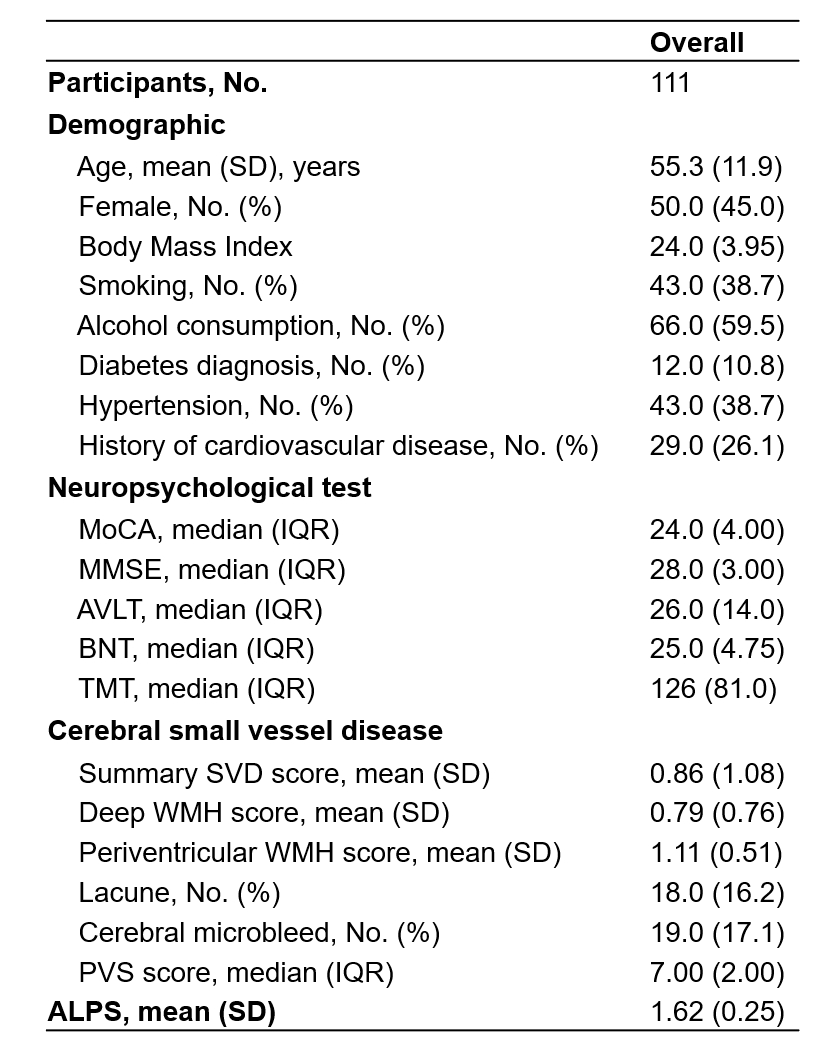
Figure 1.The characteristics of the subjects.
MoCA, Montreal cognitive assessment; MMSE, Mini Mental State Examination; AVLT, Auditory Verbal Learning Test; BNT, Boston Naming Test; TMT, Trail Making Test; SVD, small vessel disease; WMH, white matter hyperintensity; PVS, perivascular space; ALPS, Along the Perivascular Space.
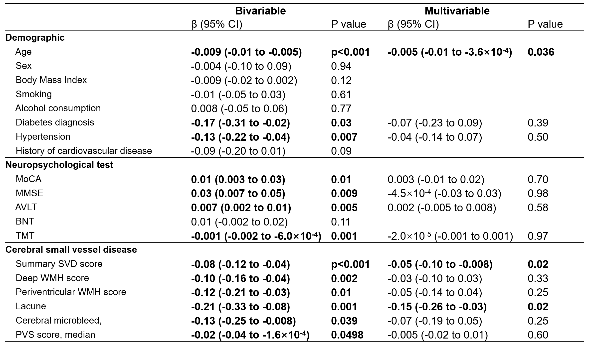
Figure 2. Association of glymphatic function with cerebrovascular risk factors, cognitive function and cerebral small vessel markers.
MoCA, Montreal cognitive assessment; MMSE, Mini Mental State Examination; AVLT, Auditory Verbal Learning Test; BNT, Boston Naming Test; TMT, Trail Making Test; SVD, small vessel disease; WMH, white matter hyperintensity; PVS, perivascular space.
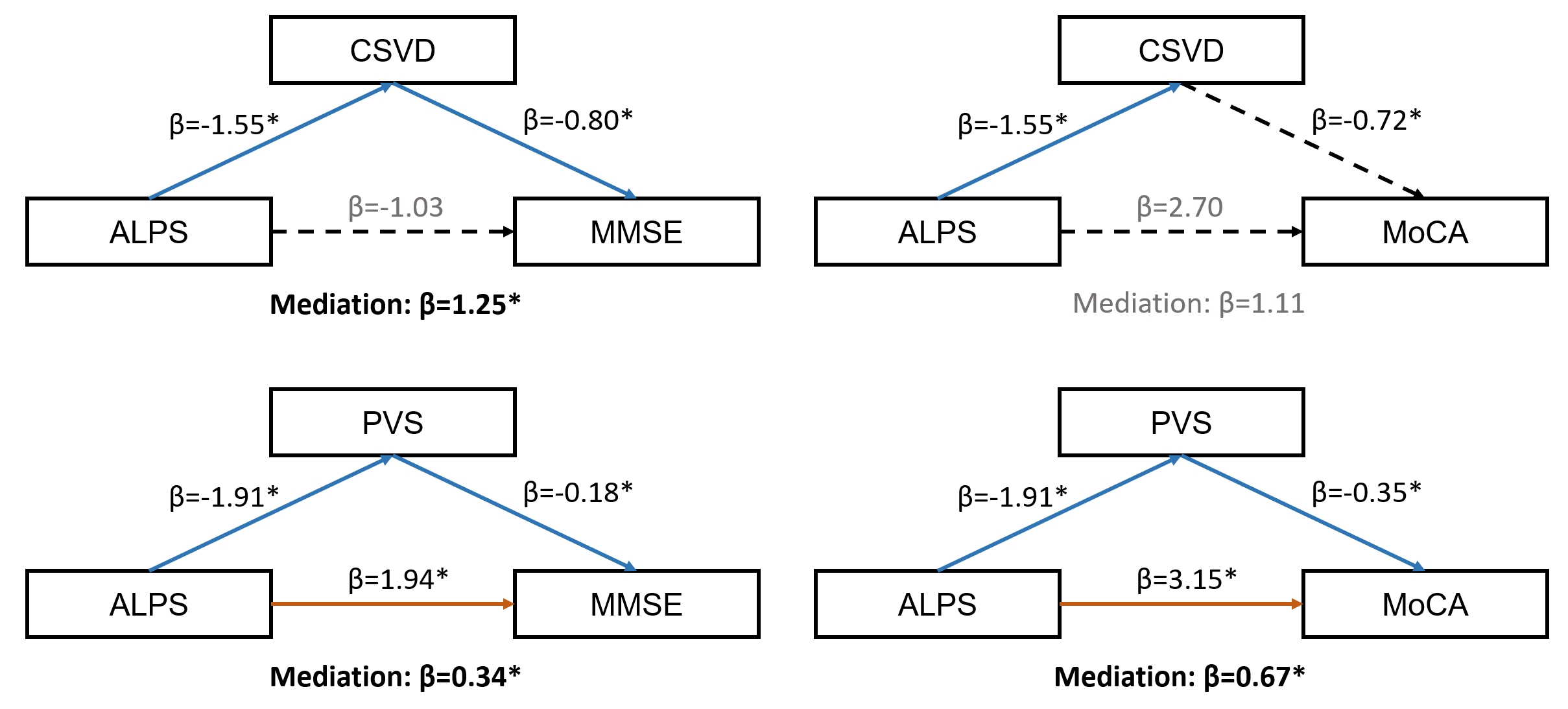
Figure 3. Mediation of cerebral small vessel disease with cognitive function and glymphatic function.
ALPS, Along the Perivascular Space; CSVD, summary small vessel disease score; MMSE, Mini Mental State Examination; MoCA, Montreal cognitive assessment; PVS, perivascular space.