4062
Cerebrospinal fluid-based spatial statistics: towards quantitative analysis of cerebrospinal fluid pseudodiffusivity1University of Cambridge, Cambridge, United Kingdom, 2Washington University, St Louis, MO, United States, 3Zhejiang University, Hangzhou, China
Synopsis
Keywords: Dementia, Diffusion/other diffusion imaging techniques, dementia, CSF, glymphatic
Motivation: Cerebrospinal fluid (CSF) circulation is crucial for removing waste from the brain, and abnormal CSF motion was associated with neurodegenerative diseases. This study aims to use low b-value diffusion MRI to assess the variance of motion of CSF, i.e., pseudodiffusivity, and investigate its association with cognitive impairment.
Goal(s): To quantify CSF pseudodiffusivity within each sulcus, cistern and ventricle.
Approach: In 93 participants from a memory clinic, Intravoxel incoherent motion (IVIM) MRI was performed to measure CSF pseudodiffusivity. Sulci and cisterns were segmented based on gray matter landmarks.
Results: In the third ventricle, CSF pseudodiffusivity was positively correlated with memory performance.
Impact: Our method of quantifying CSF pseudodiffusivity in different CSF regions in an unbiased, automatic fashion enabled discovery of potential novel non-invasive CSF-based imaging biomarkers of cognitive impairment.
Introduction
Cerebrospinal fluid (CSF) circulation is important in clearance of toxic substances and maintenance of optimal brain function (Mestre et al., 2020). Reduced CSF motion has been implicated in neurodegenerative diseases (Ringstad et al., 2017; Zhang et al., 2021). An important technique of quantifying CSF motion is intravoxel incoherent motion (IVIM), a non-invasive magnetic resonance imaging (MRI) sequence to study the motion of water molecules. IVIM is sensitive to a wide range of incoherent motion (0.1-1000 x 10-3 mm2/s) , i.e., pseudodiffusivity (Wong et al., 2020) and captures the variation of CSF diffusion pseudodiffusivity at a microscopic level (Bito et al., 2021).However, quantitative assessment of CSF pseudodiffusivity is challenging owing to extensive individual variations of the anatomy of the CSF-filled sulci, cisterns and ventricles, and the difficulty of registering low resolution IVIM signals onto a standard template for voxel-wise analysis. In this study, we introduced CSF-based spatial statistics (CBSS), a novel image analysis technique to address these two difficulties.
Method
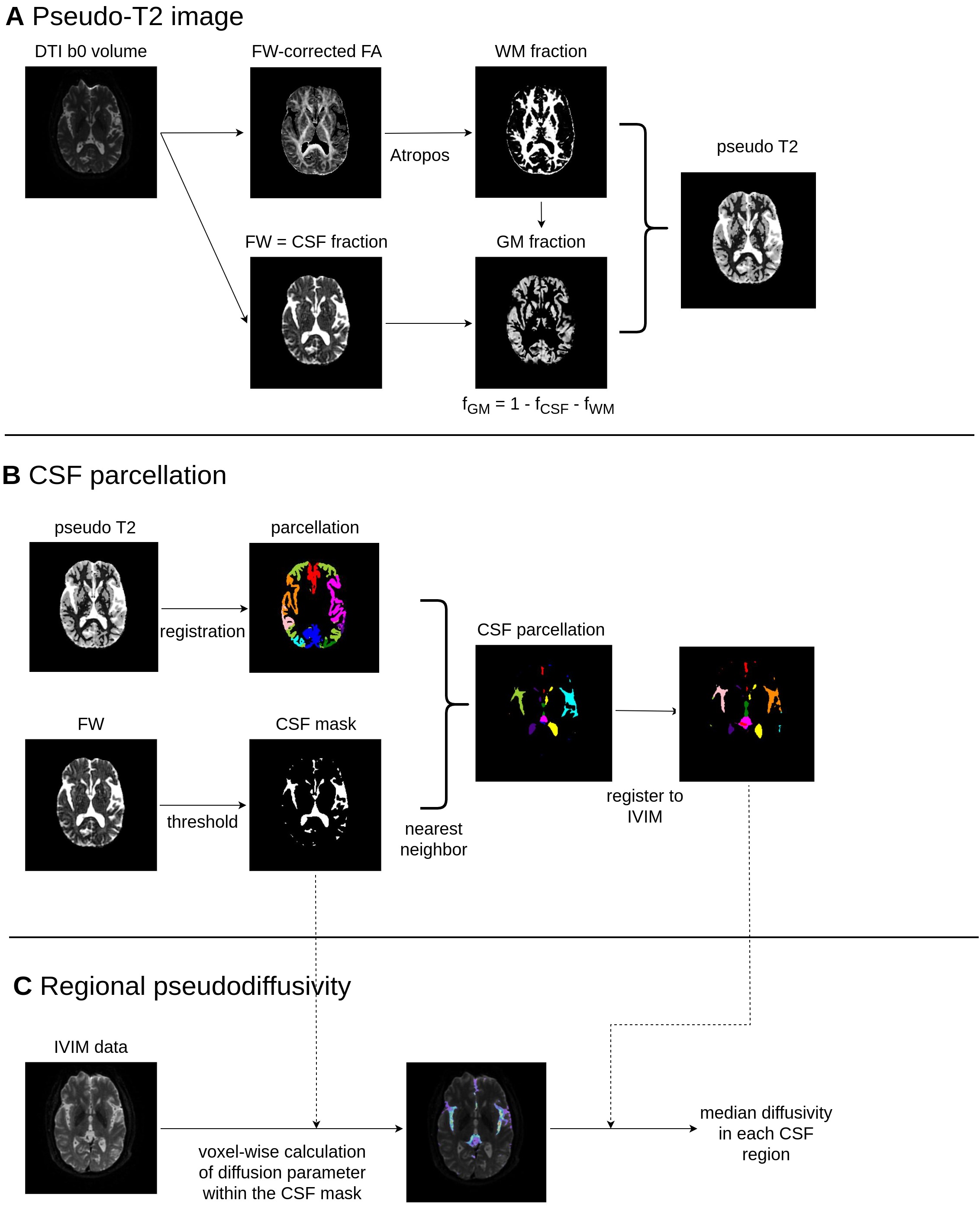
93 participants (age 68.1 ±9.0 years old, 38.3% male) with varying degrees of cognitive impairment were recruited from a memory clinic. 35 participants had normal cognitive function while 58 had clinical diagnoses of mild cognitive impairment or Alzheimer’s disease. All participants underwent comprehensive cognitive testing and MRI scanning including IVIM (16 b-values, 3 directions) and diffusion MRI (b-value = 1000 s/mm2, 30 directions). The first step of CBSS is to use diffusion MRI to create a pseudo-T2 contrast image, using a similar technique to Nazeri et al. (2017) (Figure 1). A sulcus-specific gray matter atlas was registered to the pseudo-T2 image. Voxels in the CSF regions were parcellated according to its proximity to specific gray matter regions (Figure 1). For instance, CSF regions close to the insula were labelled as “Sylvian fissure”. This CSF parcellation atlas was then registered to IVIM image. In each CSF voxel defined by the parcellation atlas, a single compartment diffusion model was fitted to IVIM data using b values ≤200 s/mm2. The resulting apparent diffusion coefficient was previously shown to correspond to the variance of CSF motion (Bito et al., 2021).Results
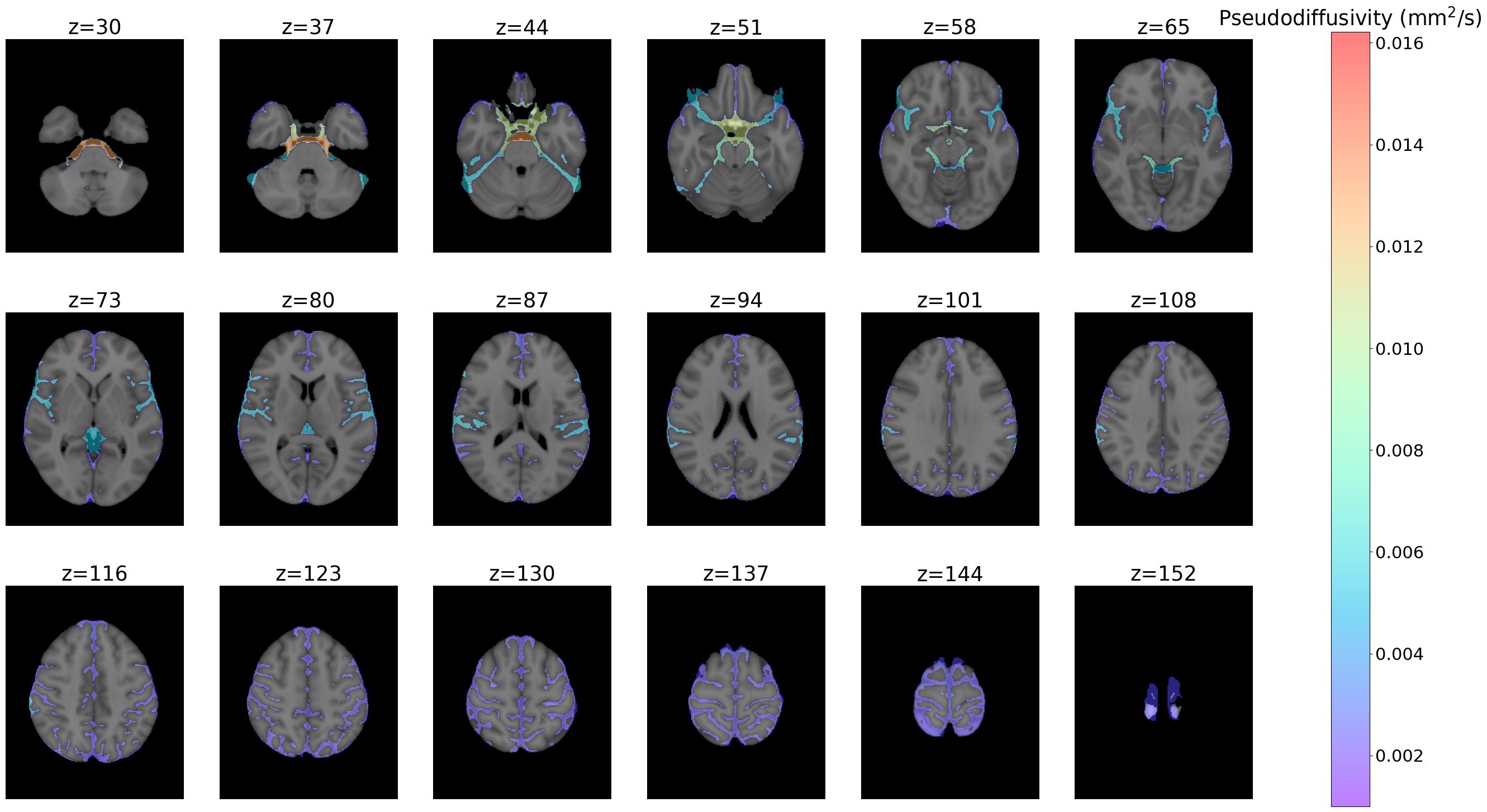
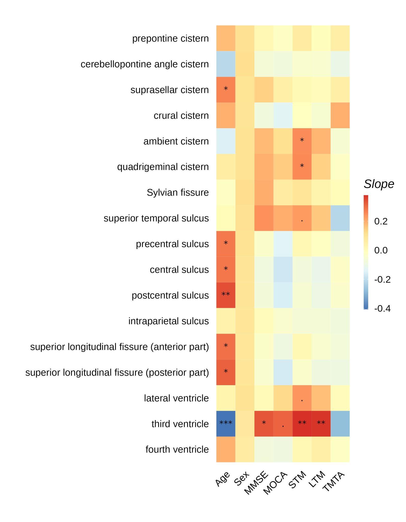
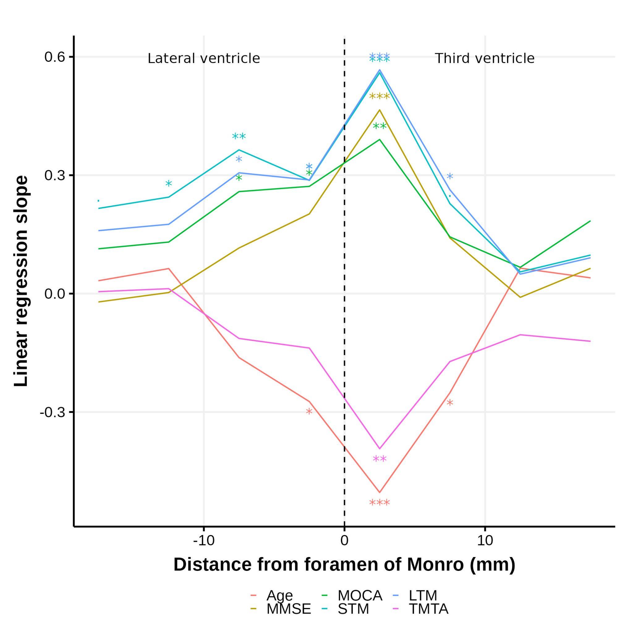
Across the entire cohort (both normal control and those with cognitive impairment), the CSF pseudodiffusivity was highest near the brainstem cisterns and decreased towards the sulci in the cerebral cortex (Figure 2). The pseudodiffusivity in the third ventricle positively correlated with short term memory (standardized slope of linear regression=0.38, adjusted p=0.0005) and long term memory (slope=0.37, adjusted p=0.005) (Figure 3). Fine mapping along the third and lateral ventricles revealed that the pseudodiffusivity in the region closest to the foramen of Monro demonstrated the highest correlation with cognitive performance (Figure 4).Discussion
Pseudodiffusivity in the third ventricle and foramen of Monro was positively correlated with short and long term memories, but was less strongly associated with processing speed and general cognition. This is consistent with mice models of Alzheimer’s disease displaying a lower CSF flow in the third ventricle compared with wild type mice (Igarashi et al., 2014a, 2014b). Future research could investigate why the pseudodiffusivity in the third ventricle correlates with cognition better than other CSF regions in humans.Conclusion
CBSS enabled quantitative spatial analysis of CSF pseudodiffusivity and suggested third ventricle pseudodiffusivity as a potential biomarker of cognitive impairment in Alzheimer’s disease. It laid the foundation for future research in the role of CSF circulation in neurodegenerative diseases.Acknowledgements
Y.C. and H.H conceived the project and created the CBSS algorithm. Y.C. implemented the algorithm and performed all statistical analysis. H.H. and X.L. created the sulcus-oriented gray matter atlas. Y.C. and H.H. wrote the manuscript under the supervision of A.N., H.M., and X.L.. All authors read and approved the manuscript.References
Bito, Y., Harada, K., Ochi, H., Kudo, K., 2021. Low b-value diffusion tensor imaging for measuring pseudorandom flow of cerebrospinal fluid. Magnetic Resonance in Medicine 86, 1369–1382. https://doi.org/10.1002/mrm.28806
Han, G., Zhou, Y., Zhang, K., Jiao, B., Hu, J., Zhang, Y., Wang, Z., Lou, M., Bai, R., 2023. Age- and time-of-day dependence of glymphatic function in the human brain measured via two diffusion MRI methods. Front Aging Neurosci 15, 1173221. https://doi.org/10.3389/fnagi.2023.1173221
Igarashi, H., Suzuki, Y., Kwee, I.L., Nakada, T., 2014a. Water influx into cerebrospinal fluid is significantly reduced in senile plaque bearing transgenic mice, supporting beta-amyloid clearance hypothesis of Alzheimer’s disease. Neurological Research 36, 1094–1098. https://doi.org/10.1179/1743132814Y.0000000434
Igarashi, H., Tsujita, M., Kwee, I.L., Nakada, T., 2014b. Water influx into cerebrospinal fluid is primarily controlled by aquaporin-4, not by aquaporin-1: 17: O JJVCPE MRI study in knockout mice. NeuroReport 25, 39. https://doi.org/10.1097/WNR.0000000000000042
Mestre, H., Mori, Y., Nedergaard, M., 2020. The Brain’s Glymphatic System: Current Controversies. Trends in Neurosciences 43, 458–466. https://doi.org/10.1016/j.tins.2020.04.003
Nazeri, A., Mulsant, B.H., Rajji, T.K., Levesque, M.L., Pipitone, J., Stefanik, L., Shahab, S., Roostaei, T., Wheeler, A.L., Chavez, S., Voineskos, A.N., 2017. Gray Matter Neuritic Microstructure Deficits in Schizophrenia and Bipolar Disorder. Biol Psychiatry 82, 726–736. https://doi.org/10.1016/j.biopsych.2016.12.005Ringstad, G., Vatnehol, S.A.S., Eide, P.K., 2017. Glymphatic MRI in idiopathic normal pressure hydrocephalus. Brain 140, 2691–2705. https://doi.org/10.1093/brain/awx191
Wong, S.M., Backes, W.H., Drenthen, G.S., Zhang, C.E., Voorter, P.H.M., Staals, J., van Oostenbrugge, R.J., Jansen, J.F.A., 2020. Spectral Diffusion Analysis of Intravoxel Incoherent Motion MRI in Cerebral Small Vessel Disease. Journal of Magnetic Resonance Imaging 51, 1170–1180. https://doi.org/10.1002/jmri.26920
Zhang, W., Zhou, Y., Wang, J., Gong, X., Chen, Z., Zhang, X., Cai, J., Chen, S., Fang, L., Sun, J., Lou, M., 2021. Glymphatic clearance function in patients with cerebral small vessel disease. NeuroImage 238, 118257. https://doi.org/10.1016/j.neuroimage.2021.118257
Figures