4060
The exploration of neurovascular coupling injury in iNPH based on BOLD and ASL: a preliminary study1Department of Radiology, The First Affiliated Hospital, Zhejiang University School of Medicine,, Hangzhou City, China, 2Department of Radiology, Huadong Hospital Affiliated to Fudan University, Shanghai, China
Synopsis
Keywords: Other Neurodegeneration, fMRI (resting state)
Motivation: Currently, it has been proposed that the occurrence and development of neurodegenerative diseases such as iNPH (idiopathic normal pressure hydrocephalus) are related to the disorder of cerebral microvascular function, neurovascular uncoupling, and blood-brain barrier dysfunction.
Goal(s): We aimed to explore the impairment of neurovascular coupling in iNPH using multi-types of CBF-BOLD coupling.
Approach: In this study, we found that the coupling of spontaneous neural function to the corresponding cerebral blood flow was changed in iNPH.
Results: The results suggest that neurovascular coupling can provide new clues for further exploring the relationship between brain disfunction and disorder progression and outcome in iNPH.
Impact: The three coupling modes can be used to evaluate the potential mechanism of neurovascular coupling injury in iNPH, which may enhance our comprehension of the mechanism underlying neurovascular coupling injury in iNPH.
Background
Idiopathic normal pressure hydrocephalus (iNPH) is a syndrome characterized by gait disorder, cognitive impairment, and incontinence with normal cerebrospinal fluid pressure, which underlying pathogenesis remains unclear[1]. There is a tight coupling between the brain perfusion and neural function, that is, neurovascular coupling, which is the physiological basis of human neural network[2]. Neurovascular uncoupling may be one of the neuropathological mechanisms contributing to brain dysfunction in neurodegenerative diseases such as Alzheimer's disease and diabetes-related mild cognitive impairment[3, 4]. Neurovascular coupling is a promising imaging biomarker that can detect abnormal neurovascular coupling and explore key brain regions affected by diseases. This study aimed to evaluate the level of neurovascular coupling injury in patients with idiopathic normal pressure hydrocephalus (iNPH) and explore the key neurovascular coupling indicators that affect the clinical symptoms of iNPH. Based on the previous studies[5, 6], we propose the following hypotheses: first, there is neurovascular coupling injury in the central nervous system of iNPH patients; Secondly, the pattern of neurovascular coupling injury in the central nervous system of iNPH patients is closely related to preoperative clinical characteristics and the efficacy of shunt surgery.Methods
Thirty-three patients with confirmed iNPH and 16 age-matched healthy controls (HC) were prospectively enrolled. Magnetic resonance Blood Oxygen Level-Dependent (BOLD) and arterial spin labeling (ASL) data were collected from all subjects. The degree centrality (DC), fractional amplitude of low-frequency fluctuations, fALFF), regional homogeneity (ReHo) and cerebral blood flow (CBF) values, The coupling patterns of CBF_DC, CBF_fALFF and CBF_ReHo were evaluated by constructing three neurovascular coupling indexes of cross-subject, cross-voxel and ratio. The differences of neurovascular coupling patterns between iNPH group and HC group were compared, and the correlation of neurovascular ratio coupling with clinical symptoms and shunt efficacy in iNPH group was analyzed.Results
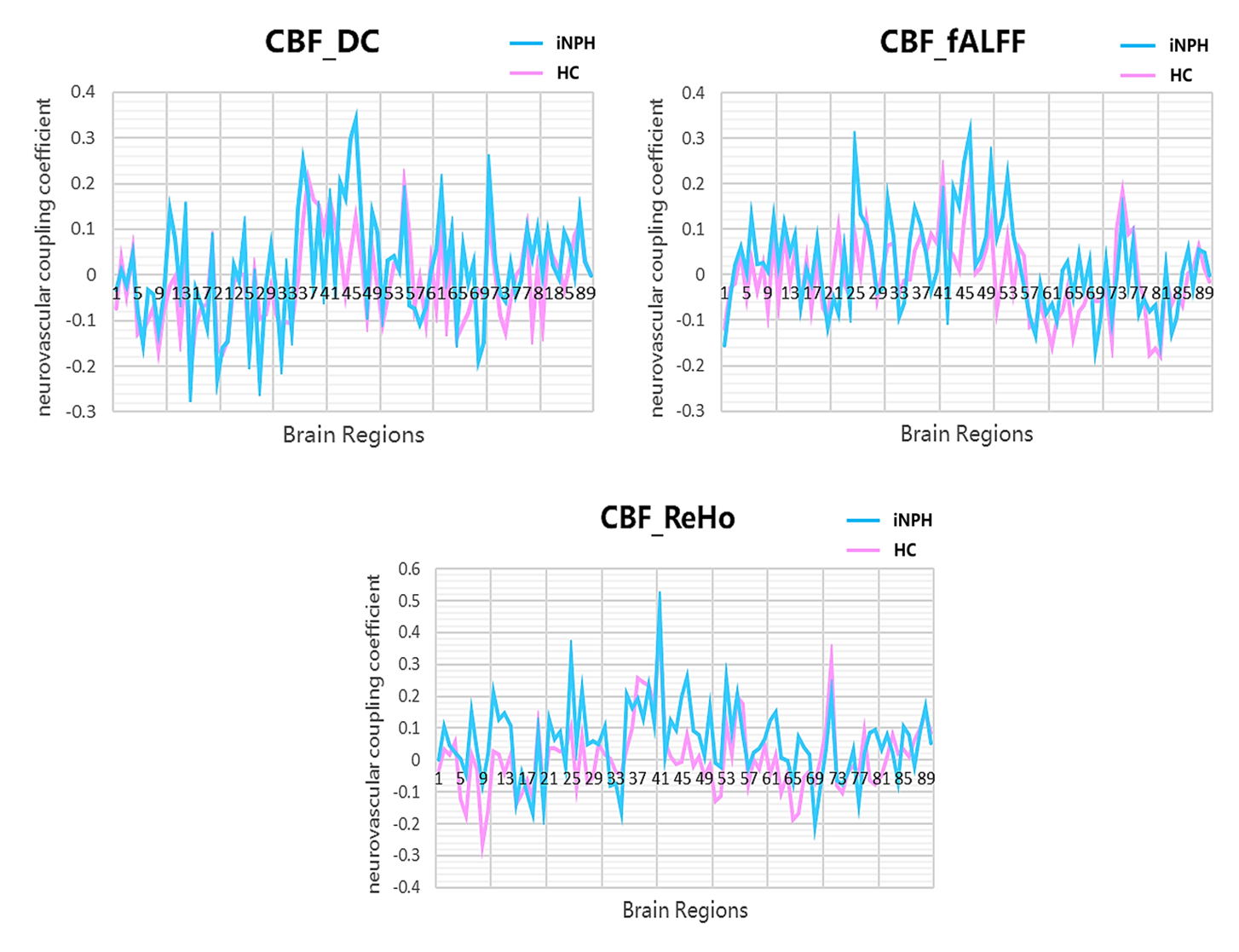
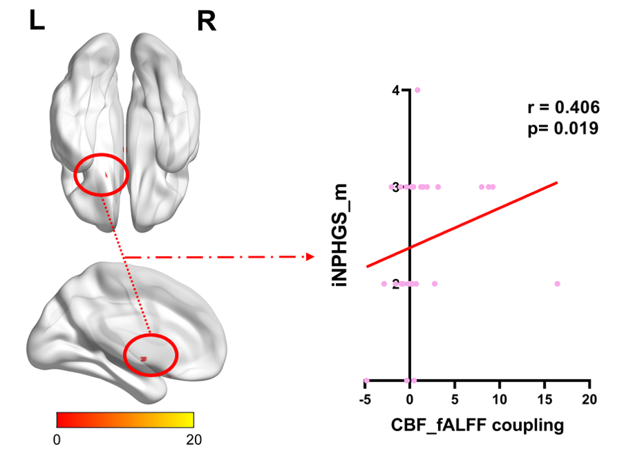
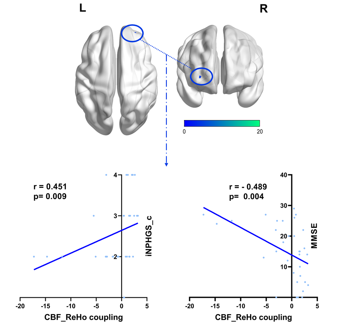
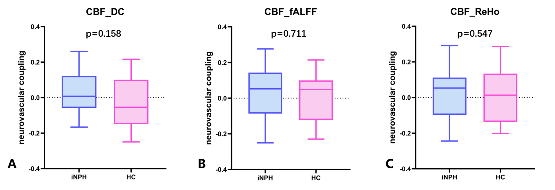
(1) There was no significant difference between iNPH and HC group in the spatial distribution of cross-subject neurovascular coupling indexes of gray voxel level. However, in iNPH group, neurovascular decoupling existed in orbital frontalis gyrus, lateral temporal and angular gyrus, precuneus and other brain regions, and neurovascular coupling levels increased in hippocampus, parahippocampal gyrus and amygdala (Figure 1). (2) Ratio coupling analysis showed that in iNPH patients, the CBF/DC ratio of the left orbital superior frontal gyrus and the right fusiform gyrus decreased, and the CBF/ReHo ratio of the right middle frontal gyrus decreased, and the decreased CBF/ReHo ratio of the right middle frontal gyrus was significantly correlated with preoperative cognitive impairment (iNPHGS cognitive score: r1 = 0.451, p1 = 0.009; MMSE score: r2 = -0.489, p2 = 0.004, Figure 2); In patients with iNPH, the CBF/DC ratio of the left middle cingulate gyrus, the right auxiliary motor area and the left precuneus increased, and the CBF/fALFF ratio of the right precuneus, the left superior orbitofrontal gyrus and the inferior gyrus increased. The coupling of CBF/fALFF ratio in the left inferior orbital frontal gyrus was positively correlated with preoperative motor function (r= 0.406, p= 0.019,Figure 3). (3) No significant difference was found in trans-voxel neurovascular coupling between iNPH and HC groups (Figure 4).Conclusion
INPH has cerebral neurovascular coupling injury, among which the decreased neurovascular coupling in the middle frontal gyrus may mediate the cognitive impairment in iNPHs, and the increased neurovascular coupling in the left orbital inferior frontal gyrus may be related to the impairment of motor function in iNPHs. This study suggests that neurovascular coupling injury may be the underlying neuropathological mechanism of iNPH, and the neurovascular coupling analysis method provides a new imaging method for exploring the brain function and perfusion injury mechanism of iNPH.Acknowledgements
This study was funded by
the National Natural Science Foundation of China (81771816), Hospital
Development Center (SHDC2022DRT025, SKLY2022CRT402) and Investigator-Initiated
Research Projects of Huadong Hospital (HDLC2022003).
References
[1] NAKAJIMA M, YAMADA S, MIYAJIMA M, et al. Guidelines for Management of Idiopathic Normal Pressure Hydrocephalus (Third Edition): Endorsed by the Japanese Society of Normal Pressure Hydrocephalus [J]. Neurologia medico-chirurgica, 2021, 61(2): 63-97.
[2] HAWKINS B T, DAVIS T P. The blood-brain barrier/neurovascular unit in health and disease [J]. Pharmacological reviews, 2005, 57(2): 173-185.
[3] YU Y, YAN L F, SUN Q, et al. Neurovascular decoupling in type 2 diabetes mellitus without mild cognitive impairment: Potential biomarker for early cognitive impairment [J]. NeuroImage, 2019, 200: 644-658.
[4] MALEKI BALAJOO S, RAHMANI F, KHOSROWABADI R, et al. Decoupling of regional neural activity and inter-regional functional connectivity in Alzheimer's disease: a simultaneous PET/MR study [J]. European journal of nuclear medicine and molecular imaging, 2022, 49(9): 3173-3185.
[5] HUANG W, FANG X, LI S, et al. Shunt Surgery Efficacy Is Correlated With Baseline Cerebrum Perfusion in Idiopathic Normal Pressure Hydrocephalus: A 3D Pulsed Arterial-Spin Labeling Study [J]. Frontiers in aging neuroscience, 2022, 14: 797803.
[6] HUANG W, FANG X, LI S, et al. Abnormal characteristic static and dynamic functional network connectivity in idiopathic normal pressure hydrocephalus [J]. CNS neuroscience & therapeutics, 2023.
Figures