4059
Associations among Glymphatic System Impairmentand its related abnormalities in Cerebral small-vessel disease patients1Department of Radiology, Affiliated Hospital of Guizhou Medical University, Guiyang, China, 2Department of Clinical and Technical Support, Philips Healthcare, Xi'an, China, 3Philips Healthcare, Shanghai, China
Synopsis
Keywords: Other Neurodegeneration, Neurodegeneration
Motivation: The emergence of glymphatic system has provided new insights into the mechanisms of cognitive impairment(CI), and cerebral small vessel disease (CSVD) is one of the most frequently diagnosed diseases impairing cognitive function.
Goal(s): We investigated the relationship among the ALPS index, white/gray matter abnormalities and structural network efficiency in CSVD patients.
Approach: The diffusion tensor imaging analysis along the perivascular space (DTI-ALPS)-index and mediation analysis were performed .
Results: Significant differences in the DTI-ALPS index were found between the HCs and CSVD_CI groups. Gray matter reserve and white matter integrity may mediates the relationship between glymphatic dysfunction and structural network efficiency in CSVD.
Impact: Our results showed distinct relationships between glymphatic dysfunction and its related abnormalities, Gray matter reserve and white matter integrity may mediate the relationship between glymphatic dysfunction and structural network efficiency.
Abstract
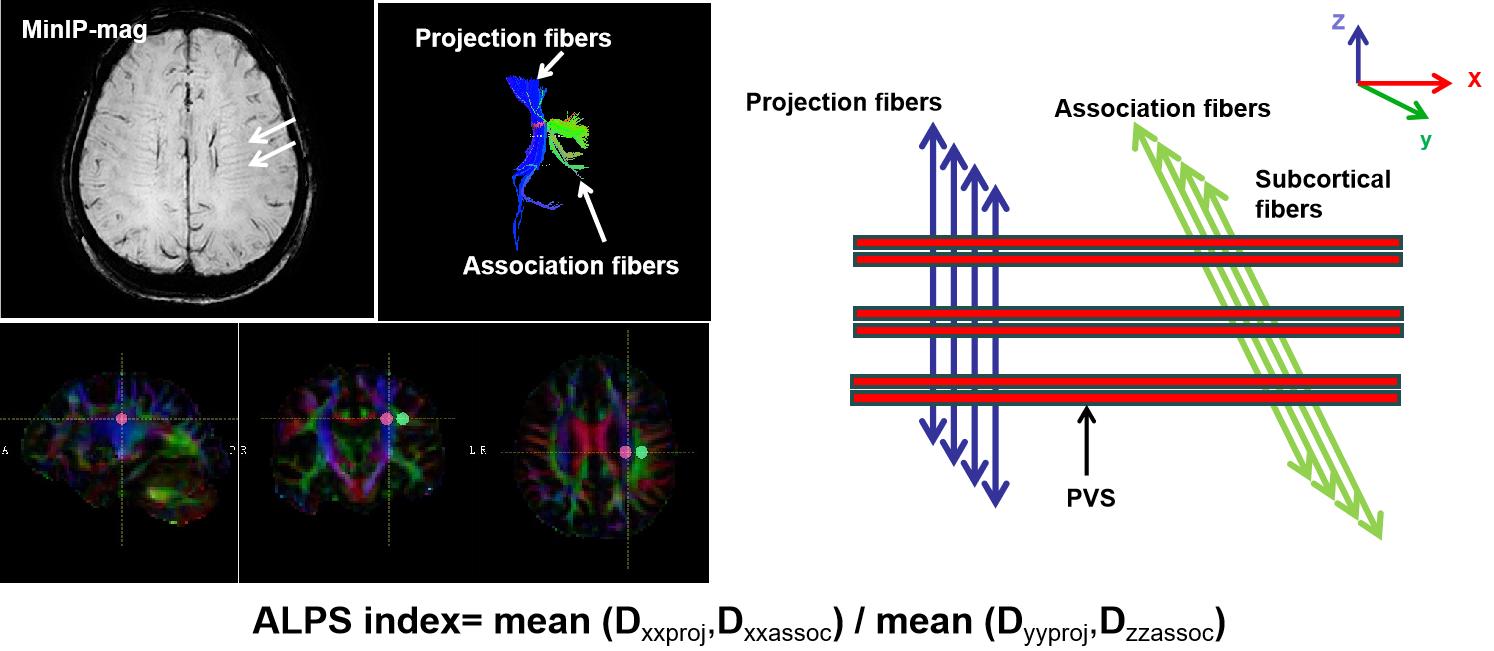
Introduction Recently, the emergence of glymphatic system has provided new insights into the mechanisms of cognitive impairment[1], and cerebral small vessel disease (CSVD) is one of the most frequently diagnosed diseases impairing cognitive function[2-4]. However, it is difficult to evaluate the glymphatic system in the human body directly, Diffusion tensor imaging analysis along the perivascular space (ALPS)-index is a widely used and a non-invasive method with good reproducibility method to evaluate glymphatic function[5]. So, in the current study, we investigated the relationship among the ALPS index, White/Gray Matter abnormalities and Structural network efficiency in CSVD patients with/without cognitive impairment, Furtherly, to explore the reason underlying cognitive impairment in patients with CSVD and identify other new imaging markers of cognitive impairment in patients with CSVD.Materials and methods Forty-two patients with CSVD (including 21 males), comprising 22 with normal cognitive status (CSVD_CN) and 20 with mild cognitive impairment (CSVD_CI); 20 age- and gender-matched healthy controls (HCs, with 9 males) were recruited from the Affiliated Hospital of Guiyang Medical University. CSVD diagnosis was based on the American Heart Association (AHA) and the American Stroke Association (ASA) in 2016[6]. They were all underwent 3 Tesla high- resolution structural and diffusion-tensor (DTI) MRI scanning, For 3D-T1-weighted sequence: repetition time (TR) = 12 ms; echo time (TE) = 6 ms; field of view (FOV) 220x220x155 mm; voxel size = 0.57x0.57x0.6 mm3; matrix size = 384x384, NSA = 1. For DTI parameters: slice thickness = 2.0 mm; pixel size = 2.0×2.0 mm2; matrix size = 128×128; gradient directions = 32 (90 cases) / 30 (14 cases); FA = 90°; TR = 7200/12,400 ms; TE = 56/95 ms. A detailed cognitive assessment was also performed for every participants. The DTI-Studio and CAT12 toolbox was applied to calculate the ALPS-index and to assess gray matter reserve (Via hippocampus and amygdala volume) and white mtter integrity (Via FA value of the mainly white matter tract). Diffusivity maps and color-coded fractional anisotropy (FA) were produced for the x, y, and z axes (Dxx, Dyy, and Dzz), and two 5 × 5 mm2 regions of interest (ROIs) were manually placed on each color-coded FA map, representative projection fibres and association fibres, the DTI-ALPS index was then calculated as [(Dxxproj + Dxxassoc)/(Dyyproj + Dzzassoc)](see Fig.1). In this study, cortical radial crown projections represent projection fibers and upper longitudinal bundles represent association fibers. And also, we obtained the global topological parameters including global shortest path length (Lp)], global efficiency (Eg), and local efficiency (Eloc) by using the GRETNA toolbox. Statistical analyses were performed using the Statistical Package for the Social Sciences (SPSS) statistical software (version 27, IBM Corp), PROCESS v3.4 software by Andrew F. Hayes was used to construct the mediation effect.
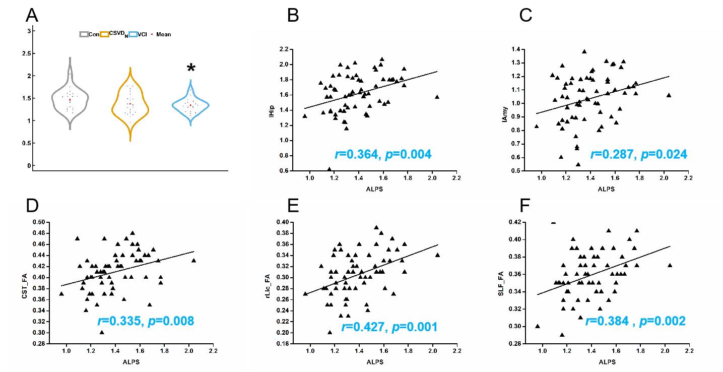
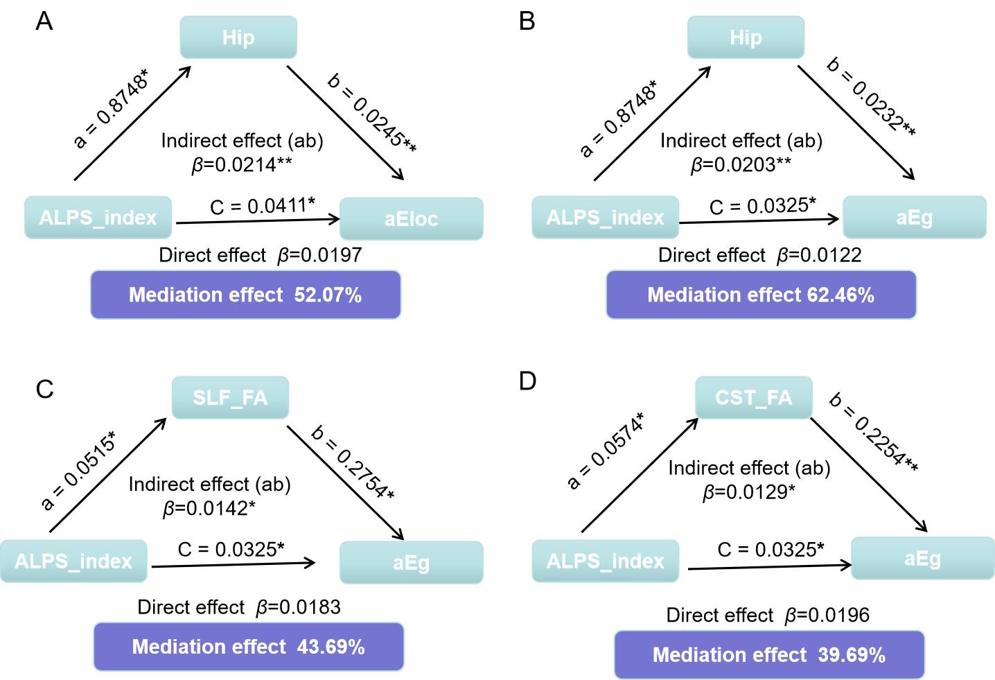
Results There were significant differences in the DTI-ALPS index (1.47 vs. 1.34, p = 0.029) between the HC and CSVD_CI groups. No significantly DTI_ALPS changes were found in CSVD_CN group. Moreover, statistically significant associations were found between the DTI-ALPS index and volume of hippocampus( left:r=0.364, p=0.004; right:r=0.342, p=0.007), volume of amygdala (left:r=0.287, p=0.024; right:r=0.342, p=0.006), and there was also significantly correlation between the DTI-ALPS index and structural network efficiency, including AUC of CP (r=0.366, p=0.003), AUC of Lp (r=-0.308, p=0.015), AUC of Eg (r=0.311, p=0.014), AUC of Eloc (r=0.351, p=0.005). Mediation analysis also suggested that gray matter reserve and white matter integrity may mediates the relationship between glymphatic dysfunction and structural network efficiency in CSVD.
Discussion Our results showed distinct relationships between glymphatic dysfunction and its related abnormalities, suggesting ALPS-index is a potential marker for early recognition of CI in CSVD. Gray matter reserve and white matter integrity may mediate the relationship between glymphatic dysfunction and structural network efficiency. These findings may provide new evidence for the influence of ALPS on cognitive decline in CSVD clinical practice.
Acknowledgements
This work is supported by grants from the Natural Science Foundation of China (8156028, 181871333, 82260340). Guizhou Province Science & Technology Innovation Talent Team (CXTD[2022]006).References
[1] Wang J, et al. Age Ageing 2023 Jul 01;52(7).
[2] Tang J , et al. Front Aging Neurosci 2022;14.
[3] Tian Y, et al.Stroke Vasc Neurol 2023 Apr 12.
[4] Ke z, et al. Brain Sci 2022 Nov 24;12(12).
[5] Taoka T, et al. Jpn J Radiol 2017 Apr;35(4).
[6] Peng D, et al. Aging Med (Milton) 2019 Jun;2(2).
Figures