4054
Diamagnetic susceptibility and relationship to amyloid burden in Alzheimer’s Disease1BioMedical Engineering and Imaging Institute, Icahn School of Medicine at Mount Sinai, New York, NY, United States, 2Cornell University, Ithaca, NY, United States, 3Urmia University of Medical Science, Urmia, Iran (Islamic Republic of)
Synopsis
Keywords: Alzheimer's Disease, Alzheimer's Disease
Motivation: Alzheimer's disease (AD) is a progressive neurological disorder affecting cognitive functions, memory, and behavior, primarily in older adults. It's marked by abnormal protein deposits, including beta-amyloid plaques and tau tangles in the brain, causing nerve cell dysfunction and death.
Goal(s): Positron Emission Tomography (PET) detects beta-amyloid but has limitations. High-resolution MRI imaging can detect beta-amyloid, focusing on diamagnetic properties associated with electron density.
Approach: Quantitative susceptibility mapping (QSM) is particularly effective at 7 Tesla MRI, offering high sensitivity and better neuroanatomy details.
Results: The study utilized 7T MRI to measure beta-amyloid's diamagnetic susceptibility in AD patients using separated QSM technique.
Impact: In this study we leveraged the high susceptible sensitivity of 7T MRI to measure the diamagnetic susceptibility of beta-amyloid aggregated in the brain of Alzheimer's disease patients using separated quantitative susceptibility mapping.
Introduction
Alzheimer's disease (AD) is a progressive neurological disorder that mainly affects cognitive functions, memory, thinking, and behavior1. AD is characterized by the accumulation of abnormal protein deposits in the brain, including beta-amyloid (Aβ) plaques and tau tangles2. Aβ can be detected using Positron Emission Tomography (PET) imaging that allows for the in-vivo visualization and quantification of Aβ plaques in the brain3. However, the primary limitations of PET, such as ionizing radiation restrict its use in routine clinical settings. Alternatively, high spatial resolution MRI imaging can be used to detect Aβ plaque. The non-invasively detection of Aβ plaques in MRI images has primarily been associated with the presence of focal iron (paramagnetic) deposition in the vicinity of these plaques4. Proteins in general, show diamagnetic properties due to their high concentration of paired electrons. Therefore, the accumulation of Aβ would result in an increased electron density, leading to noticeable alterations in local susceptibility5. When there are deposits comprising both Aβ and iron, these substances exert opposing effects on the overall magnetic susceptibility of the surrounding tissue. Studies showed that the diamagnetic susceptibility of Aβ can be observed in brain specimens using quantitative susceptibility mapping (QSM) technique2. Ultra-high field MRI at 7 Tesla (7T) provides a higher sensitivity, and thus better signal-to-noise ratio and MRI contrast than 3T or 1.5T MRI6. In this study we leveraged the high susceptible sensitivity of 7T MRI to measure the diamagnetic susceptibility of Aβ aggregated in the brain of AD patients using separated QSM.Methods
In-vivo MRI experiment was performed in AD/MCI (N=4) aged 65-80 years and who satisfied the MR safety criteria. Informed consent was obtained from each subject under approval from the Institutional Review Board. Scanning was conducted on a 7T MRI scanner (Magnetom, Siemens Healthcare, Erlangen, Germany) using 1Tx/32Rx Nova head coil. To calculate quantitative susceptibility mapping (QSM), a 3D high-resolution spoiled gradient echo (GRE) sequence was used (voxels size=0.3x0.3x1.5 mm3, TR/TE = 32/10 ms, and flip angle = 12°). QSM images were reconstructed from the same field used for DTI image correction7. The background field is removed using the projection onto dipole fields (PDF) algorithm and QSM is reconstructed using Morphology-Enabled Dipole Inversion (MEDI) with global cerebrospinal fluid (CSF) referencing. We separated magnetic sources in QSM into paramagnetic and diamagnetic sources using am algorithm developed by Sisman et al7. This technique used the diffusion tensor imaging (DTI) acquisition to deal with the fiber orientation dependent susceptibility effects. In-vivo DTI were obtained with 1.05 mm isotropic resolutions with 64 diffusion directions and 4 B0 acquisitions, each acquired twice with anterior-posterior (AP) and PA phase encode directions to allow gradient nonlinearity correction in post-processing. The estimated susceptibility values of negative sources in a set of enhancing Aβ plaque were correlated against Aβ PET distribution.Results
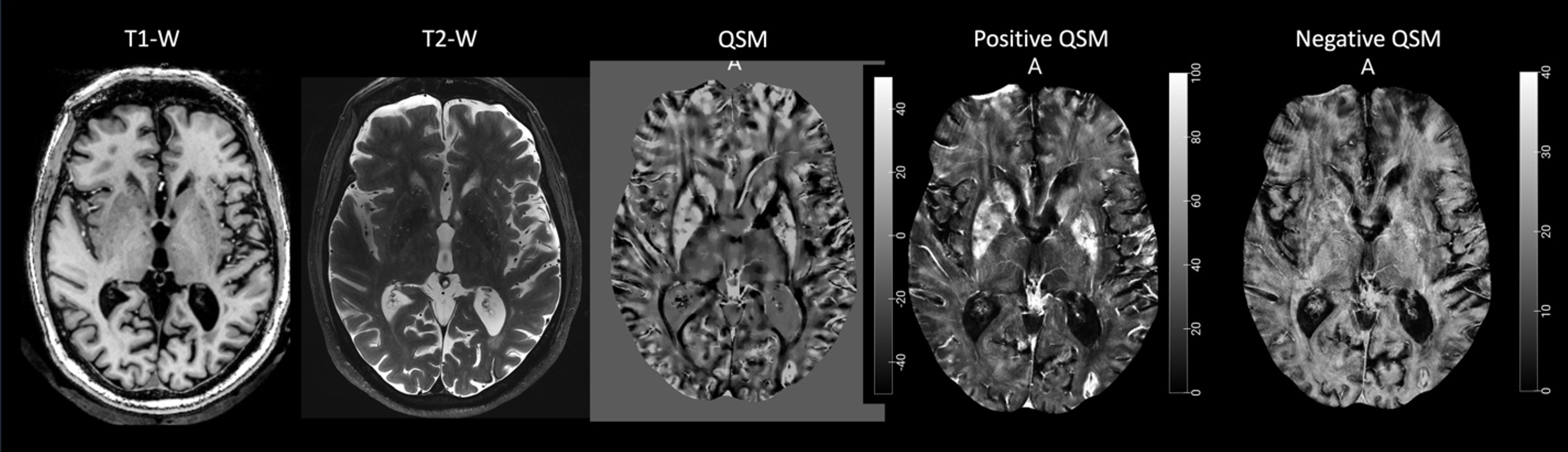
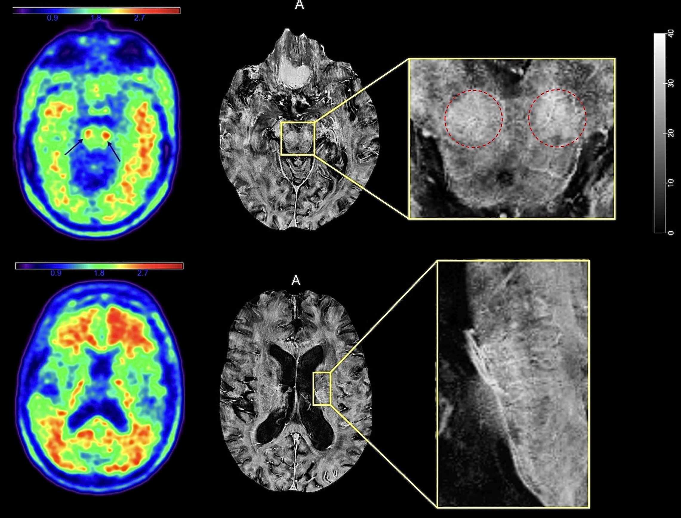
Figure 1 illustrates the processed QSM and separated paramagnetic and diamagnetic sources, showcasing notable consistency in the contrast generated and the distribution of magnetic sources across both positive and negative QSM maps. Significantly elevated overall susceptibility values were observed in regions with higher iron content, including the globus pallidus, putamen, substantia nigra, caudate, and right mesial orbitofrontal cortex. In Figure 2, the negative QSM (brighter regions) and Aβ PET images reveal a noteworthy correlation, with higher negative susceptibility values in the red nucleus displaying a strong association with Aβ levels in AD patients. A moderate correlation was observed in the proventricular white matter. However, in regions with a higher Aβ burden, a substantial correlation between Aβ and negative QSM was not observed.Discussion and Conclusion
This study represents the first implementation of separated QSM at 7T in living AD patients. The primary objective of this investigation was to assess the clinical viability of utilizing negative QSM for detecting Aβ plaque in AD patients. Our key finding revealed a strong correlation between higher Aβ levels in the red nucleus and elevated negative susceptibility values. However, we did not observe a corresponding reflection of increased Aβ burden in the frontal proventricular white matter and other white matter regions in terms of negative susceptibility levels. It's important to note that this study demonstrates the clinical feasibility of using the separated-source QSM technique. While we did not compare Aβ PET and negative QSM in AD patients with healthy controls, the global SUVR and negative susceptibility measurements may offer valuable insights into the relationship between Aβ aggregation and negative QSM. The findings presented in this study underscore the potential of the 7T separated-source QSM methodology as an alternative tool to detect Aβ imaging in AD patients.Acknowledgements
The authors would like to thank research coordinator Aislinn Diaz helping with the recruitment. This study was supported by a Developmental Project award from Mount Sinai ADRC (P30 AG066514) NIA/NIH, R21AG071179, and K01 AG075178-01 NIA/NIH grants.References
[1] Hebert LE, Scherr PA, Bienias JL, et al. Alzheimer disease in the US population: prevalence estimates using the 2000 census. Arch Neurol 2003; 60: 1119–1122.
[2] Gong NJ, Dibb R, Bulk M, van der Weerd L, Liu C. Imaging beta amyloid aggregation and iron accumulation in Alzheimer's disease using quantitative susceptibility mapping MRI. Neuroimage.2019 May 1;191:176-185. doi: 10.1016/j.neuroimage.2019.02.019. Epub 2019 Feb 7. PMID: 30739060.
[3] Jack CR, Knopman DS, Jagust WJ, Shaw LM, Aisen PS, Weiner MW, Petersen RC, Trojanowski JQ (2010) Hypothetical model of dynamic biomarkers of the Alzheimer’s pathological cascade. Lancet Neurol 9, 119–128.
[4] Exley C, House E, Polwart A, Esiri MM (2012) Brain burdens of aluminum, iron, and copper and their relationships with amyloid-beta pathology in 60 human brains. J Alzheimers Dis 31, 725-730.[5] Babaei M, Jones IC, Dayal K, Mauter MS. Computing the Diamagnetic Susceptibility and Diamagnetic Anisotropy of Membrane Proteins from Structural Subunits. J Chem Theory Comput. 2017 Jun 13;13(6):2945-2953. doi: 10.1021/acs.jctc.6b01251. Epub 2017 Apr 25. PMID: 28418668.
[6] Tiepolt S, Schäfer A, Rullmann M, Roggenhofer E; Netherlands Brain Bank; Gertz HJ, Schroeter ML, Patt M, Bazin PL, Jochimsen TH, Turner R, Sabri O, Barthel H. Quantitative Susceptibility Mapping of Amyloid-β Aggregates in Alzheimer's Disease with 7T MR. J Alzheimers Dis. 2018;64(2):393-404. doi: 10.3233/JAD-180118. PMID: 29865069.
[7] Mert Şişman, Thanh D. Nguyen, Alexandra G. Roberts, Dominick J. Romano, Alexey V. Dimov, Ilhami Kovanlikaya, Pascal Spincemaille, Yi Wang., Microstructure-Informed Myelin Mapping (MIMM) from Gradient Echo MRI using Stochastic Matching Pursuit. medRxiv 2023.09.22.23295993; doi: https://doi.org/10.1101/2023.09.22.23295993.
Figures