4053
Association of Brain Diffusion-Tensor Parameters with Risk of Alzheimer’s Disease-Related Cognitive Decline1Hainan General Hospital, Haikou, China, 2MR Research Collaboration, Siemens Healthineers, Shanghai, China, Shanghai, China
Synopsis
Keywords: Alzheimer's Disease, Alzheimer's Disease, Cognitive decline
Motivation: Diffusion-tensor parameters of projection and association neural fibers associated with glymphatic function may be able to predict the development of Alzheimer’s Disease (AD).
Goal(s): To study the association between diffusion-tensor parameters and cognition in patients with AD, and investigate whether diffusion-tensor parameters relate to cognitive decline.
Approach: Linear regression models were performed to examine associations between diffusion-tensor parameters and cognition. linear mixed-effects models were used to evaluate the association between the baseline ALPS index and cognitive decline.
Results: There was a positive association between ALPS index and cognition. Higher ALPS index levels were associated with less cognitive decline over time.
Impact: Higher ALPS index levels are associated with lower risk of AD-related changes. These findings suggest that ALPS index derived from diffusion-tensor parameters may provide useful AD progression or treatment biomarkers.
Introduction
Alzheimer’s disease (AD) is the primary cause of dementia and is becoming one of this century’s most costly, lethal, and burdensome diseases.1 Currently, the most widely accepted explanation for AD pathogenesis is the amyloid cascade hypothesis, which proposes that AD is initiated primarily by β-amyloid (Aβ) peptide accumulation into senile plaques, followed by phosphorylated tau (pTau) protein accumulation into tangles, and subsequent neuronal loss, leading to cognitive decline associated with loss of independence in the activities of daily living.2 Previous studies demonstrated that the glymphatic system is responsible to eliminate abnormal β-amyloid (Aβ) and tau proteins from the brain.3-5 Diffusion-tensor parameters of projection and association neural fibers associated with glymphatic function6, 7 may be able to predict the development of AD. We aimed to study the association between diffusion-tensor parameters and cognition in patients with AD, and investigate whether diffusion-tensor parameters relate to cognitive decline and risk of AD dementia.Methods
This study included two 3.0T magnetic resonance imaging (MRI) cohorts: the Alzheimer’s Disease Neuroimaging Initiative (ADNI) (n = 180) and our cohort (n = 127). All participants underwent brain diffusion tensor imaging (DTI) examination and neuropsychological evaluation, including the Mini-Mental State Examination (MMSE), the CDR sum of boxes (CDR-SB), and Functional Activities Questionnaire (FAQ). In ADNI cohort, participants had a median follow-up time of 5 years. The diffusivity of projection and association fibers and diffusion tensor image analysis along the perivascular space (ALPS) index were calculated. The diffusivity and ALPS index compared according to the groups with use of the Kruskal-Wallis test for both cohorts. Linear regression models adjusted for age, sex, education, and APOE4 were perform to examine associations between diffusivity and ALPS index and cognition across both cohorts. In ADNI cohort, linear mixed-effects models were used to evaluate the association between the baseline ALPS index and cognitive decline as measured by the MMSE.Results
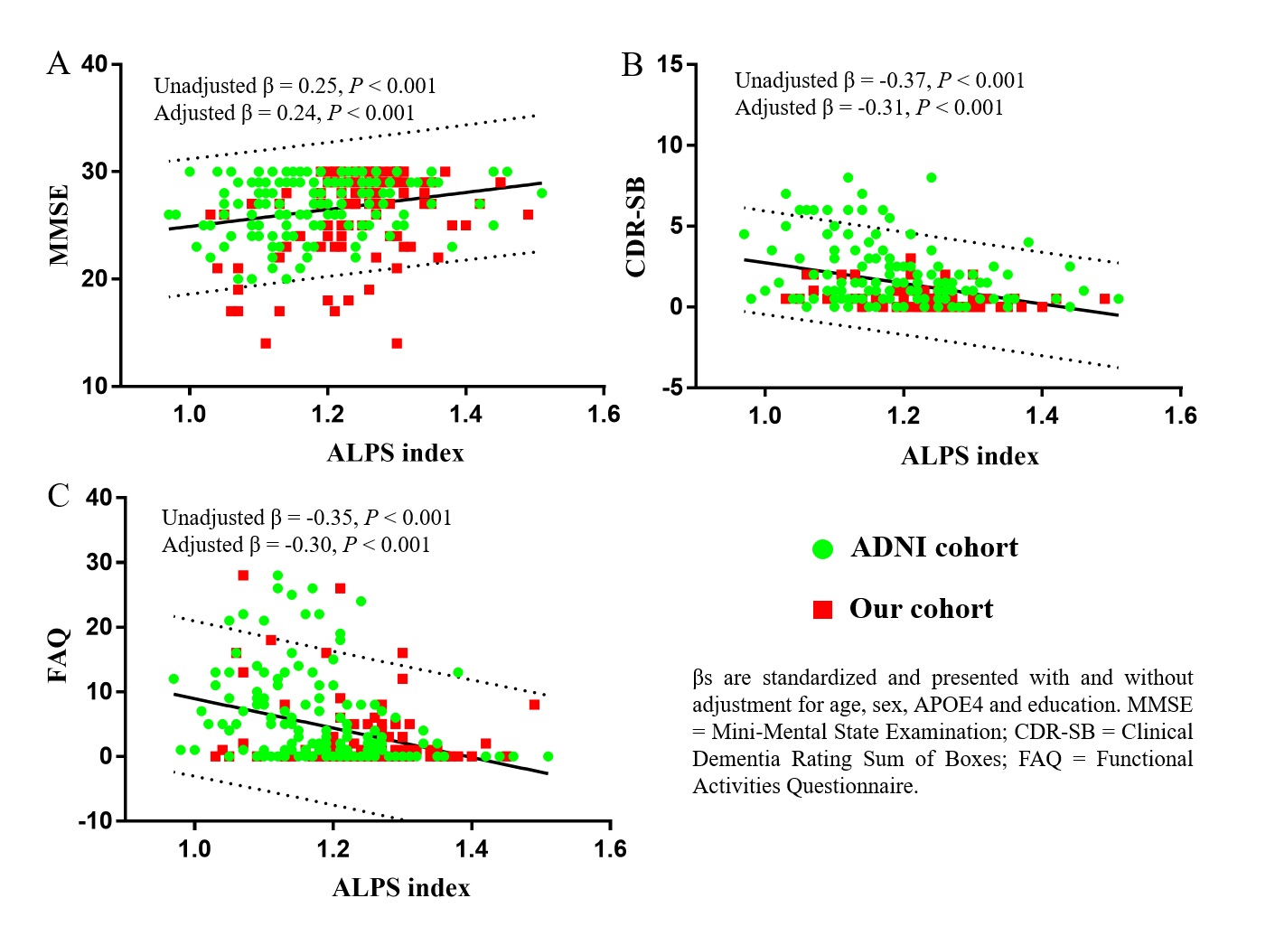
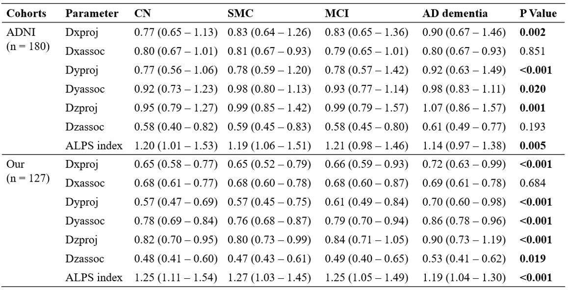
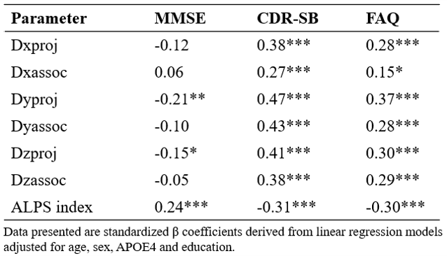
The subjects were grouped according to their clinical diagnosis at baseline into the following groups: Cognitively normal (CN, n = 35), Subjective memory concern (SMC, n = 28), mild cognitive impairment (MCI, n = 82), and AD dementia (n = 35) for ADNI cohort. In our cohort, there were CN (n = 25), SMC (n = 51), MCI (n = 32), and AD dementia (n = 19). The median follow-up time was 60 months, with 84.25% of participants having at least a 2-year visit and 65.7% having at least a 4-year visit. The averaged diffusivity and ALPS index according to the participant groups for both cohorts are summarized in Table 1 and Figure 1. The median ALPS index was lower in AD dementia group than in CN, SMC, and MCI groups for both cohorts. We tested the linear regression analysis between the diffusion-tensor parameters and neuropsychological scores for patients with AD across both cohorts (Figure 2 and Table 2), controlling for age, sex, education and APOE4. There was a positive association between the ALPS index and MMSE score (Figure 2A). The CDR-SB (Figure 2B) and FAQ (Figure 2C) scores were negatively associated with the ALPS index. Higher ALPS index levels adjusted for demographics (age, sex, and education) and APOE4 carriers were associated with less decline in the MMSE score over time (P < 0.001). Higher ALPS index levels adjusted for Aβ42 levels were also associated with less decline in the MMSE score over time (P = 0.008). Higher ALPS index levels adjusted for pTau levels were also associated with less decline in the MMSE score over time (P < 0.001). These results are displayed graphically in Figure 3.Discussion and Conclusion
This study explored the relationship between the ALPS index with cognitive function and the risk of cognitive decline. Previous studies showed that ALPS index had significant correlations with cognitive scores in patients with AD dementia, MCI individuals, and elderly CN individuals,7-9 consistent with our findings showing significant positive correlations between the ALPS index and the MMSE score. The observed association between the ALPS index, MMSE, CDR-SB, and FAQ demonstrated that the ALPS index has excellent potential to be a biomarker for the prediction of cognitive decline. Our current study showed that higher ALPS index levels were associated with less cognitive decline. The adjustment of our statistical models for core AD biomarkers reflected our attempt to manage the notion that binary cutoffs must necessarily be used in patient workflows. The results demonstrated that higher CSF Aβ42 and lower CSF pTau levels were also significantly associated with less cognitive decline. These findings demonstrate that ALPS index acts as an effective biomarker in AD progression or treatment evaluation.Acknowledgements
This project was supported by the National Natural Science Foundation of China (81971602, 82160327, and 82271977), the Key Science and Technology Project of Hainan Province (ZDYF2021SHFZ239), the Hainan Academician Innovation Platform Fund, and the Hainan Province Clinical Medical Center. Parts of the data used in preparation of this manuscript were obtained from the ADNI database (adni.loni. usc.edu). As such, the investigators within the ADNI study contributed to the design and implementation of ADNI and/or provided data but did not participate in analysis or writing of this article. Data collection and sharing for this project was funded by the Alzheimer's Disease Neuroimaging Initiative (ADNI) (National Institutes of Health Grant U01 AG024904) and DOD ADNI (Department of Defense award number W81XWH-12-2-0012). ADNI is funded by the National Institute on Aging, the National Institute of Biomedical Imaging and Bioengineering, and through generous contributions from the following: AbbVie, Alzheimer’s Association; Alzheimer’s Drug Discovery Foundation; Araclon Biotech; BioClinica, Inc.; Biogen; Bristol-Myers Squibb Company; CereSpir, Inc.; Cogstate; Eisai Inc.; Elan Pharmaceuticals, Inc.; Eli Lilly and Company; EuroImmun; F. Hoffmann-La Roche Ltd and its affiliated company Genentech, Inc.; Fujirebio; GE Healthcare; IXICO Ltd.; Janssen Alzheimer Immunotherapy Research & Development, LLC.; Johnson & Johnson Pharmaceutical Research & Development LLC.; Lumosity; Lundbeck; Merck & Co., Inc.; Meso Scale Diagnostics, LLC.; NeuroRx Research; Neurotrack Technologies; Novartis Pharmaceuticals Corporation; Pfizer Inc.; Piramal Imaging; Servier; Takeda Pharmaceutical Company; and Transition Therapeutics. The Canadian Institutes of Health Research is providing funds to support ADNI clinical sites in Canada. Private sector contributions are facilitated by the Foundation for the National Institutes of Health (www.fnih.org). The grantee organization is the Northern California Institute for Research and Education, and the study is coordinated by the Alzheimer’s Therapeutic Research Institute at the University of Southern California. ADNI data are disseminated by the Laboratory for Neuro Imaging at the University of Southern California.References
1. Scheltens P, De Strooper B, Kivipelto M, et al. Alzheimer's disease. Lancet (London, England) 2021; 397:1577-1590.
2. Karran E, Mercken M, De Strooper B. The amyloid cascade hypothesis for Alzheimer's disease: an appraisal for the development of therapeutics. Nature reviews Drug discovery 2011; 10:698-712.
3. Iliff JJ, Wang M, Liao Y, et al. A paravascular pathway facilitates CSF flow through the brain parenchyma and the clearance of interstitial solutes, including amyloid β. Science translational medicine 2012; 4:147ra111.
4. Cao X, Xu H, Feng W, et al. Deletion of aquaporin-4 aggravates brain pathology after blocking of the meningeal lymphatic drainage. Brain research bulletin 2018; 143:83-96.
5. Patel TK, Habimana-Griffin L, Gao X, et al. Dural lymphatics regulate clearance of extracellular tau from the CNS. Molecular neurodegeneration 2019; 14:11.
6. Hsu JL, Wei YC, Toh CH, et al. Magnetic Resonance Images Implicate That Glymphatic Alterations Mediate Cognitive Dysfunction in Alzheimer Disease. Annals of neurology 2023; 93:164-174. 7. Taoka T, Masutani Y, Kawai H, et al. Evaluation of glymphatic system activity with the diffusion MR technique: diffusion tensor image analysis along the perivascular space (DTI-ALPS) in Alzheimer's disease cases. Jpn J Radiol 2017; 35:172-178.
8. Siow TY, Toh CH, Hsu JL, et al. Association of Sleep, Neuropsychological Performance, and Gray Matter Volume With Glymphatic Function in Community-Dwelling Older Adults. Neurology 2022; 98:e829-e838.
9. Steward CE, Venkatraman VK, Lui E, et al. Assessment of the DTI-ALPS Parameter Along the Perivascular Space in Older Adults at Risk of Dementia. J Neuroimaging 2021; 31:569-578.
Figures