4052
Memory impairment in AD association with Dentate Gyrus atrophy: A pilot study using the Uniform Data Set Neuropsychological Battery and 7T MRI1Physics, University of Nottingham, NOTTINGHAM, United Kingdom, 2Physics, University of Abuja, Abuja, Nigeria, 3Neurology, Nottingham University Hospital, Nottingham, United Kingdom, 4Physics, University of Nottingham, Nottingham, United Kingdom
Synopsis
Keywords: Alzheimer's Disease, Alzheimer's Disease
Motivation: The research study is primarily motivated by the need to understand the relationship between Alzheimer's disease (AD), hippocampal structure, and cognitive function.
Goal(s): To evaluate whether the atrophy of specific hippocampal subfields correlates with cognitive decline in individuals with Alzheimer's disease?
Approach: 36 participants (19 with AD) were scanned at 7T. Hippocampal sub-field volumes obtained by segmentation of high-resolution TSE images were compared to the neuropsychological test scores.
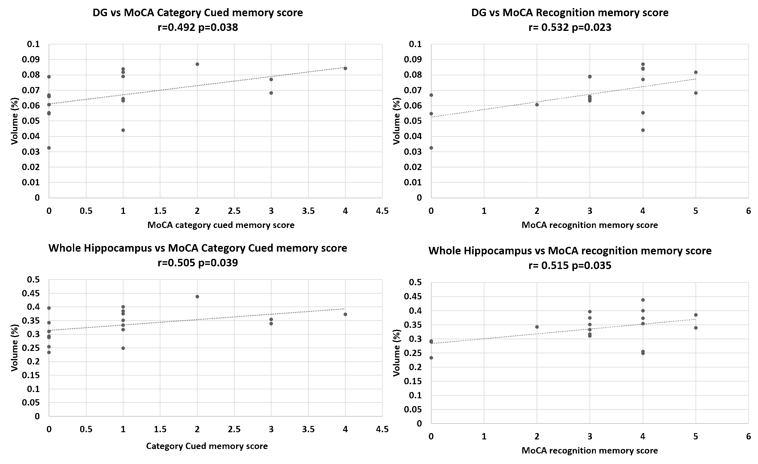
Results: In AD participants, hippocampal volume correlated with memory category cue and recognition memory scores. In analysis of hippocampal subfield volumes, the dentate gyrus volume significantly correlated with recognition and cued memory scores.
Impact: These findings matter because they provide crucial insights into the relationship between hippocampal subfield atrophy and cognitive decline in AD. It has the potential to improve the accuracy of diagnosis, targeted treatments, and the understanding of the neurobiology of AD.
BACKGROUNG
Alzheimer's disease (AD) is a progressive neurodegenerative condition characterized by a gradual decline in cognitive and functional abilities1. It particularly affects the hippocampus, a brain structure that is central cognitive processes2. Volumetric studies3 and molecular investigations4 show that the posterior hippocampus is important for memory and spatial navigation, whereas the anterior hippocampus is associated with emotional functions, including mood and anxiety-related behaviours4,5. Heterogeneous atrophy within hippocampal subfields has been observed in adults with Mild Cognitive Impairment (MCI)6,7. Notably, hippocampal atrophy across hippocampal subfields is a predictive indicator of cognitive decline as individuals progress from MCI to a dementia syndrome7. For instance, individuals with AD exhibit more pronounced atrophy in Cornu Ammonis (CA1) and subiculum (SUB) compared to age-matched controls, as evidenced in histological post-mortem studies8. Studies have also shown significant correlations between CA1 volume and delayed recall performance, particularly in individuals with amnestic MCI, which primarily affects memory. This correlation, however, is less apparent in non-amnestic MCI, where other cognitive skills are primarily affected9. Importantly, amnestic MCI is associated with the highest risk of transitioning to AD10. Furthermore, smaller volumes in various hippocampal subfields, as detected through 3T MRI, are significantly associated with lower Montreal Cognitive Assessment (MoCA) memory scores in participants with neurodegenerative diseases who exhibit cognitive complaints11. Here we used high resolution imaging at 7T to evaluate the association between cognitive decline and hippocampal atrophy in AD.METHOD
36 (19 AD, 17 HC) participants (61% female; aged 42-79) were scanned using a Philips Achieva 7T scanner equipped with a Nova Medical (Wilmington MA, USA) single-channel transmit, 32 channel receive (1Tx32Rx) head-coil. TSE images (TE/TR=119/59001ms, FA=90o, 0.38x0.39x1.50 mm3 resolution) were segmented into Cornu Ammonis (CA=CA1+CA2+CA3), Subiculum (SUB), Dentate Gyrus (DG), and Entorhinal Cortex (ERC) using ASHS 12. UDSNB3.0 Neuropsychological tests were administered by trained Assistant Psychologists. These included the MoCA and a variety of cognitive tests assessing memory (immediate/delayed recall, un-cued/cued recall, and recognition), attention, language and visuospatial abilities. Two participants (1 AD, 1 HC) withdrew from the study due to MRI intolerance, leaving 16 HC and 18 AD participants included in the final analysis. The study was approved by the Research Ethics Committee (IRAS: 276174) and the Health Research Authority. Written informed consent was provided by all participants.RESULT
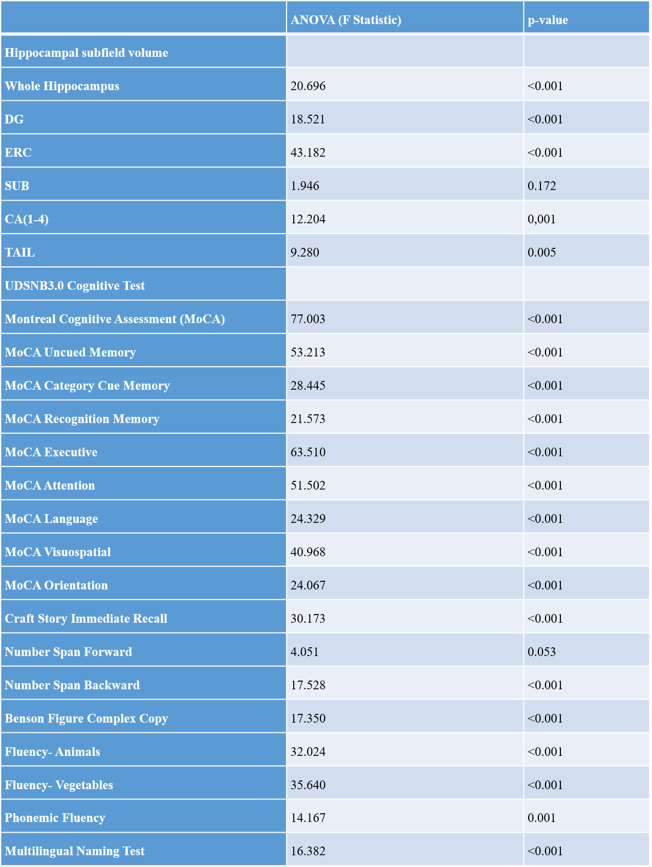
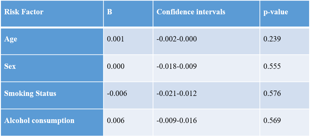
In the whole analysed sample (n=34), there were strong positive correlations between UDSNB3.0 memory test scores and hippocampal subfield volumes, as can be seen in Figure 1 and Table 1. Significant differences were found between HC and AD cognitive test scores (Table 1), so control and AD participants were analysed separately. In AD participants, the whole hippocampal volume correlated with MoCA memory category cue scores (r2=0.515, p<0.05) and MoCA recognition memory scores (r2=0.505, p<0.05); in the analysis of the hippocampal subfield volumes, only DG volume was found to be significantly correlated with the MoCA recognition and cued memory scores (category cue: r2=0.559, p<0.05, recognition memory: r2=0.543, p<0.05, Figure 1). Age, sex, smoking status, and alcohol status were not significant predictors of the relationships between the DG volume and the significant neuropsychological scores in a regression analysis (Table 2).DISCUSSION
We present evidence demonstrating correlation between cognitive test scores, particularly the scores from the MoCA, administered as part of the UDSNB3.0, with both whole hippocampal and DG volumes in individuals with AD. The DG, an area within the hippocampus, is responsible for adult brain neurogenesis 13. There have been indications that DG-specific neurogenesis plays a significant role in learning and memory processes dependent on the hippocampus 14, with dysregulation of the adult hippocampal neurogenesis being associated with cognitive decline. In this study we report an association between atrophy of the DG and short-term cued recall and recognition memory in an AD participant group, which is of particular interest. The DG has been implicated as having a key role in pattern separation15, a process that distinguishes different yet similar inputs into more independent, differentiated outputs to store information separately into distinct, though similar, memories16.CONCLUSION
The results of this study confirm that hippocampal atrophy in AD is associated with cognitive decline. Additionally, our results suggest that 7T MRI may be able to detect subtle in vivo DG atrophy in AD that is related to cued memory. The 7T scans appear to reveal DG changes in AD that to date have been confirmed only in neuropathological studies.Acknowledgements
No acknowledgement found.References
1. Gold CA, Budson AE. Memory loss in Alzheimer’s disease: Implications for development of therapeutics. Expert Rev Neurother. 2008;8(12):1879-1891. doi:10.1586/14737175.8.12.1879
2. Frisoni GB, Fox NC, Jack CR, Scheltens P, Thompson PM. The clinical use of structural MRI in Alzheimer disease. Nat Rev Neurol. 2010;6(2):67-77. doi:10.1038/nrneurol.2009.215
3. Strange BA, Witter MP, Lein ES, Moser EI. Functional organization of the hippocampal longitudinal axis. Nat Rev Neurosci. 2014;15(10):655-669. doi:10.1038/nrn3785
4. Genon S, Bernhardt BC, La Joie R, Amunts K, Eickhoff SB. The many dimensions of human hippocampal organization and (dys)function. Trends Neurosci. 2021;44(12):977-989. doi:10.1016/j.tins.2021.10.003
5. Fyhn M, Molden S, Witter MP, Moser EI, Moser MB. Spatial representation in the entorhinal cortex. Science (80- ). 2004;305(5688):1258-1264. doi:10.1126/science.1099901
6. Izzo J, Andreassen OA, Westlye LT, van der Meer D. The association between hippocampal subfield volumes in mild cognitive impairment and conversion to Alzheimer’s disease. Brain Res. 2020;1728(December 2019):146591. doi:10.1016/j.brainres.2019.146591
7. Jack CR, Dickson DW, Parisi JE, et al. Antemortem MRI findings correlate with hippocampal neuropathology in typical aging and dementia. Neurology. 2002;58(5):750-757. doi:10.1212/WNL.58.5.750 8. West MJ, Kawas CH, Stewart WF, Rudow GL, Troncoso JC. Hippocampal neurons in pre-clinical Alzheimer’s disease. Neurobiol Aging. 2004;25(9):1205-1212. doi:10.1016/j.neurobiolaging.2003.12.005
9. Broadhouse KM, Mowszowski L, Duffy S, et al. Memory Performance Correlates of Hippocampal Subfield Volume in Mild Cognitive Impairment Subtype. Front Behav Neurosci. 2019;13. doi:10.3389/fnbeh.2019.00259
10. Petersen RC, Roberts RO, Knopman DS, et al. Mild cognitive impairment: Ten years later. Arch Neurol. 2009;66(12):1447-1455. doi:10.1001/archneurol.2009.266
11. Ritter A, Hawley N, Banks SJ, Miller JB. The Association between Montreal Cognitive Assessment Memory Scores and Hippocampal Volume in a Neurodegenerative Disease Sample. J Alzheimer’s Dis. 2017;58(3):695-699. doi:10.3233/JAD-161241
12. Wisse LEM, Kuijf HJ, Honingh AM, et al. Automated hippocampal subfield segmentation at 7T MRI. Am J Neuroradiol. 2016;37(6):1050-1057. doi:10.3174/ajnr.A4659
13. Abbott LC, Nigussie F. Adult neurogenesis in the mammalian dentate gyrus. J Vet Med Ser C Anat Histol Embryol. 2020;49(1):3-16. doi:10.1111/ahe.12496
14. Li B, Yamamori H, Tatebayashi Y, et al. Failure of neuronal maturation in Alzheimer disease dentate gyrus. J Neuropathol Exp Neurol. 2008;67(1):78-84. doi:10.1097/nen.0b013e318160c5db
15. Clelland CD, Choi M, Romberg C, et al. A functional role for adult hippocampal neurogenesis in spatial pattern separation. Science (80- ). 2009;325(5937):210-213. doi:10.1126/science.1173215
16. Santoro A. Reassessing pattern separation in the dentate gyrus. Front Behav Neurosci. 2013;7(JUL):1-4. doi:10.3389/fnbeh.2013.00096
Figures