4048
Exploring Pathology Along the White Matter Tract near Hyperintense Vasogenic Edema through Multiparametric MRI and Diffusion1Radiology, NYU Grossman School of Medicine, New York, NY, United States, 2Bernard and Irene Schwartz Center for Biomedical Imaging & Center for Advanced Imaging Innovation and Research (CAI2R), NYU Grossman School of Medicine, New York, NY, United States, 3Neurology, NYU Grossman School of Medicine, New York, NY, United States, 4Small Animal Imaging Facility, MD Anderson Cancer Center, Houston, TX, United States, 5Michael E. Keeling Center for Comparative Medicine and Research, MD Anderson Cancer Center, Bastrop, TX, United States
Synopsis
Keywords: Alzheimer's Disease, Alzheimer's Disease, Vasogenic edema, white matter hyperintensities.
Motivation: There is a critical need for neuroimaging methodologies that can characterize the neuropathological changes associated with cerebral amyloid angiopathy (CAA) in Alzheimer’s disease (AD).
Goal(s): Our goal was to explore CAA-associated pathology using multiparametric MRI including diffusion MRI.
Approach: A combination of MRI techniques and histopathology was employed to evaluate and characterize the underlying neuropathological changes in a squirrel monkey model of AD.
Results: Unique pathology was found to be present along normal appearing white matter tracts using conventional MRI near areas of hyperintense vasogenic edema.
Impact: Given the prominence of CAA in human AD cases and the critical need for a more proximate model of AD pathology, our study evaluated the feasibility of advanced neuroimaging methodologies characterizing neuropathological changes in squirrel monkeys that develop spontaneous CAA.
Introduction
Cerebral amyloid-related angiopathy (CAA) is a prominent factor in Alzheimer's disease (AD) and poses significant challenges to current immunotherapeutic approaches, including the two recently FDA-approved Alzheimer's drugs1,2. These challenges manifest as amyloid-related imaging abnormalities (ARIA), characterized by vasogenic edema and cerebral microhemorrhages. Given the prevalence of CAA in virtually all AD cases, addressing this issue has become paramount3.To tackle this clinical need, our research focuses on the use of a squirrel monkey (SQM) non-human primate (NHP) model with sporadic amyloid pathology. Unlike other NHP species, SQMs develop abundant CAA in all aged animals4,5.Previously, we demonstrated the effectiveness of quantitative R2* mapping to discern age-related differences in SQM neuropathology, both cross-sectionally and longitudinally6,7. This study now offers a valuable opportunity to assess brain tissue integrity in vivo, particularly focusing on white matter (WM) microstructural changes, by combining conventional MRI including T2-weighted TSE, FLAIR, quantitative R2* mapping and multi-shell diffusion-weighted MRI.Methods
In vivo MRI was performed at UT/MD Anderson Cancer Center on a 7-Tesla Bruker 7030 Biospec scanner interfaced to an Avance 3-HD console. A CP Tx/Rx birdcage RF coil (ID=86mm) accommodated the SQM body and enabled full head coverage. 3 groups of SQMs with ages ranging from 5 years old (n=4, young), 16-17 years old (n=5, middle-aged) and 20-23 years old (n=12, old) were examined. Scan protocols included: 1) multigradient echo sequence (MGE, 8 Echoes, TE/ES=2.96/4.0ms TR=41ms, FA=13°, FOV=38.4 × 25.6 × 25.6mm, Matrix=384 × 256 × 256; Acq. Time=45min, NRep=3, Total scan=2hr30min); 2) 2D T2-w Turbo-spin-echo (TSE, 1TFac=10, TE=36ms, TR=3200ms); 3) 2D FLAIR (TFac=10, TE=35ms, TR=3200ms, TI=1557ms; and 4) Diffusion MRI using 2D Monopolar PGSE EPI (TE/TR=34/5500ms, Spatial resolution=0.8 x 0.8mm, slice thickness=0.8mm 5 non-DWI, b-values=250, 500, 1000, 2500, 5000s/mm2, with 60 directions each). Immunohistochemistry was subsequently performed to characterize the neuropathological underpinnings of observed MRI abnormalities. Diffusion MRI data was preprocessed using Designer prior to computing fractional anisotropy and mean diffusivity using Diffusion Tensor Imaging. The DTI fit was limited to data with b < 1500s/mm2.Results and Discussion
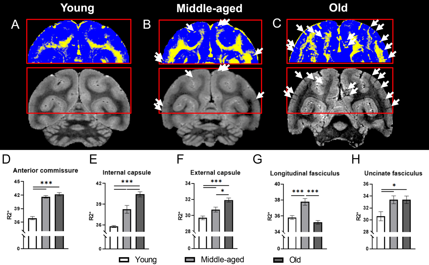
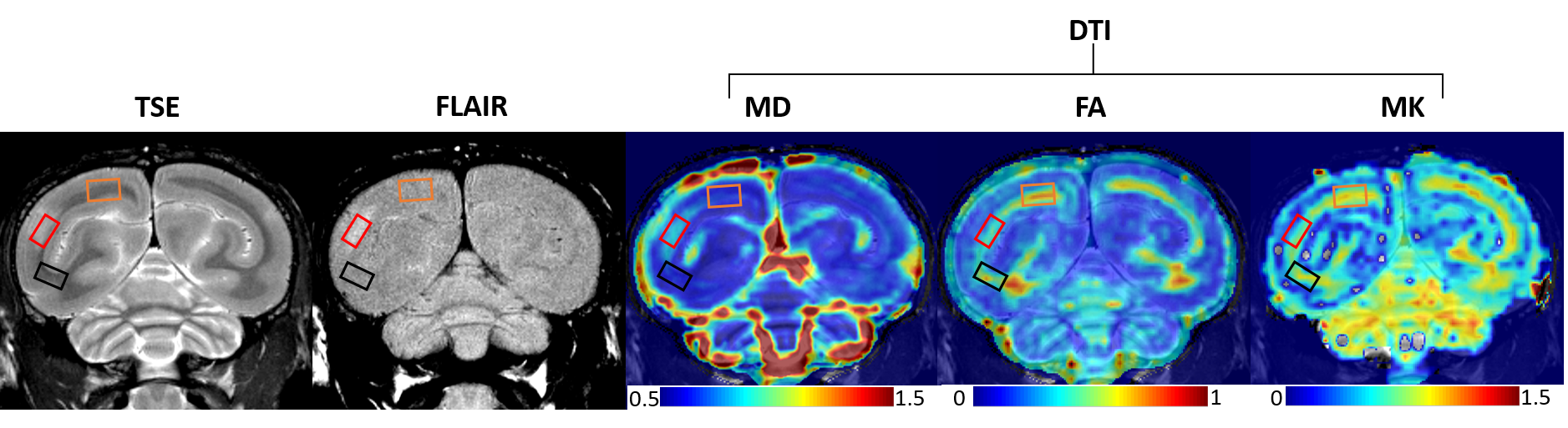
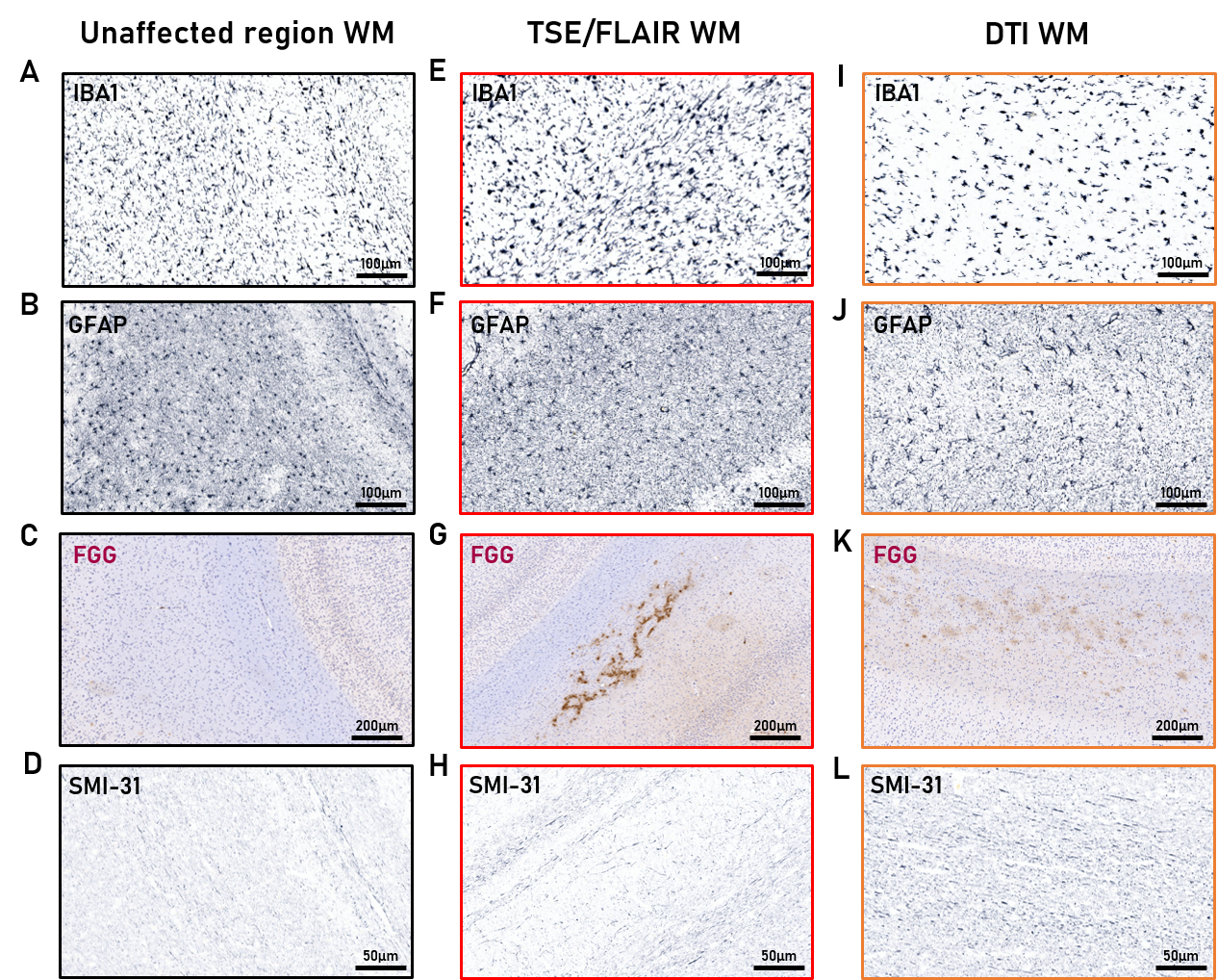
The current study combines a comprehensive examination of NHP subjects at distinct stages of aging using quantitative R2* mapping (Figure 1) and conventional ARIA MRI (Figure 2) including T2-weighted TSE and FLAIR as well as multi-shell diffusion-weighted MRI.As shown previously, R2* mapping proved very useful for monitoring pathological event that includes iron deposits associated with the presence of amyloid plaques, the presence of micro-hemorrhaging and the eventual alteration of cerebral flow resulting in local hypoxia. Importantly, these R2* alterations are steadily increasing with aging in various anatomical regions with varying intensities both in the GM and WM. However, some of the WM regions such as the example shown in Figure1.G exhibited R2* reversal suggesting the predominant presence of edema shown in the example outlined in red as WM hyper-intensities through the combination of T2-weighted TSE for anatomical identification and FLAIR. Furthermore, multi-shell diffusion-weighted MRI (outlined in orange) helped reveal the presence of abnormal WM which goes undetected using ARIA-E criteria. Figure 3 are representative sections to characterize histopathological features of the brain region that exhibited ARIA-E signal abnormalities by conventional and diffusion MRIs. Pronounced extravascular fibrinogen immunoreactivity (FGG) was detected in the lesion area, both in the WM and (to a lesser degree) the gray matter. Microglia dystrophy and/or hyperactivity were observed in the same region (IBA1), as were similarly dense clusters of astrocytes (GFAP). In addition, immunostaining for phosphorylated neurofilaments (SMI-31) showed evidence of axonal abnormalities in the involved area.Conclusion
The current study promotes the combination of advanced MRI biomarkers as a neuroimaging tool for a more sensitive and specific interpretation of imaging abnormalities, potentially linked to age-associated cerebrovascular dysfunction. Evaluating the interaction between MRI metrics and neuropathological correlates will further assess the use of diffusion MRI as a sensitive and clinically relevant biomarker of disease progression.Acknowledgements
This work was supported, in part, by Alzheimer’s Association: AARG-16-440596 (HS), NIH grants: R01NS102845 (HS), R21AG073305 (WH, HS), R21NS127091 (HS), R01NS128190 (HS), R01NS088040, and P41EB017183. MRI imaging was performed both at UT-MD Anderson Cancer Center and at the NYU Langone Health Preclinical Imaging Core. The latter is a shared resource partially supported by the NIH/SIG 1S10OD018337-01, the Laura and Isaac Perlmutter Cancer Center Support Grant NIH/NCI 5P30CA016087 and the NIBIB Biomedical Technology Resource Center Grant NIH P41 EB017183.References
- Qiao Y, et al. Safety and efficacy of lecanemab for Alzheimer's disease: a systematic review and meta-analysis of randomized clinical trials. Front Aging Neurosci. 2023;15:1169499. Published 2023 May 5. doi:10.3389/fnagi.2023.1169499
- Yadollahikhales G, et al. Anti-Amyloid Immunotherapies for Alzheimer's Disease: A 2023 Clinical Update. Neurotherapeutics. 2023 Jul;20(4):914-931. doi: 10.1007/s13311-023-01405-0. Epub 2023 Jul 25. PMID: 37490245; PMCID: PMC10457266.
- Banerjee G, et al. The increasing impact of cerebral amyloid angiopathy: essential new insights for clinical practice. Journal of neurology, neurosurgery, and psychiatry. 2017;88(11):982-94. Epub 2017/08/28. PMID: 28844070
- Patel AG, et al. Innate immunity stimulation via CpG oligodeoxynucleotides ameliorates Alzheimer’s disease pathology in aged squirrel monkeys. Brain : a journal of neurology. 021;144(7):2146-65.
- Heuer E, et al. Amyloid-Related Imaging Abnormalities in an Aged Squirrel Monkey with Cerebral Amyloid Angiopathy. Journal of Alzheimer's disease : JAD. 2017;57(2):519-30. doi: 10.3233/JAD-160981. PMID: 28269776.
- Murray S, et al.. Development of neuroimaging biomarkers across disease continuum in non-human primate model of sporadic Alzheimer’s disease pathology. The 26th Alzheimer's Association International Conference, Amsterdam, Netherlands, 16-20 July, 2023
- Zaim Wadghiri Y, et al. Multi-parametric MRI Characterization of the Aging Squirrel Monkey as a Model of Sporadic Cerebral Amyloid Angiopathy. Proceedings 32ndSci. Meeting, Int’l Society for Magnetic Resonance in Medicine, Toronto, Ontario-Canada, 03-08 June, 2023.
Figures