4047
Cerebral Blood Flow in Midlife Obesity: Associations with Visceral and Subcutaneous Abdominal Adipose Tissue1Washington University in St. Louis, Saint Louis, MO, United States, 2Computer Science, State University of New York at Binghamton, Binghamton, NY, United States, 3Missouri state university, Columbia, MO, United States, 4University of California San Diego, La Jolla, CA, United States
Synopsis
Keywords: Alzheimer's Disease, Perfusion, adiposity, obesity
Motivation: Understanding the role of midlife obesity in Alzheimer disease (AD) risk is key to AD prevention.
Goal(s): We aimed to investigate the association between obesity and abdominal visceral and subcutaneous adipose tissue (VAT, and SAT) and brain perfusion, altered early in AD.
Approach: For this aim, we performed brain and abdominal MRI scans to compare absolute cerebral blood flow (CBF), derived from pCASL sequence, between the obese vs. non-obese, the high- vs. low-VAT and high- vs. low-VAT groups.
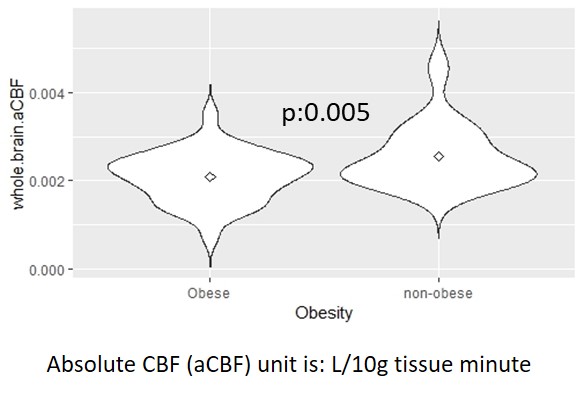
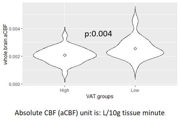
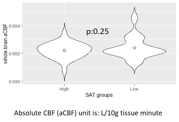
Results: A lower whole-brain CBF was observed in the obese vs. non-obese and high-VAT vs. low-VAT, but not between high- and low-SAT groups.
Impact: Lower brain perfusion in individuals with obesity and higher VAT, especially in AD-related areas like middle temporal cortex, highlights midlife visceral obesity’s role in AD development. Future studies should explore the association of AD neuroimaging markers with body mass components.
Introduction
Obesity and higher adiposity in midlife are increasingly recognized as contributors to the risk of Alzheimer disease (AD) 1. Neurodegeneration in AD is at least partly mediated by vascular compromise and brain hypoperfusion 2. On the other hand, the association of adipose tissue with cortical thickness and brain vascular changes has been observed 3. In this study, we aimed to investigate the associations between body mass index (BMI), as well as abdominal visceral and subcutaneous adipose tissue (VAT, SAT) and brain cerebral blood flow (CBF) in cognitively normal midlife individuals.Methods
A total of 66 middle-aged cognitively normal adults (age: 49.86 ± 5.99 years, females: 66.7%, obesity (BMI of 30 kg/m2 or higher): 51.5 %, BMI: 31.72 ± 6.96 kg/m2) underwent abdominal and brain MRI. Using an in-house Matlab program, abdominal VAT and SAT were automatically segmented followed by manual editing. A 3D Pseudo-Continuous Arterial Spin Labeling (pCASL) sequence, with a single post-labeling delay of 2.025 s, was used for assessing CBF. Statistical Parametric Mapping (SPM) 12 was used to generate ASL difference and absolute CBF (aCBF) maps with a single compartment model, co-registered to the gray matter segmentations, and normalized to MNI space, followed by spatial smoothing with a 6mm FWMH Gaussian kernel 4. Using AAL3 atlas and Matlab, region of interest masks were created for amygdala, hippocampus, posterior cingulate, precuneus, parahippocampal, medial orbitofrontal, middle temporal, and Calcarine cortices, and applied to absolute CBF (aCBF) maps. Based on VAT and SAT volumes, the sample was dichomotized to low- and high-VAT, and low- and high-SAT group. The aCBF differences between the obese vs. non-obese, high-VAT vs. low-VAT, and high-SAT vs. low-SAT was assessed, with age and sex as covariates. Also, BMI, VAT, and SAT as separate predictor variables, with age and sex as covariates, were used for voxel-wise analysis.Results
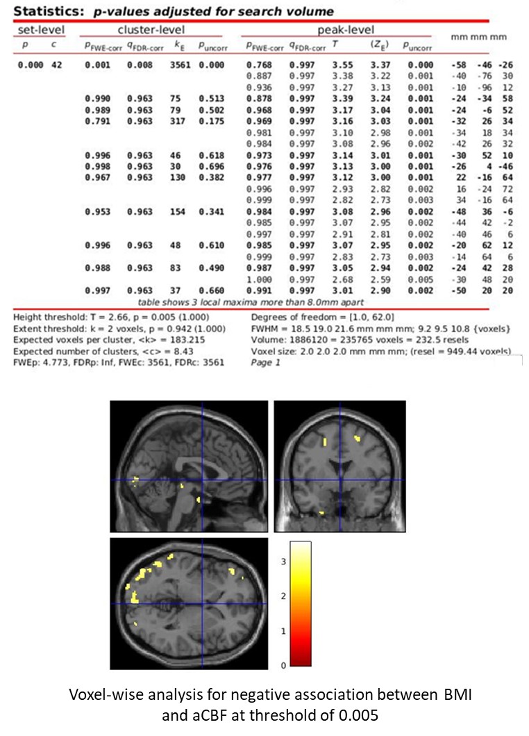
The high-VAT group showed lower whole-brain aCBF (p=0.004), particularly in the right and left Calcarine gyri (p=0.001, 0.002). A lower whole-brain aCBF was found in the obese group (p=0.005), which was more prominent in the left middle temporal lobe (p=0.002). No significant difference was observed in global and regional aCBF in the high-SAT vs. low-SAT groups. Voxel-wise analyses showed significantly lower aCBF in association with BMI in temporal, occipital, and frontal lobe clusters after false discovery rate correction.Conclusion
Obesity and increased visceral abdominal fat are associated with a lower cerebral blood flow, with a more prominent decrease in the middle temporal cortex, as an AD-signature area, in cognitively normal midlife individuals. These findings, in line with previous studies, highlight the role of obesity, especially visceral obesity, in brain hypoperfusion and potentially Alzheimer disease risk, as early as midlife 2,3. The role of other body mass components, like muscle mass, and the effectiveness of interventions aimed at modifying body mass components in modifying brain perfusion should be explored by future studies.Acknowledgements
This study was funded by the National Institute of Health (NIH), under the project number 1RF1AG072637-01 entitled “Neuroinflammation and Alzheimer's Disease Imaging Biomarkers in Midlife Obesity” and was performed at Washington University in Saint Louis. Enrollment and assessment of Alzheimer Disease Research Center (ADRC) participants were funded by these projects: Alzheimer’s Disease Research Center (ADRC, P30AG066444), Antecedent Biomarkers for Alzheimer Disease: The Adult Children Study (ACS, P01AG026276), and Healthy Aging and Senile Dementia (HASD, P01AG003991).References
1. Li J, Joshi P, Ang TFA, Liu C, Auerbach S, Devine S, et al. Mid- to Late-Life Body Mass Index and Dementia Risk: 38 Years of Follow-up of the Framingham Study. Am J Epidemiol. 2021;190(12):2503-10. 2. Zhou TD, Zhang Z, Balachandrasekaran A, Raji CA, Becker JT, Kuller LH, et al. Prospective Longitudinal Perfusion in Probable Alzheimer's Disease Correlated with Atrophy in Temporal Lobe. Aging Dis. 2023.
3. Knight SP, Laird E, Williamson W, O'Connor J, Newman L, Carey D, et al. Obesity is associated with reduced cerebral blood flow - modified by physical activity. Neurobiol Aging. 2021;105:35-47.
4. Dai W, Garcia D, de Bazelaire C, Alsop DC. Continuous flow-driven inversion for arterial spin labeling using pulsed radio frequency and gradient fields. Magn Reson Med. 2008;60(6):1488-97.
Figures