4042
Preliminary Diffusion MRI Findings in Hippocampal Subregions of People with Subjective Cognitive Decline1New York University School of Medicine, New York, NY, United States, 2Massachusetts General Hospital, Boston, MA, United States, 3Harvard University, Cambridge, MA, United States
Synopsis
Keywords: Alzheimer's Disease, Data Analysis, hippocampus, subjective cognitive decline
Motivation: Hippocampal subregions are differentially impacted by early Alzheimer’s pathology and may differ in their roles in information processing. However, differences in microstructure in hippocampal subregions have not been investigated in SCD.
Goal(s): To investigate how hippocampal subregions (anterior, medial, and posterior) are affected in SCD.
Approach: Anatomical and diffusion imaging was used to extract diffusion tensor and diffusional kurtosis imaging metrics from hippocampal subregions.
Results: Reduced fractional anisotropy, decreased kurtosis, and increased mean diffusivity were found in SCD compared with a control group for the examined regions. The pattern of change varied across hemispheres and along the anterior-posterior axis.
Impact: These data suggest heterogeneous pathological changes in the hippocampal microstructure in SCD indicative of neurodegeneration. These findings may inform future research on biomarker development for early AD detection.
Introduction
Patients who complain of memory difficulties but score normally on cognitive testing have subjective cognitive decline, or SCD(1). These patients are more likely to have Alzheimer’s Disease pathology(2). They are twice as likely to develop dementia(3). Among people who are amyloid positive and cognitively normal, those with SCD are five times more likely to develop dementia(4). This increased risk of dementia is of interest because early Alzheimer’s detection allows for prevention, life planning(5), and therapeutic interventions(6).The entorhinal cortex followed by anterior hippocampus is the most common starting point of Alzheimer’s pathological changes, which then spread to nearby regions(7). While the whole hippocampus is important for memory formation, hippocampal subregions may differ in their role in information processing. For example, the anterior hippocampus is implicated in emotional memory and anxiety (8), while posterior hippocampus is implicated in spatial learning(9). Prior research shows that anterior hippocampus presents atrophy and reduced metabolism earlier in AD(10). People with SCD have higher rates of anxiety than people without SCD(11). Thus, the microstructure of hippocampal subregions is of particular interest in this population. However, there has been little research studying hippocampal subregions in SCD. Therefore, the aim of this study was to examine microstructural changes in hippocampal subregions in SCD using diffusional kurtosis (DKI) and diffusion tensor (DTI) imaging.
Methods
Anatomical and diffusion images were collected on a 3T Siemens Prisma for 163 healthy participants. Participants were included in the SCD group (N=78) if they had a rounded average score of 2 on the Cognitive Change Index(12). Cognitively normal controls (N=85) had a rounded average Cognitive Change Index of 1.Data acquisition included: a) 1mm isotropic 3D sagittal MPRAGE T1w images (TR=2100ms, TE=4.9ms, TI=900ms, acceleration=2, echo spacing=8.9ms); b) 1mm isotropic 3D sagittal FLAIR T2w images (TR=6000ms, TE=374ms, TI=2100ms, 7/8 partial Fourier, acceleration=2, echo spacing=3.46ms) and c) 2.5mm isotropic EPI diffusion-weighted images (DWI). The DWI were acquired for five b=0 s/mm2, four b=250s/mm2, 20 b=1000s/mm2, and 60 b=2000s/mm2. The b=0 s/mm2 images were obtained for opposite polarities (AP and PA) to allow estimation of the B0 field map.
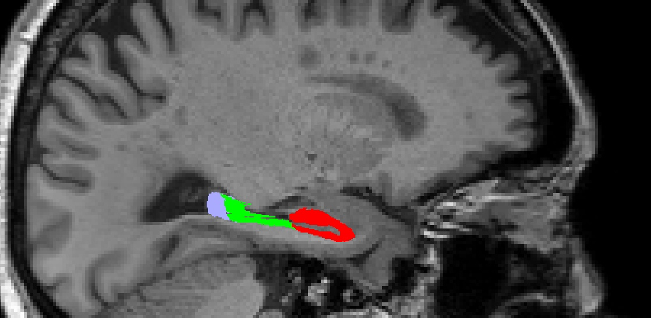
Bilateral hippocampal subregions (anterior, medial, and posterior) were segmented using T1w and T2w images with FreeSurfer 7.4.1(13) and co-registered to the diffusion space (Figure 1). Diffusion images were first denoised, then corrected for motion and B0 field distortions using PyDesigner 1.0.0(14). The diffusion and kurtosis tensors were obtained by fitting the diffusion signal using the diffusional kurtosis approximation and employed to obtained three-dimensional maps of fractional anisotropy (FA), mean diffusivity (MD), mean kurtosis (MK) and kurtosis fractional anisotropy (KFA). Mean values for each metric of interest were obtained for each hippocampal subregion under investigation. Between-group comparisons (SCD versus controls) were conducted using Wilcoxon rank-sum test in R 4.3.1(15).
Results
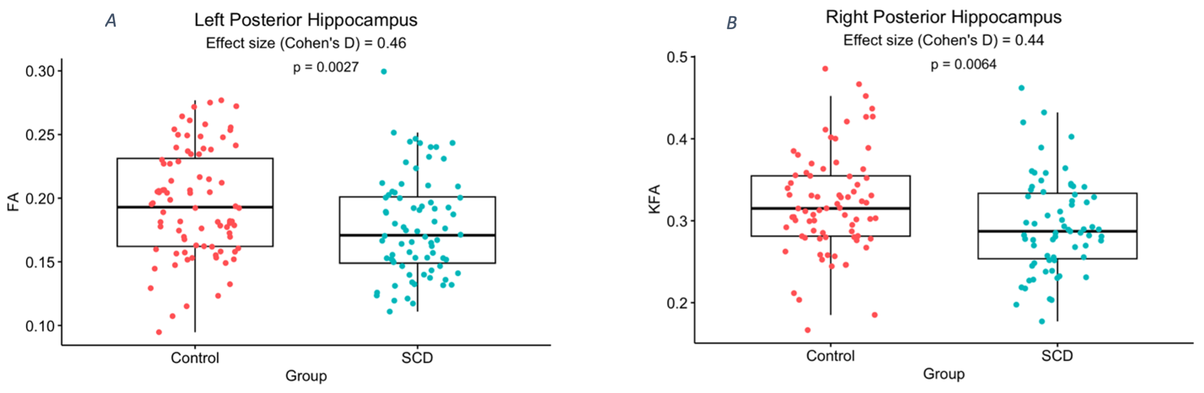
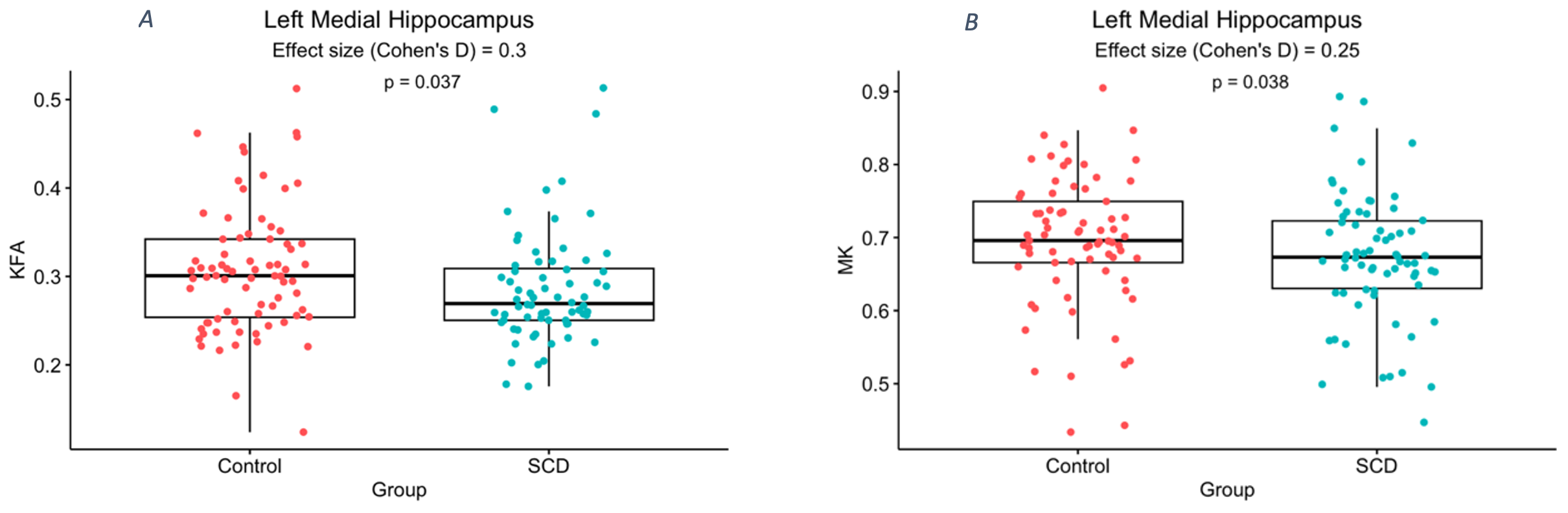
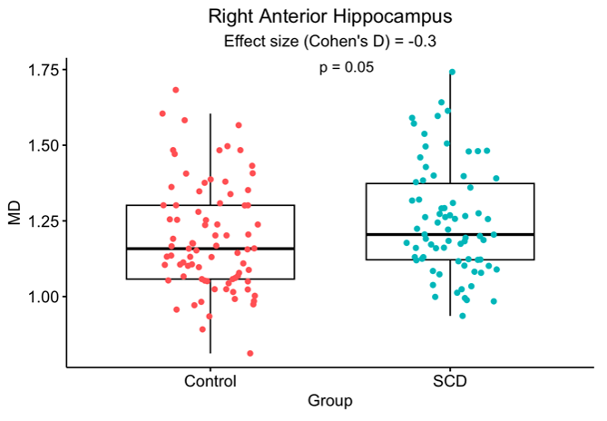
The posterior hippocampus showed reduced FA on the left and reduced KFA on the right in SCD versus controls (Figure 2). The medial hippocampus showed reduced KFA, and MK on the left in SCD, but no significant differences on the right (Figure 3). The anterior hippocampus showed no significant differences on the left but did show increased MD on the right in SCD (Figure 4).Discussion
Hippocampal microstructural changes were noted in SCD compared with the control group for all of the three examined subregions. While the accumulation of amyloid or tau protein aggregates are expected to restrict diffusion or increase kurtosis(16, 17), neither effect was observed in this study. Further, right versus left hippocampus showed different microstructural results. The findings on the right showed increased diffusion with no change in kurtosis in the anterior hippocampus, suggestive of increased extracellular space (18-20). However, findings in the left medial hippocampus showed decreased kurtosis with no change in diffusion, which is suggestive of decreased microstructural complexity potentially from demyelination or axonal or neuronal loss. Mouse models of amyloidosis show the opposite, increased kurtosis with no change in diffusion (16). Reduced FA and KFA, seen in the bilateral posterior hippocampus, may be also indicative of demyelination and potential neuronal and axonal loss (21).Conclusion
These data suggest heterogeneous pathological changes in the hippocampal microstructure in SCD indicative of neurodegeneration. Findings may inform future research on biomarker development for early AD detection.Acknowledgements
We thank all the participants who contributed their time and effort in the study. We acknowledge the NYU Langone Alzheimer’s Disease Research Center for curating and providing the dataset and funding this research (NIA P30AG066512 and P30AG008051).This work was supported in part by the NYU Alzheimer’s Disease Research Center Research Education Component. It was performed under the rubric of the Center for Advanced Imaging Innovation and Research (CAI2R, www.cai2r.net) an NIBIB Biomedical Technology Resource Center (NIH P41 EB017183).References
1. Jessen F, Amariglio RE, Buckley RF, van der Flier WM, Han Y, Molinuevo JL, et al. The characterisation of subjective cognitive decline. The Lancet Neurology. 2020;19(3):271-8.
2. Jonker C, Geerlings MI, Schmand B. Are memory complaints predictive for dementia? A review of clinical and population‐based studies. International journal of geriatric psychiatry. 2000;15(11):983-91.
3. Mitchell AJ, Beaumont H, Ferguson D, Yadegarfar M, Stubbs B. Risk of dementia and mild cognitive impairment in older people with subjective memory complaints: meta-analysis. Acta Psychiatrica Scandinavica. 2014;130(6):439-51.
4. Buckley RF, Maruff P, Ames D, Bourgeat P, Martins RN, Masters CL, et al. Subjective memory decline predicts greater rates of clinical progression in preclinical Alzheimer's disease. Alzheimer's & Dementia. 2016;12(7):796-804.
5. The Need for Early Detection and Treatment in Alzheimer's Disease. EBioMedicine. 2016;9:1-2.
6. Shi M, Chu F, Zhu F, Zhu J. Impact of Anti-amyloid-beta Monoclonal Antibodies on the Pathology and Clinical Profile of Alzheimer's Disease: A Focus on Aducanumab and Lecanemab. Front Aging Neurosci. 2022;14:870517.
7. Braak H, Braak E. Staging of Alzheimer's disease-related neurofibrillary changes. Neurobiology of aging. 1995;16(3):271-8.
8. Ciocchi S, Passecker J, Malagon-Vina H, Mikus N, Klausberger T. Selective information routing by ventral hippocampal CA1 projection neurons. Science. 2015;348(6234):560-3.
9. Bannerman DM, Rawlins JN, McHugh SB, Deacon RM, Yee BK, Bast T, et al. Regional dissociations within the hippocampus--memory and anxiety. Neurosci Biobehav Rev. 2004;28(3):273-83.
10. Masurkar AV. Towards a circuit-level understanding of hippocampal CA1 dysfunction in Alzheimer’s disease across anatomical axes. Journal of Alzheimer's disease & Parkinsonism. 2018;8(1).
11. Perrotin A, La Joie R, La Sayette V, Barré L, Mézenge F, Mutlu J, et al. Subjective cognitive decline in cognitively normal elders from the community or from a memory clinic: Differential affective and imaging correlates. Alzheimer's & Dementia. 2017;13(5):550-60.
12. Rattanabannakit C, Risacher SL, Gao S, Lane KA, Brown SA, McDonald BC, et al. The Cognitive Change Index as a Measure of Self and Informant Perception of Cognitive Decline: Relation to Neuropsychological Tests. J Alzheimers Dis. 2016;51(4):1145-55.
13. Iglesias JE, Augustinack JC, Nguyen K, Player CM, Player A, Wright M, et al. A computational atlas of the hippocampal formation using ex vivo, ultra-high resolution MRI: Application to adaptive segmentation of in vivo MRI. Neuroimage. 2015;115:117-37.
14. Dhiman S, Teves JB, Thorn KE, McKinnon ET, Moss HG, Adisetiyo V, et al. PyDesigner: A pythonic implementation of the DESIGNER pipeline for diffusion tensor and diffusional kurtosis imaging. bioRxiv. 2021:2021.10. 20.465189.
15. Kassambara A. ggpubr:“ggplot2” based publication ready plots (R package version 0.4. 0)[Computer software]. ggpubr:‘ggplot2’based publication ready plots (R Package Version 0.4. 0 …; 2020.
16. Vanhoutte G, Pereson S, Delgado YPR, Guns PJ, Asselbergh B, Veraart J, et al. Diffusion kurtosis imaging to detect amyloidosis in an APP/PS1 mouse model for Alzheimer's disease. Magn Reson Med. 2013;69(4):1115-21.
17. Wolf D, Fischer FU, Scheurich A, Fellgiebel A, Alzheimer's Disease Neuroimaging I. Non-Linear Association between Cerebral Amyloid Deposition and White Matter Microstructure in Cognitively Healthy Older Adults. J Alzheimers Dis. 2015;47(1):117-27.
18. Hui ES, Du F, Huang S, Shen Q, Duong TQ. Spatiotemporal dynamics of diffusional kurtosis, mean diffusivity and perfusion changes in experimental stroke. Brain Res. 2012;1451:100-9.
19. Lodygensky GA, West T, Stump M, Holtzman DM, Inder TE, Neil JJ. In vivo MRI analysis of an inflammatory injury in the developing brain. Brain Behav Immun. 2010;24(5):759-67.
20. Alves R, Henriques RN, Kerkela L, Chavarrias C, Jespersen SN, Shemesh N. Correlation Tensor MRI deciphers underlying kurtosis sources in stroke. Neuroimage. 2022;247:118833.
21. Pasternak O, Westin CF, Dahlben B, Bouix S, Kubicki M. The extent of diffusion MRI markers of neuroinflammation and white matter deterioration in chronic schizophrenia. Schizophr Res. 2015;161(1):113-8.
Figures