4041
Whole-brain high-resolution myelin water fraction mapping within 6 min using the accelerated BMC-mcDESPOT (aBMC-mcDESPOT) method.1National Institute on Aging, National Institute of Health, Baltimore, MD, United States
Synopsis
Keywords: Alzheimer's Disease, Relaxometry, Accelerated imaging, Myelin water fraction, BMC-mcDESPOT
Motivation: Our previous works demonstrate the unique capability of BMC-mcDESPOT MRI method which provided myelin water fraction(MWF) mapping to infer myelination. However, the BMC-mcDESPOT protocol consists of multiple imaging sequences, leading to the total acquisition time of 17min.
Goal(s): In this study, we test the feasibility of accelerating the acquisition by under-sampling in both the Fourier and in the data domain.
Approach: We acquired fully-sampled BMC-mcDESPOT dataset, followed by under-sampled BMC-mcDESPOT dataset with compressed sensing. Further acceleration was achieved retrospectively in the data domain by selecting two flip-angles (aBMC-mcDESPOT).
Results: Our results showed that derived MWF maps using BMC-mcDESPOT and aBMC-mcDESPOT were virtually similar.
Impact: We proposed the accelerated BMC-mcDESPOT (aBMC-mcDESPOT) analysis, which drastically reduces acquisition time for accurately measuring MWF. This critical advancement in multicomponent relaxometry will allow greater integration of MWF imaging in clinical investigations for cerebral myelination in aging and neurodegenerative diseases.
Introduction
Myelin water fraction (MWF) is an important imaging biomarker for noninvasively studying cerebral development and neurodegeneration1-3. Among different MR imaging methods for MWF mapping, the Bayesian Monte Carlo analysis of multicomponent-driven equilibrium observation of T1 and T2 (BMC-mcDESPOT) has demonstrated the unique capability of isotropic high-resolution whole-brain MWF mapping within 20 min4, 5, and has been successfully applied to provide new insights on brain aging [6-8], cognitive and motor declines9-11, and various risk factors influencing cerebral myelination12-17. Due to its capability for high-resolution mapping, BMC-mcDESPOT has also provided seminal insights into the myelination of the brainstem and its implication in motor dysfunction18-20. Nevertheless, any attempt to improve the temporal resolution would assist to further integrate this MRI method in clinical studies and trials. In this study, we introduced the aBMC-mcDESPOT method, an accelerated version of BMC-mcDESPOT, through combined under-samplings of the Fourier domain using compressed sensing (CS), and the data domain, allowing a drastic reduction in the total acquisition time for whole-brain, high-resolution, MWF imaging.Methods
Data AcquisitionReference data (no-acceleration): Our subject underwent BMC-mcDESPOT protocol consisting of SPGR and bSSFP images at 10 different FAs each on a 3T Philips MRI system. bSSFP images were acquired with two different RF phase increment of 0° and 180° to correct for the off-resonance frequency artifacts4, 5, 21. For the sake of assessing reproducibility, three sets of fully-sampled datasets were collected without any under-sampling with an acquisition time of 17 min per dataset.
Fourier domain acceleration: Three sets of accelerated scans were acquired using compressed sensing (CS) with an acceleration factor of 2, resulting in the total scan time of 9 min per dataset.
Image domain acceleration: The under-sampling in the data domain were retrospectively performed on the bSSFP images acquired with phase increment of 0° by selecting only 2 images at flip angles of 2° and 50° from the total of 10 flip angle bSSFP images. In conjunction with the 10 bSSFP images obtained with phase increment of 180°, these images were used to calculate the off-resonance (B0) map using DESPOT222. Note that the B0 map is required for the MWF calculation using BMC-mcDESPOT to account for the spatial variations in the off-resonance frequencies4, 5, 21. This strategy results in a total scan time of 12.3 min.
Combined accelerations: The CS under-sampling provided a 48% acceleration factor, while combining CS under-sampling with the data domain under-sampling resulted in a 63% acceleration factor. For the sake of clarity, the combined acceleration method is referred to as aBMC-mcDESPOT with a drastically reduced acquisition time of ~6 min.
Data Processing
We divided our datasets into four categories: 1) Fully-sampled in both Fourier and data domains, 2) fully-sampled Fourier domain and under-sampled data domain, 3) under-sampled Fourier domain and fully-sampled data domain, and 4) under-sampled Fourier and data domains (aBMC-mcDESPOT). For each dataset, we estimated T1, T2 and B0 maps, then utilized the estimated B0 maps to generate the corresponding MWF maps according to the BMC-mcDESPOT protocol4, 5, 23 or the aBMC-mcDESPOT analysis described above. All maps were registered to the first reference datasets using FSL to correct for motion across scans24. White matter segmentation was also performed using FSL to examine the accuracy and precision of the estimates in different brain regions.
Results & Discussion
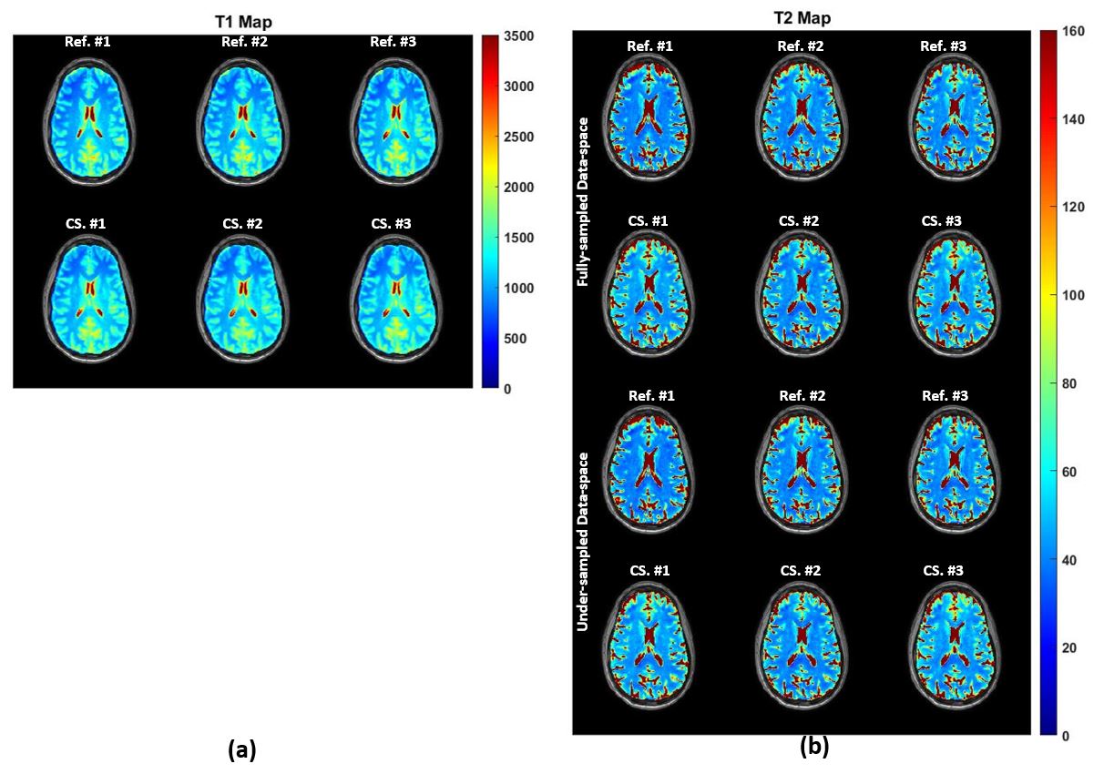
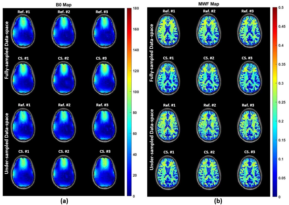
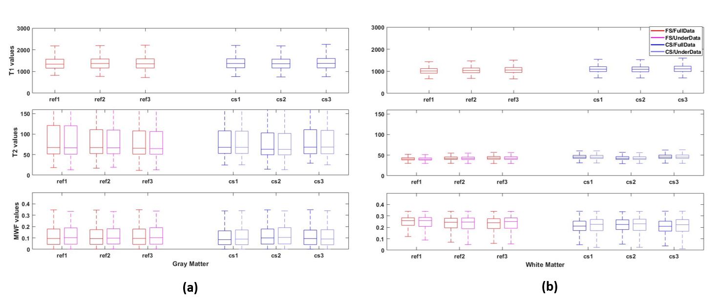
Figure 1a and 1b show the estimated T1 and T2 maps for a representative slice. Visual inspection of T2 maps with different under-sampling patterns revealed virtually identical T2 maps across all these datasets. Figure 2a displays the estimated B0 maps indicating that the acceleration in the Fourier domain did not impact the accuracy in estimating regional off-resonance frequency values. Even in the case of under-sampled data space, the B0 maps were virtually identical to those derived from the fully-sampled reference datasets. Figure 2b displays the estimated MWF maps of each dataset, which demonstrate accurate MWF estimation regardless of acceleration in either domain. Finally, Figure 3 shows the repeatability of these estimates across 3 different acquisitions, where we observe similar values across different acquisitions.Conclusion
aBMC-mcDESPOT approach leads to accurate determination of MWF within 6 min, offering unique opportunities to further integrate myelin imaging in clinical studies and trials.Acknowledgements
This work was supported by the Intramural Research Program of the National Institute on Aging of the National Institutes of Health.References
1. Bouhrara, M., et al., Evidence of demyelination in mild cognitive impairment and dementia using a direct and specific magnetic resonance imaging measure of myelin content. Alzheimer's & Dementia, 2018. 14(8): p. 998-1004.
2. Dean III, D.C., et al., Alterations of myelin content in Parkinson’s disease: a cross-sectional neuroimaging study. PloS one, 2016. 11(10): p. e0163774.
3. MacKay, A.L. and C. Laule, Magnetic resonance of myelin water: an in vivo marker for myelin. Brain plasticity, 2016. 2(1): p. 71-91.
4. Bouhrara, M. and R.G. Spencer, Improved determination of the myelin water fraction in human brain using magnetic resonance imaging through Bayesian analysis of mcDESPOT. Neuroimage, 2016. 127: p. 456-471.
5. Bouhrara, M. and R.G. Spencer, Rapid simultaneous high-resolution mapping of myelin water fraction and relaxation times in human brain using BMC-mcDESPOT. NeuroImage, 2017. 147: p. 800-811.
6. Bouhrara, M., et al., Adult brain aging investigated using BMC-mcDESPOT based myelin water fraction imaging. Neurobiology of Aging, 2019.
7. Qian, W., et al., Nonlinear associations of neurite density and myelin content with age revealed using multicomponent diffusion and relaxometry magnetic resonance imaging. NeuroImage, 2020. 223: p. 117369.
8. Kiely, M., et al., Insights into human cerebral white matter maturation and degeneration across the adult lifespan. NeuroImage, 2022. 247: p. 118727.
9. Akhonda, M.A., et al., The effect of the human brainstem myelination on gait speed in normative aging. The Journals of Gerontology: Series A, 2023: p. glad193.
10. Faulkner, M.E., et al., Lower myelin content is associated with lower gait speed in cognitively unimpaired adults. The Journals of Gerontology: Series A, 2023. 78(8): p. 1339-1347.
11. Gong, Z., et al., Lower myelin content is associated with more rapid cognitive decline among cognitively unimpaired individuals. Alzheimers Dement, 2023. 19(7): p. 3098-3107.
12. Bouhrara, M., et al., Association of cerebral blood flow with myelin content in cognitively unimpaired adults. BMJ Neurol Open, 2020. 2(1): p. e000053.
13. Bouhrara, M., et al., Evidence of association between obesity and lower cerebral myelin content in cognitively unimpaired adults. International Journal of Obesity, 2021. 45(4): p. 850-859.
14. Kiely, M., et al., Evidence of An Association Between Cerebral Blood Flow and Microstructural Integrity in Normative Aging Using a Holistic MRI Approach. J Magn Reson Imaging, 2022.
15. Laporte, J.P., et al., Hypertensive Adults Exhibit Lower Myelin Content: A Multicomponent Relaxometry and Diffusion Magnetic Resonance Imaging Study. Hypertension, 2023.
16. Triebswetter, C., et al., Differential associations between apolipoprotein E alleles and cerebral myelin content in normative aging. Neuroimage, 2022. 251: p. 118988.
17. Walker, K.A., et al., MRI and fluid biomarkers reveal determinants of myelin and axonal loss with aging. Ann Clin Transl Neurol, 2023.
18. Bouhrara, M., et al., Maturation and degeneration of the human brainstem across the adult lifespan. Aging (Albany NY), 2021. 13(11): p. 14862.
19. Bouhrara, M., et al., Quantitative age-dependent differences in human brainstem myelination assessed using high-resolution magnetic resonance mapping. Neuroimage, 2020. 206: p. 116307.
20. Akhonda, M., et al., The effect of the human brainstem myelination on gait speed in normative aging. J Gerontol A Biol Sci Med Sci, 2023.
21. Deoni, S.C., Correction of main and transmit magnetic field (B0 and B1) inhomogeneity effects in multicomponent‐driven equilibrium single‐pulse observation of T1 and T2. Magnetic resonance in medicine, 2011. 65(4): p. 1021-1035.
22. Deoni, S.C., Transverse relaxation time (T2) mapping in the brain with off‐resonance correction using phase‐cycled steady‐state free precession imaging. Journal of Magnetic Resonance Imaging: An Official Journal of the International Society for Magnetic Resonance in Medicine, 2009. 30(2): p. 411-417.
23. Bouhrara, M., et al., Adult brain aging investigated using BMC-mcDESPOT–based myelin water fraction imaging. Neurobiology of aging, 2020. 85: p. 131-139.
24. Jenkinson, M., et al., Fsl. Neuroimage, 2012. 62(2): p. 782-790.
Figures