4028
Examining the changes of default-mode network in the developmental brain of transgenic monkeys with Huntington’s disease using rsfMRI1EPC Imaging Center, Emory National Primate Research Center, Emory University, Atlanta, GA, United States
Synopsis
Keywords: Other Neurodegeneration, Genetic Diseases, monkey
Motivation: The dysfunction of the default-mode network (DMN) in Huntington Disease (HD) patients has been reported in previous studies, but the results remain inconclusive.
Goal(s): This study aimed to investigate the dysfunction of the DMN during the evolution of the disease using a transgenic monkey model.
Approach: Four HD and 4 age-matched wild-type monkeys were utilized and functional connectivity (FC) data at 3, 4 and 5 years of age were analyzed.
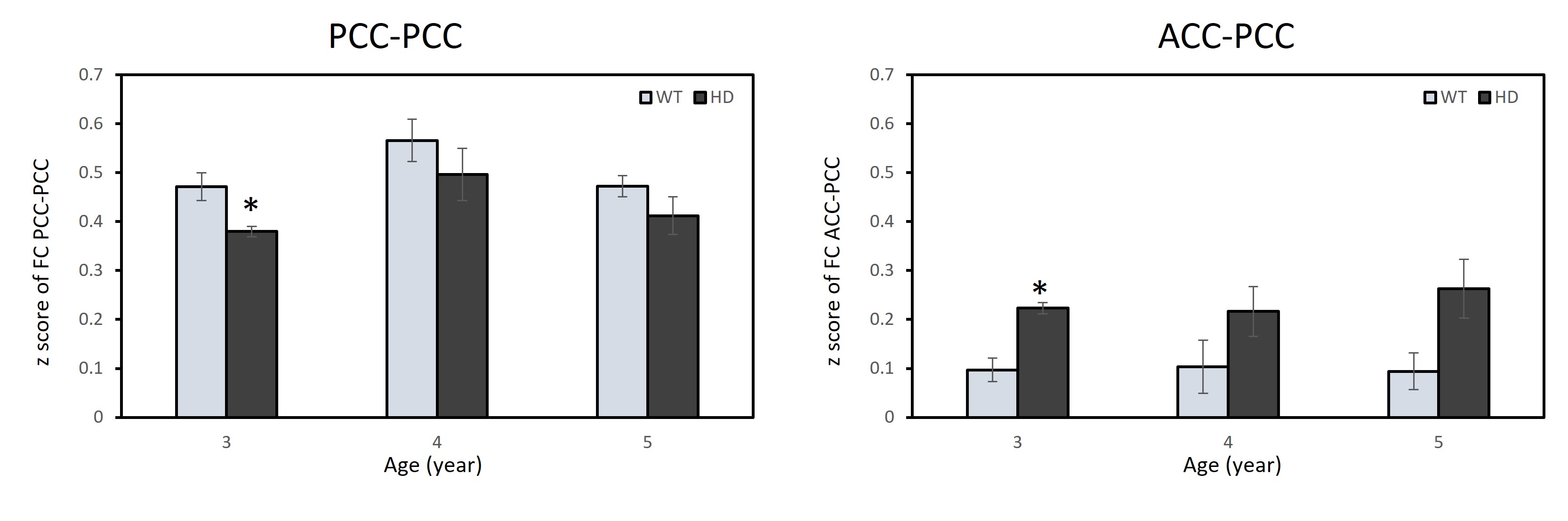
Results: Decreased FC in posterior cingulate cortex (PCC) - PCC and increased FC in anterior cingulate cortex (ACC) - PCC in HD monkeys were observed.
Impact: The findings suggest divergent FC alteration patterns in different DMN area exist concurrently with the evolution of the disease, and the monkey models could provide a unique platform for preclinical studies of dysfunctionality and cognitive decline in HD.
Introduction
Huntington’s disease (HD) is a neurodegenerative disorder caused by mutated huntingtin gene (HTT). Dysfunction of the default-mode network (DMN) has been seen in previous studies of HD patients and rodent models [1], but it is still remained inconclusive [2]. A transgenic HD monkey model has been established and resembles most aspects in anatomical, neurochemical and micro-structural progression seen with HD patients [3]. In the present study, the transgenic HD monkey model was employed to investigate the longitudinal changes of functional connectivity (FC) in DMN of the developing monkey brain.Methods
Four transgenic HD rhesus monkeys (n=4, male, HD) and four age-matched wild-type rhesus macaques (WT, n=4, 2 males and 2 females) were used as control. The monkeys were scanned under 1% isoflurane at 3, 4 and 5 years old on a Siemens 3T scanner with an 8-channel Tx/Rx volume coil. The rsfMRI data were acquired using the single-shot EPI sequence with TR/TE=2190ms/25ms, 300 volumes, FOV = 96 mm × 96 mm, spatial resolution= 1.5×1.5×1.5mm3, and 34 contiguous slices. The 3D T1 weighted images and field map images were acquired also.The rsfMRI data were preprocessed for image distortion correction using the FSL software. Slice timing correction, rigid body registration, nuisance signal removal, temporal filtering (0.009 Hz ~0.0237 Hz), spatial smoothing (FWHM = 2.5mm) and normalize individual brains to a template brain were conducted with AFNI. The averaged time courses of rsfMRI signals in posterior cingulate cortex (PCC) was used for seed-based correlation analysis. Z transformation was applied to the individual correlation maps to show normalized correlation. The averaged z values of FC between PCC and anterior cingulated cortex (ACC) and PCC were examined for statistical analysis. Multivariate analysis of variance (MANOVA) was conducted to compare FC results with LSD (Least significant difference) Post Hoc tests were conducted to perform pair-wise comparison. P-values less than 0.05 were considered statistically significant.
Results
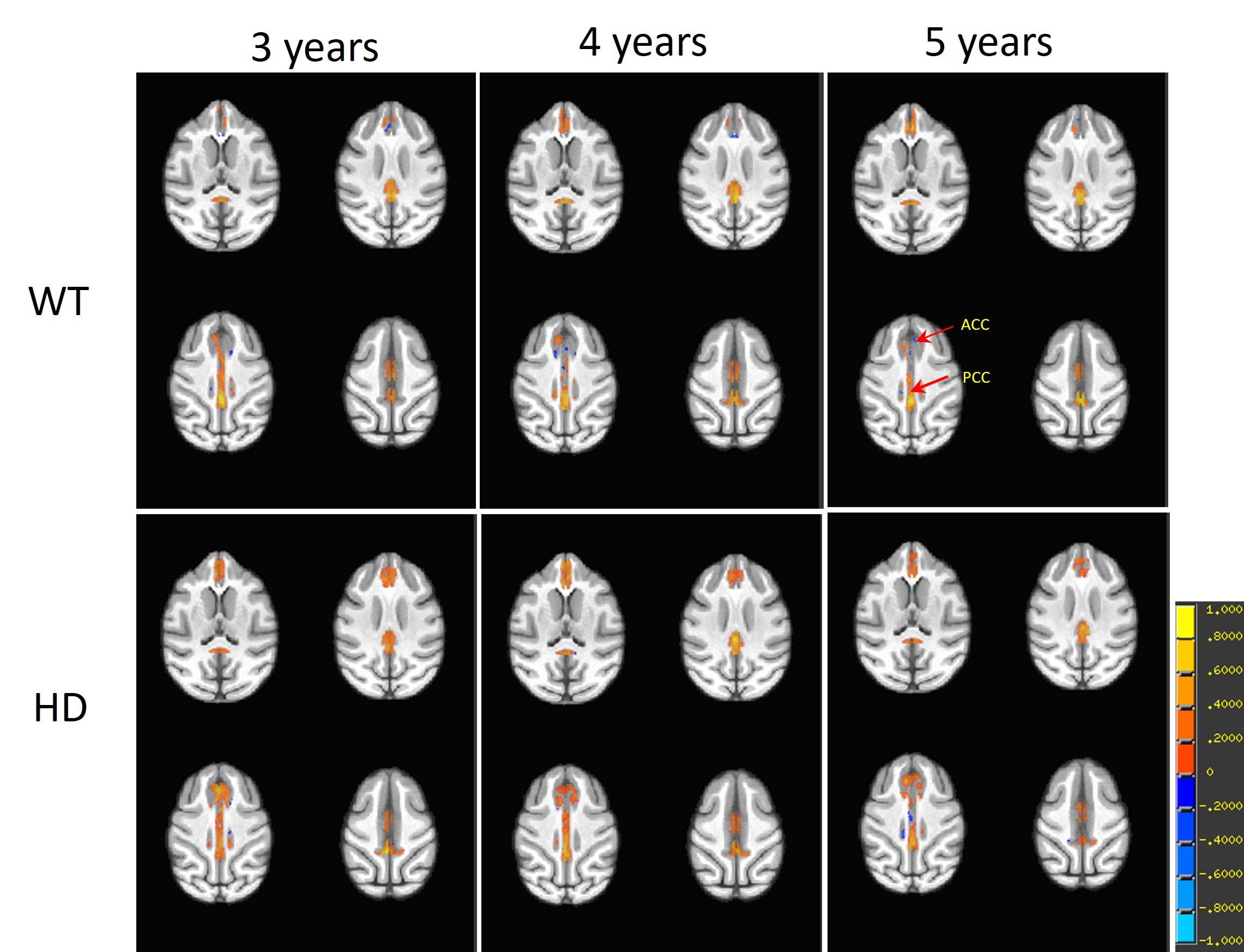
Averaged z maps in DMN, derived by seed-based analysis, of WT and HD monkeys are illustrated in Figure 1. Decreased FC in PCC - PCC and increased FC in ACC-PCC were observed in HD monkeys from 3 to 5 years old (significance was reached at 3 years old) (Fig.2).Discussion and conclusion
HD results in progressive cognitive decline has been examined by using rsfMRI previously. However, controversial results were seen in the assessment of DMN abnormality of HD patients [2], and both reduced FC [4, 5] and increased FC in DMN [6] were reported in different studies. We hypothesized such discrepancy may be associated with the evolution of HD. Accordingly, a longitudinal study could be useful to investigate the FC changes during disease evolution.Our preliminary findings revealed that divergent patterns of FC (such as hypo- PCC-PCC interaction and hyper- ACC-PCC coupling) could exist concurrently in DMN during the progression of the disease. Decreased PCC-PCC interaction in HD monkeys might indicate the dependent disturbance of the normal connection upon disease stages since FC reduction in DMN was correlated with cognitive disturbances of symptomatic HD patients [5]. Increased FC in ACC-PCC was also reported in patients with premanifest gene carriers (preHD) [7], which could reflect the functional compensation in order to maintain or optimize cognitive performance. Further studies would be conducted to elucidate the potential relationship between functional compensation and the disease progression of HD.
In conclusion, the preliminary results suggest that the HD induced FC changes may be associated with the evolution of the disease. HD monkeys could be used to evaluate the FC dysfunction seen in HD patients and provide a unique platform for preclinical HD studies for long-term assessment of translational outcome measurements.
Acknowledgements
This project was funded by awarded by the ORIP/NIH (OD010930 to Dr Anthony Chan) and the office of Research Infrastructure Programs/OD P51OD011132.References
1. Vasilkovska, T., et al., Resting-state fMRI reveals longitudinal alterations in brain network connectivity in the zQ175DN mouse model of Huntington?s disease. Neurobiology of Disease, 2023. 181.
2. Pini, L., et al., Aberrant brain network connectivity in presymptomatic and manifest Huntington's disease: A systematic review. Human Brain Mapping, 2020. 41(1): p. 256-269.
3. Chan, A.W.S., et al., Progressive Cognitive Deficit, Motor Impairment and Striatal Pathology in a Transgenic Huntington Disease Monkey Model from Infancy to Adulthood. Plos One, 2015. 10(5).
4. Dumas, E.M., et al., Reduced functional brain connectivity prior to and after disease onset in Huntington's disease. Neuroimage Clin, 2013. 2: p. 377-84.
5. Quarantelli, M., et al., Default-Mode Network Changes in Huntington's Disease: An Integrated MRI Study of Functional Connectivity and Morphometry. Plos One, 2013. 8(8).
6. Sanchez-Castaneda, C., et al., Resting-state connectivity and modulated somatomotor and default-mode networks in Huntington disease. CNS Neurosci Ther, 2017. 23(6): p. 488-497.
7. Wolf, R.C., et al., Default-mode network changes in preclinical Huntington's disease. Exp Neurol, 2012. 237(1): p. 191-8.
Figures