4026
Cerebral hemodynamics in the ageing marmoset monkeys using ECG-synchronized PCMRI.1CerCo, CNRS-Toulouse University, Toulouse, France, 2U1214 ToNIC, Inserm-Toulouse University, Toulouse, France, 3Image Processing, Hospital University Center of Amiens-Picardie, Amiens, France
Synopsis
Keywords: Neurofluids, Aging
Motivation: Some cerebrovascular function alterations and cognitive changes are related to ageing. We investigated this issue in marmoset monkeys, offering human-like physiological models.
Goal(s): We aimed at describing age-related changes in brain hemodynamics and their potential interest as early markers of normal/pathological ageing.
Approach: We analyzed blood flow in intracranial arteries and sinuses by using ECG-synchronized PCMRI.
Results: The arterial (basilar trunk and carotids) inflow and venous (inferior and superior sinuses ) outflow sequence is earlier and shorter along the cardiac cycle in old versus young adult marmoset brains. Consequently, neurofluid oscillations appear different during the cardiac cycle as a function of age.
Impact: Our study highlights the importance of nonhuman primate models in studying age-related cerebrovascular changes, offering insights into the link between vascular health, brain ageing, and cognitive decline, with implications for early neurodegenerative diseases.
Introduction
Ageing is associated with declining cerebrovascular function, which is closely linked to brain function alterations among older people. Besides, age-related cardiovascular deterioration increases the risk of cerebrovascular diseases, significantly impacting cerebral activity and cognitive abilities. Investigating these relationships requires intensive exploration of hemodynamic processes in the cardio-cerebral system. Nonhuman primates, owing to their closer phylogenetic proximity to humans compared to commonly used rodents, serve as invaluable models for studying human health and diseases. In this study, we analyzed blood flow dynamics in intracranial arteries and (venous) sinuses in young and old adult marmoset monkeys, Callithrix jacchus, a species with an approximate lifespan of 10 years. We used dedicated MRI sequences, coupled with ECG monitoring, to examine blood flow dynamics along the cardiac cycle.Methods
Nineteen marmosets were categorized into two groups: 10 young adults (YA, median age 3.9 years) and 9 old adults (OA, median age: 8.9 years). MRI acquisitions were performed using a 7T MRI unit on anaesthetized animals (1-2% Isoflurane/O2). Initially, a TOF acquisition was conducted to optimize the localization and orientation of two adequate brain slices to further record blood flow velocity in carotid and basilar trunk arteries and sagittal and inferior sinuses, respectively (Figure 1). High-resolution Phase Contrast (PC) MRI images were acquired with prospective cardiac gating, featuring a voxel size of 150µm x 150µm x 1000µm. VENC was set at 50 cm/s for the arteries and 20 cm/s for sinuses, with data acquired at intervals of 2.7 to 6.9% of the cardiac cycle (CC). Vessel segmentation within the PC images involved delineating regions of interest (ROIs) through automated and manual methods, facilitating the determination of vessel blood velocity and section area. Raw data were linearly adjusted to provide blood flow values at regular intervals of the complete CC, consistent across all marmosets and all vessels/sinuses. Blood flow data were distributed across 32 steps of the CC, each representing 3.125% CC. Moreover, T2-W morphological images were normalized for intensity and segmented into probability maps for gray matter (GM), white matter (WM), and cerebrospinal fluid (CSF using the SPM software and incorporating the marmoset MRI Standard Brain probabilistic tissue templates (1).Results
Intracerebral arterial (basialry+carotids) and venous (both sagittal sinuses) blood flow dynamics exhibited some discrepancies between OA and YA marmosets (see Figure 2). The chronological sequence of ECG-synchronized detection of the maximal flow value in the different compartments? remained consistent in both young and old marmosets. The established sequence for arterial inlets included the basilary trunk, the left carotid and the right carotid. For venous outlets, the sequence comprised the inferior sinus followed by the superior sinus. Notably, in old marmosets, this sequence occurred significantly earlier during the cardiac cycle and had a shorter duration (see Figure 3). The total blood volume entering the brain during a complete cardiac cycle (Total arteries/CC) was more significant higher in old marmosets (69±18 µl) than in young ones (48 ±11 µl), p=0.02. However, this significant difference disappeared when considering perfusion, i.e., the blood volume delivered per minute, as typically calculated (Total arteries/CC x Heart rate), resulting in approximately 10 ml/min in both age groups. Similarly, with a further individual normalization by brain volume, with a brain density of 1.1, we found that the mean cerebral blood flow (CBF, expressed as ml/min/100g of brain tissue) was statistically similar in both age groups. The dephasing between arterial blood inflow and venous blood outflow led to a modulation of cerebral blood volumes during the cardiac cycle, which would be balanced by the movement of cerebrospinal fluid (CSF) between the intra and extra cranial compartments (2). Oscillation of the cerebral blood volume, resulting from the difference between inflow and normalized outflow along the cardiac cyle is presented in Figure 4. The absolute value of this volume, corresponding to less than 1 % of the total CSF volume, with no significant age difference.Discussion conclusion
This cross-sectional study originally demonstrates that intracranial blood flow parameters differ in several ways between young and old adult marmosets: arterial blood flow peaks earlier in old monkeys, suggesting age-related changes in vessel structural properties such as vessel wall rigidity. Notably, these findings have not been observed in human studies, where blood flow dynamics are typically retrospectively synchronized with peripheral pulse measurements taken from the fingertip rather than prospectively using the ECG signal. Additionally, the kinetics of blood flow input-output in brain tissue differs in old monkeys, possibly reflecting alterations in both brain vessels and tissue mechanical.Acknowledgements
Non-Invasive Exploration service of the US006/CREFRE-Anexplo Inserm/UT3/ENVT is gratefully acknowledged. This work was supported by an ANR grant (ANR-18-CE45-0014).References
(1) Liu C, Chern-Chyi Yen C, Szczupak D et al.. Marmoset brain mapping V3: population multi-modal standard volumetric and surface-based templates. NeuroImage, 2021, 226, 117620.
(2) Lokossou A, Metanbou S, Gondry-Jouet C and Balédent O. Extracranial versus intracranial hydro-hemodynamics during aging: a PC-MRI pilot cross-sectional study. Fluids Barriers CNS. 2020; 17: 1.
Figures
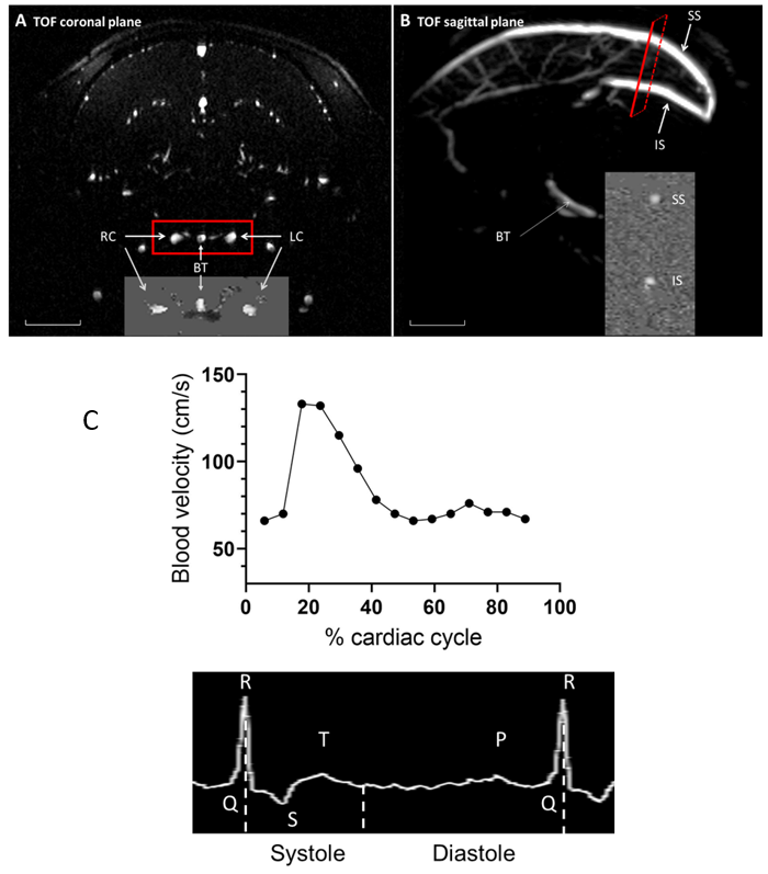
Blood velocity measurements by PC-MRI. A : On the same coronal section, identified by TOF, measurements were taken simultaneously for the basilar trunk (BT), the right (RC) and left (LC) carotid arteries. B : Venous blood velocity was acquired in the superior (SS) and inferior (IS) sagittal sinuses on a same section. TOF images : scale bar = 1 cm. C : One example of blood velocity data obtaine by PC MRI (top) in function of cardiac activity (bottom : ECG signal). Blood velocity data were obtained at regular intervals during the cardiac cycle between two successive P waves.
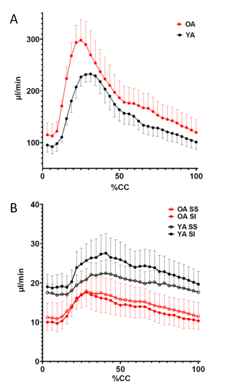
Time course of arterial and venous blood flows during the cardiac cycle (cc) in young (YA) and old (OA) adult marmosets. A : sum of the blood flows in the basilary trunk and the two carotids. B : blood flow in the inferior sagittal –IS- and superior sagittal -SS- sinus. Mean ± SEM (n=7 for each age group).
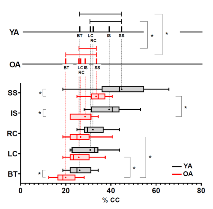
Comparative arrival times of the maximal blood flow in the different vessels
Lower part: comparison of different inflow or outflow timings, in YA and OA. In OA, BT maximal flow preceeds the RC and the LC. Upper part : Comparison of arterial input and venous output timecourse . Duration between the arterial and the venous (SS) is shorter in OA.
cc: cardiac cycle, BT : basilar trunk, RC : right carotid, LC : left carotid, IS : inferior sagittal, SS : superior sagittal. YA : young adult marmoset group, OA : old adult marmoset group, *p<0.05
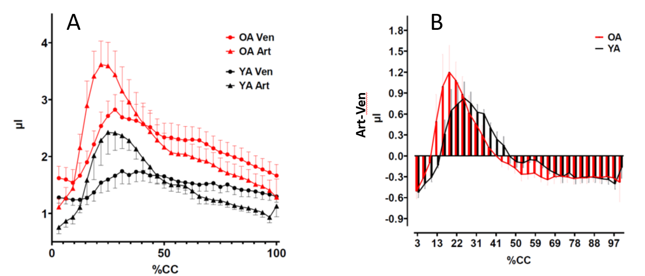
Timecourse of fluids in the marmoset brain during the cardiac cycle.
A : Inflow (Art) and outflow (Ven) of blood during the cardiac cycle (cc) (Art = sum of BT, RC and LC) and venous (Ven = sum of IS and SS) in YA and OA marmosets. Mean ± SEM (n=7 for each age group). B : Evolution of the balance between arterial and venous blood volumes (arterial -venous blood volumes) during the CC. The excess of blood (A-V>0) inside the brain vascular compartment (positive values) comes earlier for OA than YA marmosets, in coherence with the earlier peak of arterial blood flow in the OA group.