4025
Comprehensive Assessment of Age-Related Hippocampal Changes in Murine Models Through Multimodal MRI and Spectroscopy1Zuckerman Institute, Columbia University, New York, NY, United States, 2Department of Taub Institute for Research on Alzheimer’s Disease and the Aging Brain, Columbia University, New York, NY, United States, 3Department of Neurology, Columbia University, New York, NY, United States, 4Department of Psychiatry, Columbia University, New York, NY, United States
Synopsis
Keywords: Aging, Aging, hippocampus, CBV, TBM, glutamate, semi-LASER
Motivation: Analyzing aging mice and controls reveals insights into structural, functional, and neurochemical alterations linked to cognitive decline, and offers potential therapeutic leads.
Goal(s): To comprehensively analyze age-related alterations in the hippocampus of mice with MRI, and to elucidate the underlying mechanisms of aging.
Approach: We utilized a multimodality MRI protocol to investigate age-related changes in the mouse hippocampus. Aging mice and a control group were compared.
Results: Structural MRI revealed a significant reduction in hippocampal size in the aging mice. Cerebral blood volume (CBV) MRI showed reduced CBV, indicating hypometabolism. Proton MRS identified significant glutamate (GLU) reductions, suggesting neuronal function alterations.
Impact: Our study of age-related hippocampal changes in mice, revealing reduced hippocampal volume, metabolism and GLU levels. This translational multimodality approach enhances understanding of age-related deficits in the hippocampus, and promotes potential therapies interventions for both preclinical and human studies.
Introduction
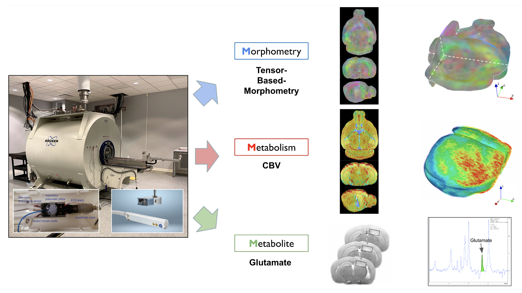
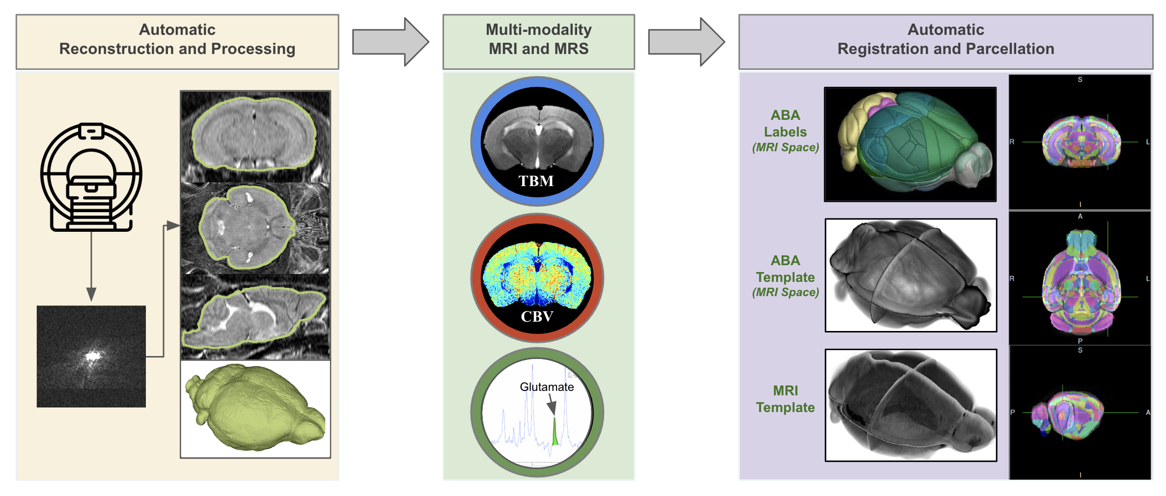
Aging is often associated with significant alterations in the hippocampus, a key brain region for memory and cognitive function. In our study, we utilized a multimodality MRI protocol to investigate age-related alterations in the mouse hippocampus. Aging mice and a control group were compared. We used structural MRI to measure hippocampal volume, finding significant changes in aging mice. CBV MRI assesses hemodynamics, detecting aging-induced alterations in brain metabolism. Proton magnetic resonance spectroscopy (1H-MRS) examines the hippocampal neurochemical profile, revealing GLU changes associated with age-related hippocampal alterations (Figure 1).This multimodality approach offers age-induced hippocampal changes in mouse models, encompassing structural and CBV along with neurochemical correlates. These findings contribute to our understanding of the mechanisms underlying age-related cognitive decline, offering insights for potential therapeutic interventions. Additionally, this mouse model serves as a valuable platform for preclinical studies aimed at investigating aging-associated hippocampal changes (Figure 2).
Methods
In our study, we utilized C57BL/6J mice at 4 and 20 months respectively, consisting of 10 male mice in each age group.MRI Data Acquisition and Processing
T2w Structure MRI: T2-weighted MRI images were obtained using a fast spin echo sequence, which was acquired before intraperitoneal injections of contrast agent gadolinium (10 mmol/kg)1,2. Four pre-contrast acquired for 5 mins. We segmented brain tissue using DL-BET (https://github.com/SAIL-GuoLab/DL-BET). Scans were up-sampled to 80×80×80 µm³. ANTs was performed between-subject coregistration to a group-wise template.
CBV MRI: T2-weighted MRI images were also acquired after intraperitoneal injections of contrast agent gadolinium. 50 post-contrast scans acquired for 40 mins. CBV MRI processing, as in prior studies2, includes registering CBV images to the template space using a derived transformation.
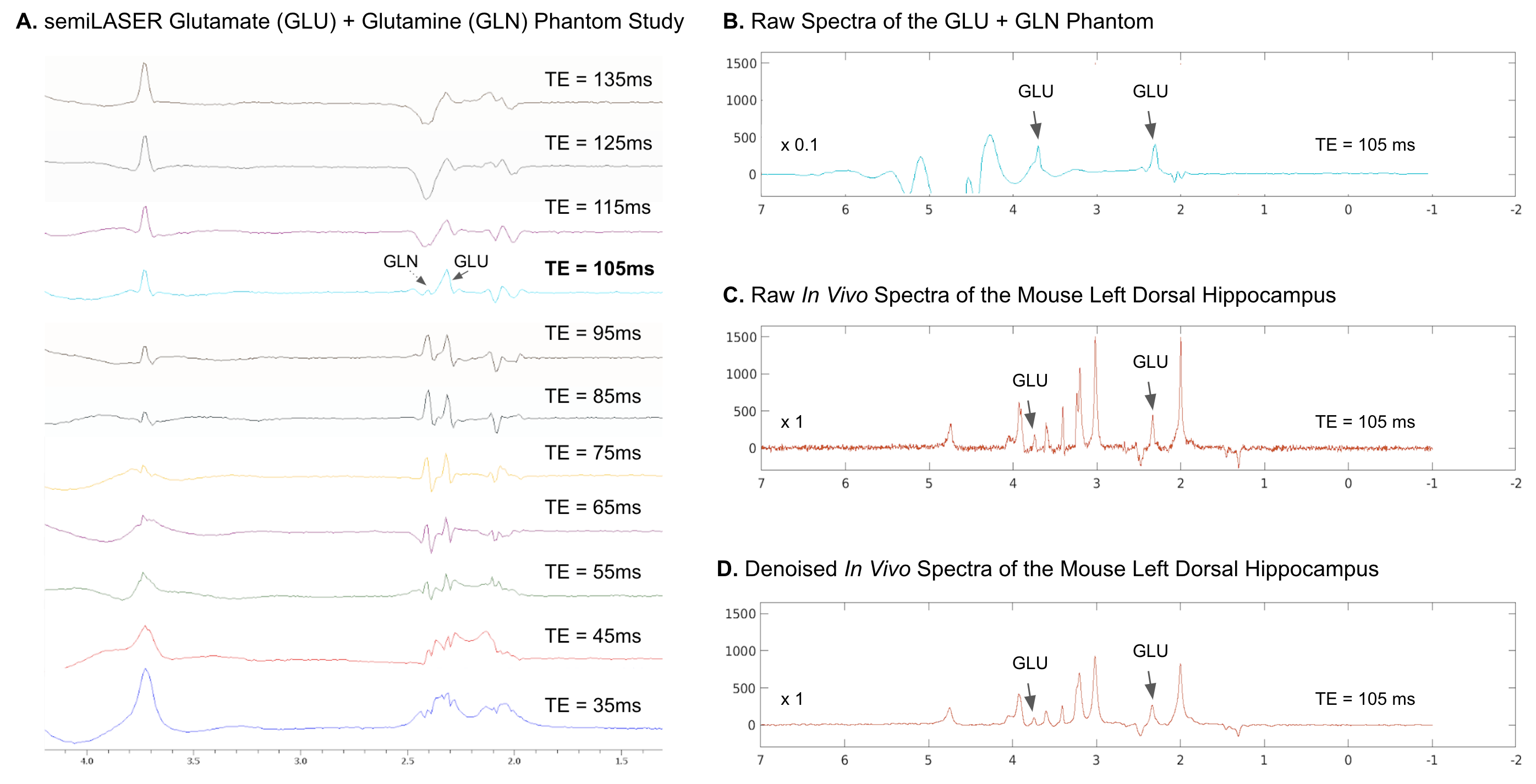
Glutamate semi-LASER MRS: We examined a 3x2x2 mm³ voxel in the right hippocampus, ensuring no ventricular interference. Magnetic field uniformity was adjusted with MAPSHIM and iterative shims, with a water peak linewidth < 20 Hz. GLU was measured using an optimized semi-LASER method (TR=2000 ms, optimized TE=105 ms) at 9.4 T, as shown in Figure 3, taking 17 min 4 sec3,4. We used the JET toolkit (http://doi.org/10.5281/zenodo.3967565) for automated MRS spectra processing and quantification. We quantified metabolites including Creatine(Cr), N-acetylaspartate(NAA), Choline, and GLU, and calculated GLU/Cr to normalize acquisition variations (Figure 3).
Data Analysis
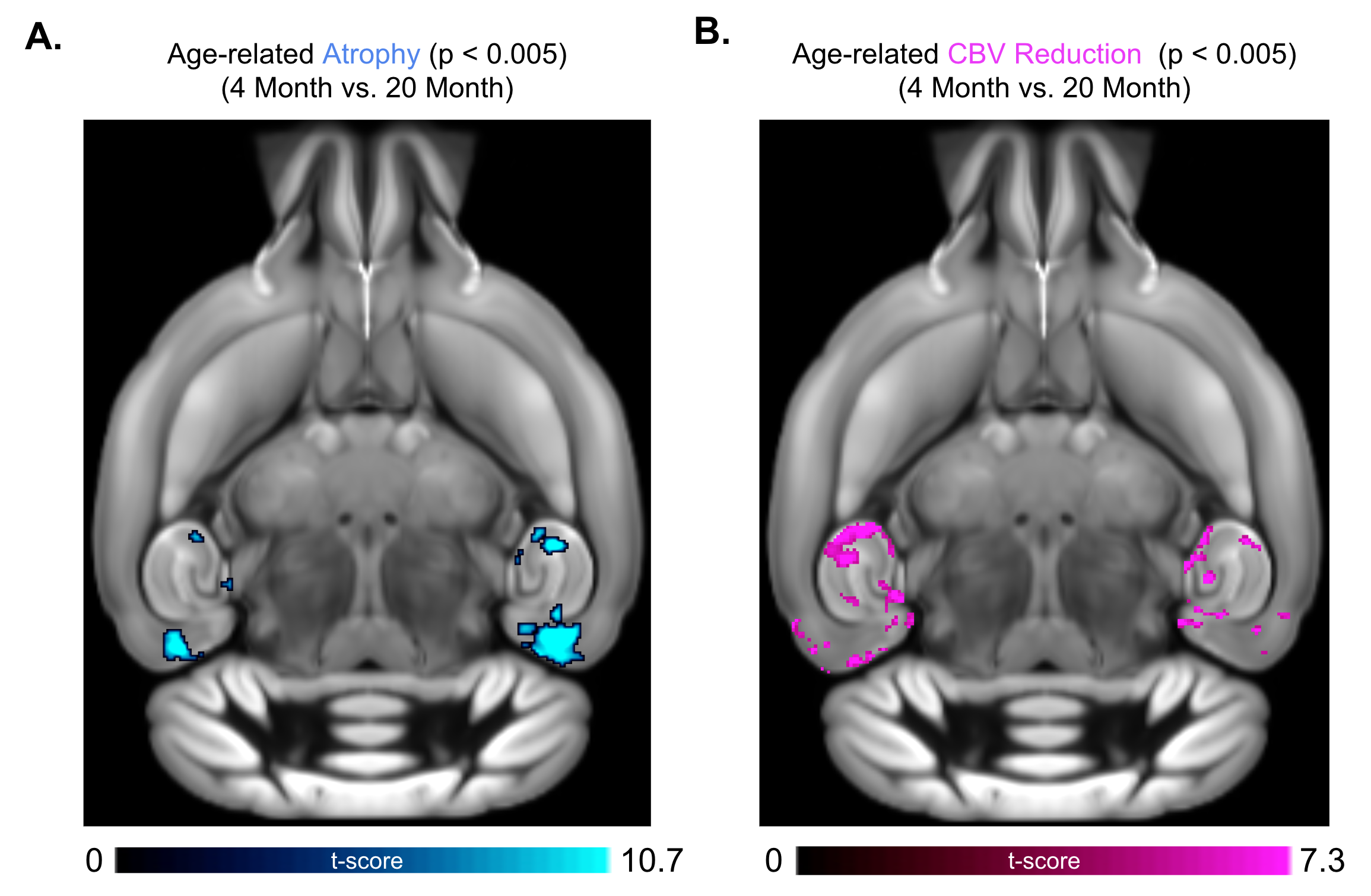
Hippocampus voxel-based-analysis (VBA) and region-of-interest (ROI) analysis were conducted to assess the quantitative changes in volume and CBV. VBA was conducted using a general linear model in the statistical parametric mapping contrasted using a two-sample Student’s t-test. Results were corrected for multiple comparisons using 3dFWHMx and 3dClustSim, to achieve a voxel-wise p < 0.005 and cluster-wise p < 0.05. We also conducted modality-specific ROI analyses of the hippocampus measurements. We used a two-sample Student’s t-test comparing the hippocampal volume, CBV and GLU/Cr levels between the young and old groups. The ROI significance level was set at 0.05.
Results
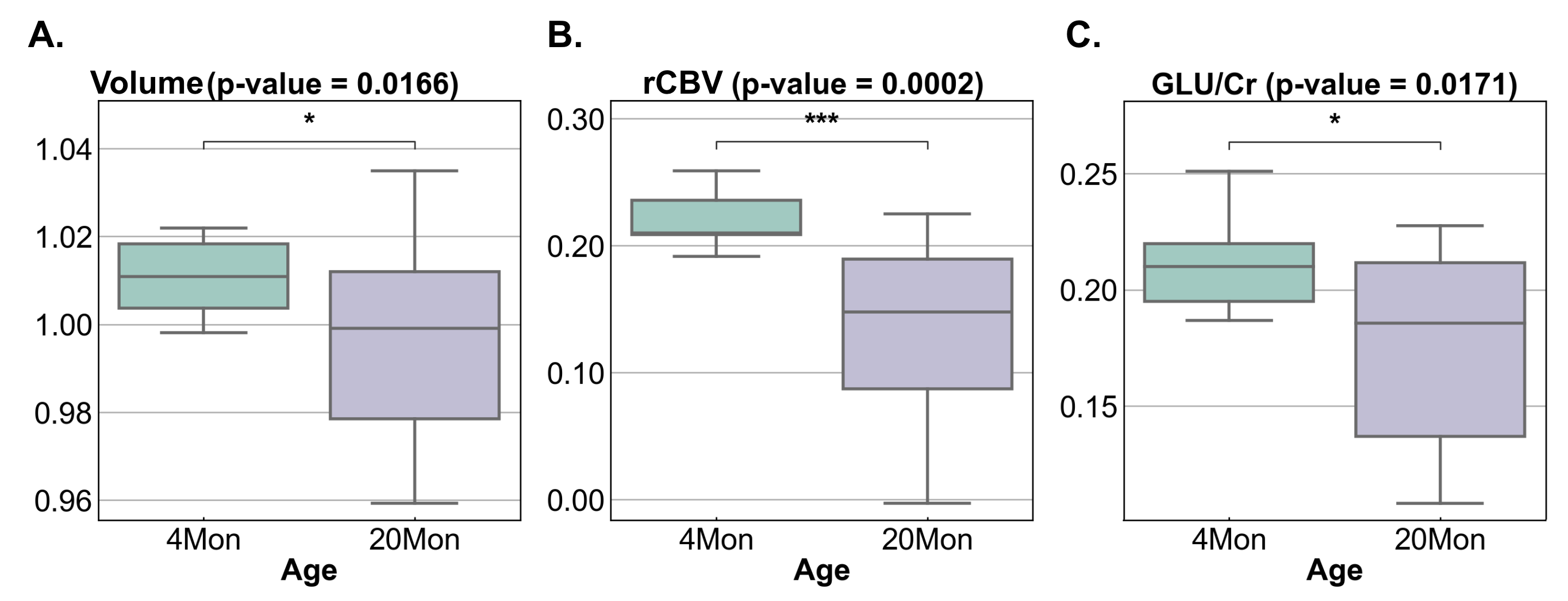
The VBA results are summarized in Figure 4. The results of our ROI analysis for hippocampal MRI/MRS measurements are as follows: A. Comparison of hippocampal volume between young and old individuals revealed a statistically significant reduction in volume associated with aging. B. Evaluation of hippocampal relative cerebral blood volume (rCBV) between young and old subjects demonstrated a significant reduction in CBV related to aging. C. Examination of the hippocampal glutamate to creatine ratio (GLU/Cr) between young and old subjects indicated a significant reduction linked to the aging process (Figure 4,5).Discussion and Conclusion
In this study, we used multimodal MRI to investigate age-related changes in the mouse hippocampus, a crucial region for memory and cognition. We observed significant volume reductions, altered cerebral blood volume, and metabolic shifts in aging mice. This approach provides a comprehensive understanding of aging-related alterations in the hippocampus.These findings enhance our grasp of the aging hippocampus and hold promise for interventions against aging. Moreover, the mouse model employed here serves as a valuable tool for deeper exploration of hippocampal aging mechanisms, opening avenues for further research in aging neuroscience.
Acknowledgements
This study was performed at the Zuckerman Mind Brain Behavior Institute MRI Platform, a shared resource and Columbia MR Research Center site.References
- Moreno, H., Hua, F., Brown, T. and Small, S., 2006. Longitudinal mapping of mouse cerebral blood volume with MRI. NMR in Biomedicine: An International Journal Devoted to the Development and Application of Magnetic Resonance In vivo, 19(5), pp.535-543.
- Simoes, S., Guo, J., Buitrago, L., Qureshi, Y.H., Feng, X., Kothiya, M., Cortes, E., Patel, V., Kannan, S., Kim, Y.H. and Chang, K.T., 2021. Alzheimer's vulnerable brain region relies on a distinct retromer core dedicated to endosomal recycling. Cell reports, 37(13).
- Guo, J., Gang, Z., Sun, Y., Laine, A., Small, S.A. and Rothman, D.L., 2018. In vivo detection and automatic analysis of GABA in the mouse brain with MEGA‐PRESS at 9.4 T. NMR in Biomedicine, 31(1), p.e3837.
- Ma, D.J., Yang, Y., Harguindeguy, N., Tian, Y., Small, S.A., Liu, F., Rothman, D.L. and Guo, J., 2022. Magnetic Resonance Spectroscopy Spectral Registration Using Deep Learning. Journal of Magnetic Resonance Imaging.
Figures