4021
The Utility of Gadoxetic acid-Enhanced MRI in Quantitative Assessment of Transplanted Liver: Functional T1 Mapping1Department of Radiology, Tongji Hospital Tongji Medical College Huazhong University of Science and Technology, Wuhan, China
Synopsis
Keywords: Liver, Liver
Motivation: Gadoxetic acid-enhanced MRI is a promising tool for assessing liver function, especially in the context of liver transplants.
Goal(s): This study aims to examine the relationship between functional T1 mapping from gadoxetic acid-enhanced MRI and transplanted liver function.
Approach: Liver transplant recipients underwent liver MRI, including T1 mapping and gadoxetic acid-enhanced MRI, to evaluate the relationship between MRI parameters and MELD.
Results: Liver transplant recipients at low or moderate risk had significantly higher ΔT1% values compared to high-risk recipients.
Impact: Gadoxetic acid-enhanced MRI offers dynamic and hepatocyte-specific imaging, making it a comprehensive "one-stop" method for post-transplant complication and liver injury evaluation.
Introduction and Purpose:
Gadoxetic acid -enhanced MRI has shown promise as a valuable tool for assessing liver function. It is considered one of the most promising imaging techniques for evaluating transplanted livers, as it can effectively detect post-transplant complications and assess the severity of liver injury. The purpose of this study is to investigate the correlation between functional T1 mapping derived from gadoxetic acid-enhanced MRI and transplanted livers function, and to evaluate whether this method helps identify recipients at high risk.Methods:
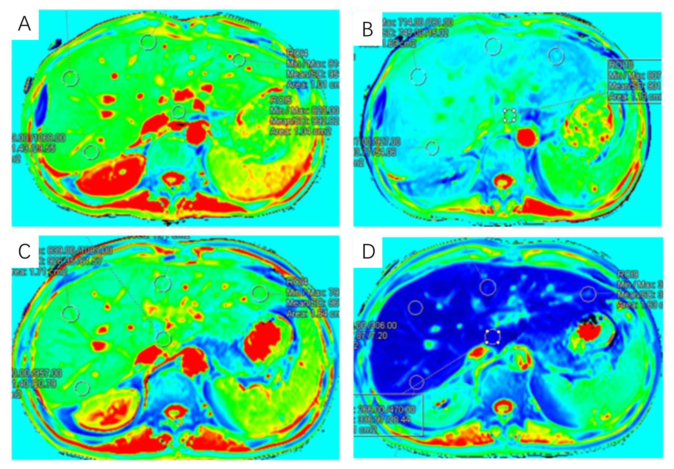
Between January 2021 and September 2022, 101 liver transplant recipients underwent liver MRI at 3T system (MAGNETOM Skyra, Siemens Healthcare, Erlangen, Germany) , including T1 mapping and gadoxetic acid-enhanced MRI. transplanted livers function was quantitatively assessed using the Model for End-Stage Liver Disease (MELD) model. T1 mapping images were obtained before and 20 minutes after the injection of gadoxetic acid (0.025 mmol/kg). The relationship between the MELD model and the following MRI parameters was evaluated: pre- and post-contrast liver T1 values, percentage reduction (ΔT1%), and biliary enhancement. The diagnostic performance of ΔT1% for identifying high-risk patients was compared with the area under the receiver operating characteristic curve.Results
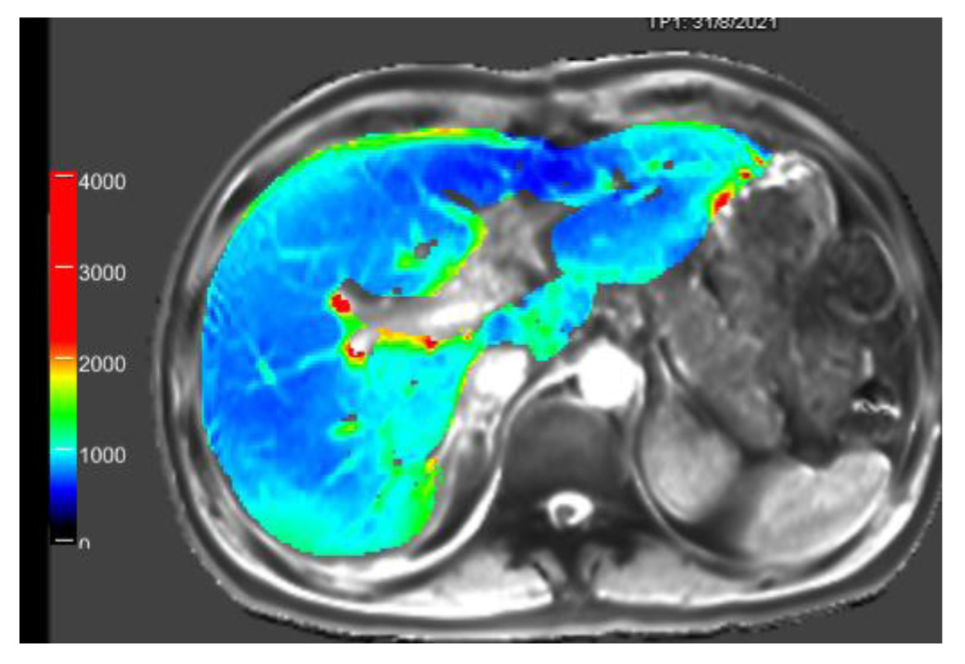
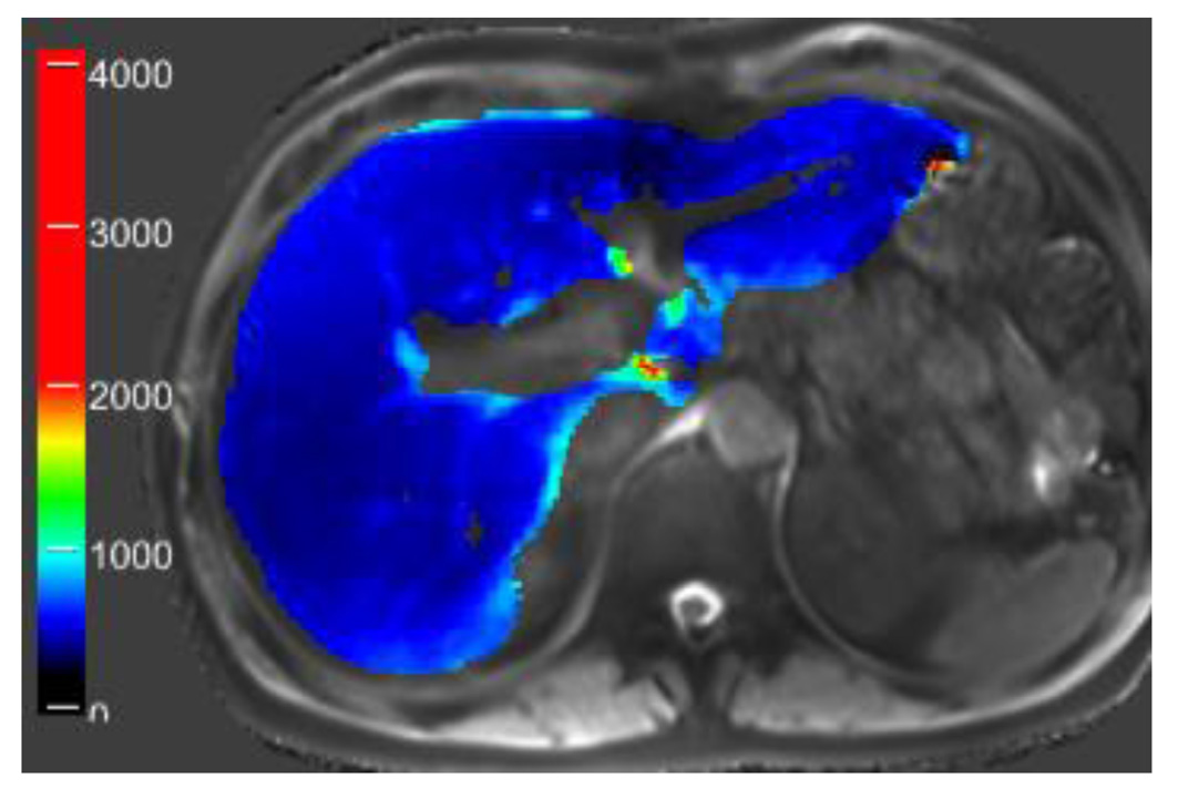
ΔT1% was negatively correlated with the MELD model, with an r value of -0.61 (P < 0.001). In liver transplant recipients at low or moderate risk, their ΔT1% was higher than in high-risk recipients (P < 0.001).Discussion
The use of gadoxetic acid-enhanced MRI in assessing liver function in transplant recipients has shown promising results. This study aimed to investigate the correlation between functional T1 mapping derived from gadoxetic acid-enhanced MRI and transplanted liver function, as well as to evaluate its potential in identifying recipients at high risk. The study provides valuable insights into the potential of gadoxetic acid-enhanced MRI in assessing transplanted liver function and identifying high-risk recipients. Further research and clinical application of this imaging technique could enhance the management and care of liver transplant patients.Conclusion
ΔT1% is negatively correlated with transplanted liver function measured by the MELD model and provides acceptable diagnostic performance in identifying high-risk recipients. Furthermore, gadoxetic acid-enhanced MRI enables dynamic imaging and hepatocyte-specific imaging in a single examination, providing a comprehensive approach for evaluating post-transplant complications and liver injury. Therefore, gadoxetic acid-enhanced MRI represents a "one-stop" method for liver assessment.Acknowledgements
We thank Qiuxia Wang,Lifen Zhou, and Hao Wu whose important contributions to this study were indispensable to its success.References
1.Yang L, Ding Y, Rao S, Chen C, Zeng M. T1 Mapping on Gd-EOB-DTPA-Enhanced MRI for the Prediction of Oxaliplatin-Induced Liver Injury in a Mouse Model. J Magn Reson Imaging. 2021 Mar;53(3):896-902. doi: 10.1002/jmri.27377. Epub 2020 Sep 26. PMID: 32979019. 2.Yoon JH, Lee JM, Kang HJ, Ahn SJ, Yang H, Kim E, Okuaki T, Han JK. Quantitative Assessment of Liver Function by Using Gadoxetic Acid-enhanced MRI: Hepatocyte Uptake Ratio. Radiology. 2019 Jan;290(1):125-133. doi: 10.1148/radiol.2018180753. Epub 2018 Oct 30. PMID: 30375932.
Figures