4017
Deep-learning-based phase correction during reconstruction of high-resolution, multi-shot reduced-FOV pancreatic DWI.1Radiology, Stanford, Stanford, CA, United States, 2Stanford, Stanford, CA, United States, 3GE Healthcare, Waukesha, WI, United States
Synopsis
Keywords: Pancreas, Diffusion/other diffusion imaging techniques, Pancreas, Deep-learning, Phase Correction, DWI, rFOV, multishot, SNR
Motivation: Diffusion weighted imaging (DWI) is valuable in pancreatic imaging but suffers from artifacts and low SNR. The combination of reduced FOV imaging with a multishot data sampling strategy (rFOV-msDWI) improves artifacts from susceptibility and allows higher achievable resolution but still suffers from low SNR.
Goal(s): Here we report early findings from an ongoing pilot study
Approach: Evaluate a DL-based phase correction algorithm for improved SNR in patients undergoing rFOV-msDWI of the pancreas.
Results: DL-based phase correction subjectively improves image quality.
Impact: DL-based phase correction may improve rFOV-msDWI of the pancreas. Further evaluation is warranted.
Goal: To determine if DL-phase correction during image reconstruction can improve image quality in rFOV-msDWI of the pancreas.
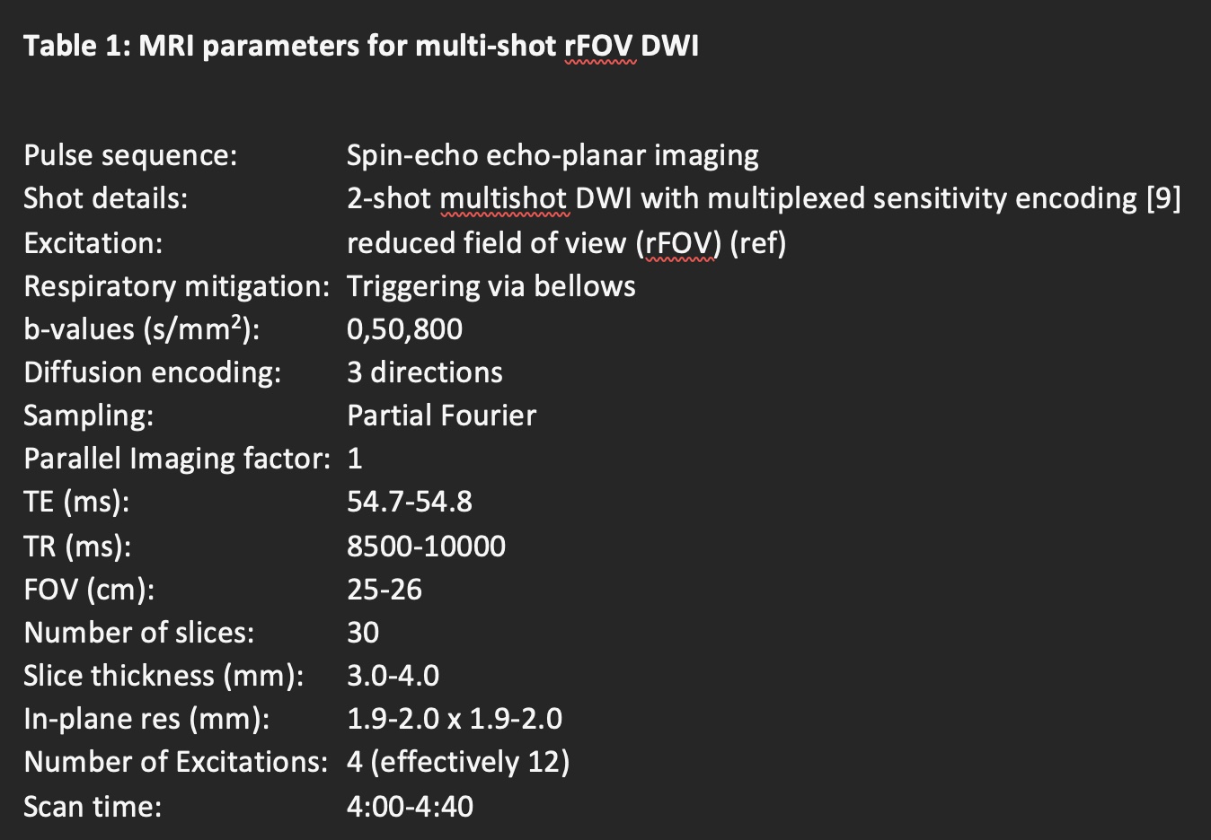
Approach: This single-center prospective pilot study was performed with Institutional Review Board approval and an informed consent waiver. The initial three patients undergoing pancreatic DWI with rFOV-msDWI (Parameters in Table 1) were included in this preliminary assessment. Raw data was collected and processed through DL Phase Correction (DLPC), which aims to generate a high-quality phase (high resolution and SNR) for phase correction using a deep-learning based network, which was trained from a database of over 10,000 images with various SNR levels and background phases [7]. This correction step was combined with a commercially available DL-based denoising tool [8] to further improve in-plane resolution and SNR of the DL phase corrected image. Two sets of images from the same set of raw MR data were produced: original image and DL Phase Corrected (DLPC) images. High b-value datasets and ADC maps were reviewed by a radiologist with subspecialty training in abdominal MRI and 10 years of experience and a descriptive summary presented. Additional DWI sequences included a full FOV respiratory triggered msDWI in all cases (2-shots, slice thickness 5mm, in-plane resolution 2.2 x 2.2 mm, TE~55ms, TR~8500ms).
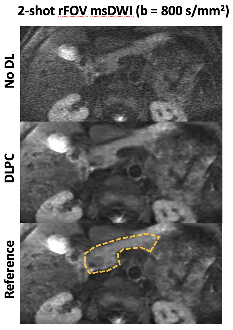
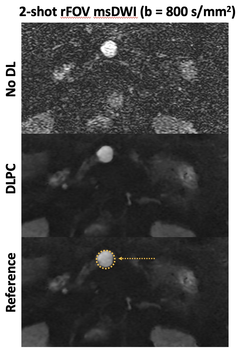
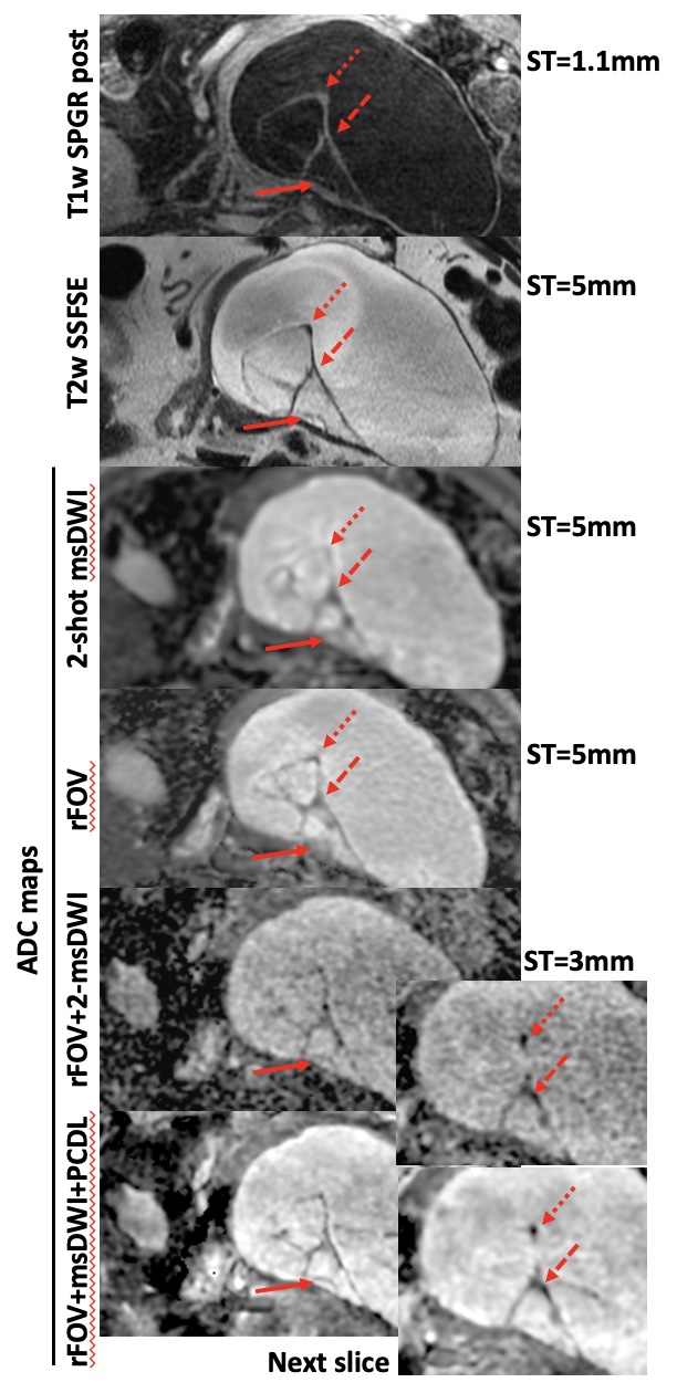
Results: In all three patients, DLPC subjectively improved perceived SNR and boundary sharpness on the high b-value images from rFOV-msDWI, both in the pancreatic tissue (Case 1: Figure 1) and on a cystic pancreatic lesion (Case 2: Figure 2). Similar results were noted on the ADC maps, which showed superior perceived resolution and improved SNR relative to the no-DL corrected images. In one patient with a 12 cm pancreatic cystic lesion (Case 3: Figure 3), high-resolution rFOV-msDWI with DLPC resolved both thin internal linear structures (solid arrow) and tiny 2-3mm nodular structures (dashed and dotted arrows) better than msDWI or rFOV DWI alone.
Conclusion: DL-based phase correction may improve rFOV-msDWI of the pancreas. Further evaluation is warranted.
Acknowledgements
No acknowledgement found.References
[1] M. Zhu et al., “Accuracy of quantitative diffusion-weighted imaging for differentiating benign and malignant pancreatic lesions: a systematic review and meta-analysis,” Eur Radiol, vol. 31, no. 10, pp. 7746–7759, Oct. 2021, doi: 10.1007/s00330-021-07880-3.
[2] S. Ichikawa, M.-L. Kromrey, U. Motosugi, and H. Onishi, “Optimal target b-value on computed diffusion-weighted magnetic resonance imaging for visualization of pancreatic ductal adenocarcinoma and focal autoimmune pancreatitis,” Abdom Radiol, vol. 46, no. 2, pp. 636–646, Feb. 2021, doi: 10.1007/s00261-020-02695-0.
[3] H. Fn et al., “Computed high-b-value high-resolution DWI improves solid lesion detection in IPMN of the pancreas,” European radiology, May 2023, doi: 10.1007/s00330-023-09661-6.
[4] F. Donati, C. Casini, R. Cervelli, R. Morganti, and P. Boraschi, “Diffusion-weighted MRI of solid pancreatic lesions: Comparison between reduced field-of-view and large field-of-view sequences,” European Journal of Radiology, vol. 143, p. 109936, Oct. 2021, doi: 10.1016/j.ejrad.2021.109936.
[5] Y. Bai et al., “MRI: Evaluating the Application of FOCUS-MUSE Diffusion-Weighted Imaging in the Pancreas in Comparison With FOCUS, MUSE, and Single-Shot DWIs,” Journal of Magnetic Resonance Imaging, vol. n/a, no. n/a, doi: 10.1002/jmri.28382.
[6] H. Rhee and M.-S. Park, “The Role of Imaging in Current Treatment Strategies for Pancreatic Adenocarcinoma,” Korean J Radiol, vol. 22, no. 1, pp. 23–40, Jan. 2021, doi: 10.3348/kjr.2019.0862.
[7] Wang X, Litwiller D, Guidon A, Lan PS, Sprenger T., “Robust complex signal averaging for diffusion weighted imaging.,” presented at the ISMRM Annual Meeting Proceedings., 2023.
[8] R. M. Lebel, “Performance characterization of a novel deep learning-based MR image reconstruction pipeline,” arXiv:2008.06559 [cs, eess], Aug. 2020, Accessed: Nov. 04, 2021. [Online]. Available: http://arxiv.org/abs/2008.06559
[9] N. Chen, A. Guidon, H.-C. Chang, and A. W. Song, “A robust multi-shot scan strategy for high-resolution diffusion weighted MRI enabled by multiplexed sensitivity-encoding (MUSE),” Neuroimage, vol. 72, pp. 41–47, May 2013, doi: 10.1016/j.neuroimage.2013.01.038.
Figures