4009
High-fidelity Breath-hold Liver DWI Through Self-referenced and Collaborative PROPELLER-EPI Reconstruction Based on POCSMUSE (SCOPUSE)1The Department of Biomedical Engineering, The Chinese University of Hong Kong, Hong Kong, China, 2Multi-Scale Medical Robotics Center, Hong Kong, China, 3The Department of Diagnostic Radiology, The University of Hong Kong, Hong Kong, China
Synopsis
Keywords: Liver, Liver, Liver Diffusion Acquisition & Reconstruction
Motivation: Diffusion-weighted PROPELLER-EPI (periodically rotated overlapping parallel lines with enhanced reconstruction using EPI as signal readout) can enable high-fidelity free-breathing liver DWI.
Goal(s): It is challenging to acquire liver DW-PROPELLER-EPI with breath hold for improving the acquisition efficiency.
Approach: In this study, we proposed a self-referenced and collaborative PROPELLER-EPI reconstruction based on POCSMUSE (SCOPUSE) framework that can 1) correct the Nyquist ghost phase errors, 2) minimize the streaking artifacts, and 3) enable breath hold for liver DWI.
Results: This method can accelerate the acquisition of DW-PROPELLER-EPI data and provide improved image quality compared with conventional PROPELLER-EPI reconstruction pipeline.
Impact: Breath-hold acquisition can reduce the respiratory artifact in liver DWI with high scan efficiency, however, the attainable image quality is often limited by the breath-hold time. The proposed SCOPUSE can enable breath-hold liver DW-PROPELLER-EPI acquisition for achieving high-fidelity liver DWI.
Introduction
Diffusion-weighted imaging (DWI) has been shown to be useful in the detection and characterization of focal liver lesions1,2 by using single-shot echo-planar imaging (ss-EPI) for data acquisition. Nonetheless, the image quality of liver DWI can be severely deteriorated by EPI-related artifacts, such as Nyquist ghosts and geometric distortion3,4,5. In addition, the respiratory and cardiac motions make liver DWI more challenging and less robust.Several multi-shot EPI techniques, such as PROPELLER-EPI and interleaved-EPI with multiplexed sensitivity encoding (MUSE), have demonstrated prominent improvements in geometric fidelity and image resolution for brain DWI6,7,8. Their potential in improving liver DWI was also preliminarily tested9,10,11, of which the DW-PROPELLER-EPI could enable high-fidelity free-breathing (FB) liver DWI. However, despite improved feasibility of FB liver DWI with inherent motion compensation, the multi-blade acquisition of DW-PROPELLER-EPI can considerably increase the scanning time (e.g., 6.5mins), making it less attractive for routine use. Therefore, an efficient and robust breath-hold (BH) multi-shot liver DWI technique with high fidelity is desirable for routine practice. To this end, we proposed a self-referenced and collaborative PROPELLER-EPI reconstruction based on POCSMUSE12 (SCOPUSE) that can enable BH liver DW-PROPELLER-EPI with accelerated data acquisition.
Methods
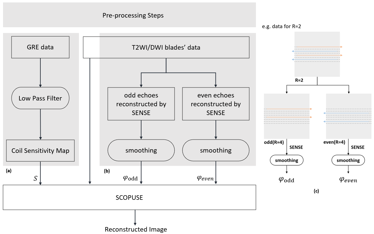
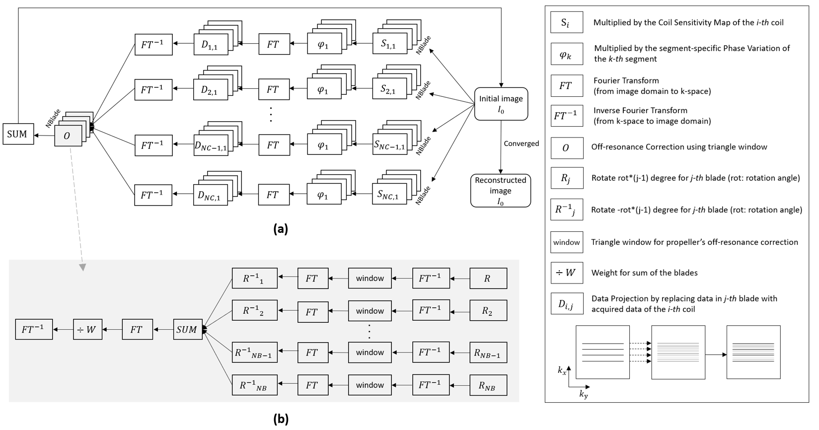
SCOPUSE framework:Fig.1 summarises the flowchart for the proposed method. In the pre-processing stage, the SENSE13 was used for estimating 1) the 2D Nyquist ghost phase errors and 2) the phase variations from b=0 and b>0 s/mm2 data, for each blade respectively (Fig.1b). Fig.2 illustrates the proposed SCOPUSE framework. The SCOPUSE also incorporated triangle weighting14,15 into the iterative joint reconstruction procedure to reduce the off-resonance effects (Fig.2b).
Data acquisition, hybrid simulation, and reconstruction:
A 2-shot long-axis DW-ROPELLER-EPI sequence was used for acquiring the phantom DWI data and the BH liver DWI from a healthy subject, with extended blade size along the phase-encoding (PE) direction (i.e., blade size=128x64). Both phantom and BH liver data were collected on a 1.5T MRI scanner (Artist, GE Healthcare), with three orthogonal diffusion directions at b-value of 500 s/mm2 and reconstructed resolution of 128x128. Different scan accelerations were simulated from the acquired data with the details given in the following subsections. All acquired and simulated data were reconstructed with either conventional PROPELLER-EPI pipeline15,16 or proposed SCOPUES framework for comparison.
1) Phantom data
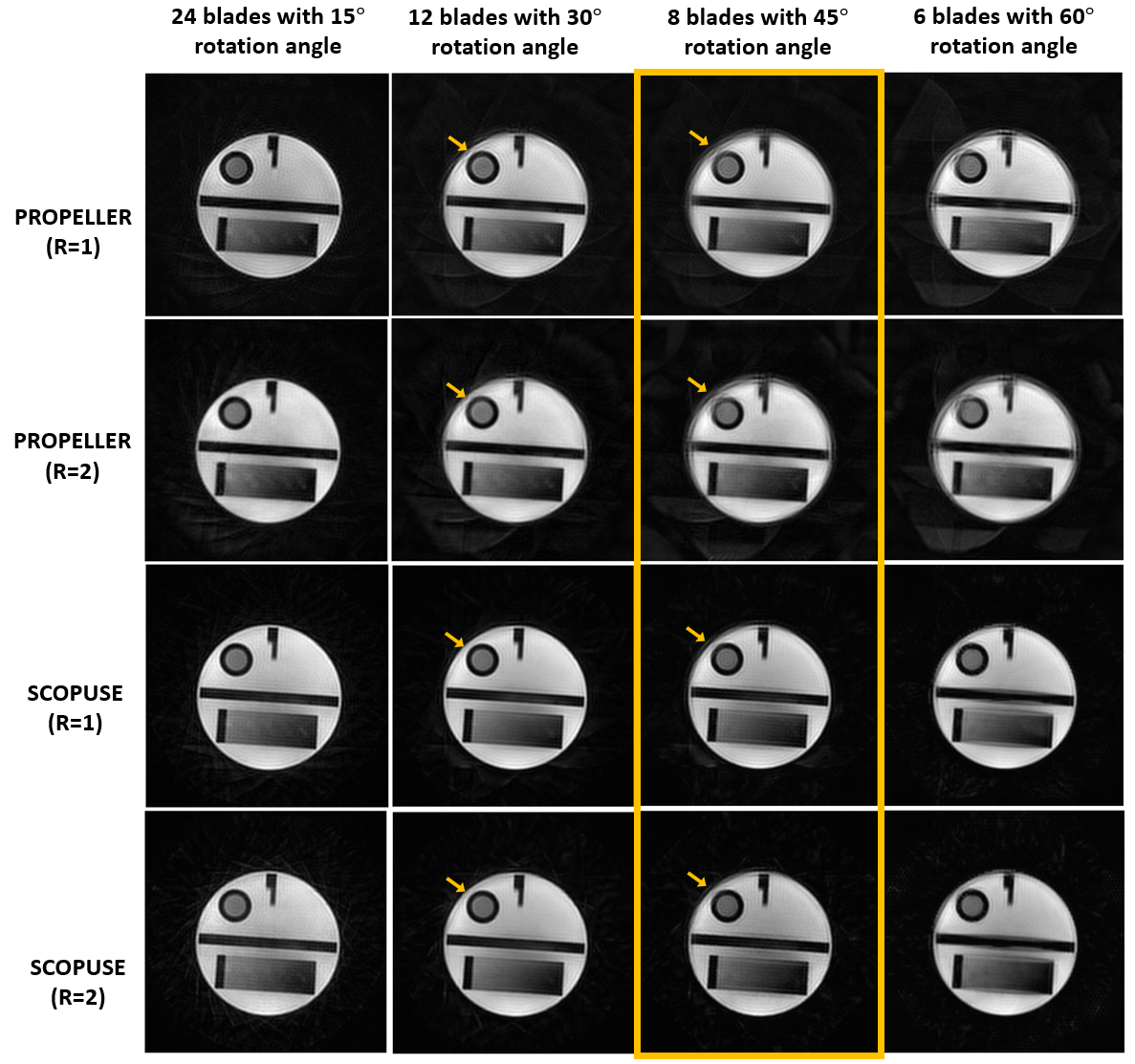
Data was collected using a 12-channel head coil with the following scan parameters: TE/TR=92/2000ms, FOV=280mm, 24 blades with 15° rotation angle for 360° k-space coverage, slice thickness=8mm, number of slices=5, and scantime=6.5mins. The data acquired with different scan accelerations were simulated by selecting 24, 12, 8, and 6 blades out of 24 blades, with either fully-sampled (i.e., R=1) or under-sampled (i.e., R=2) for each blade data.
2) BH liver DWI
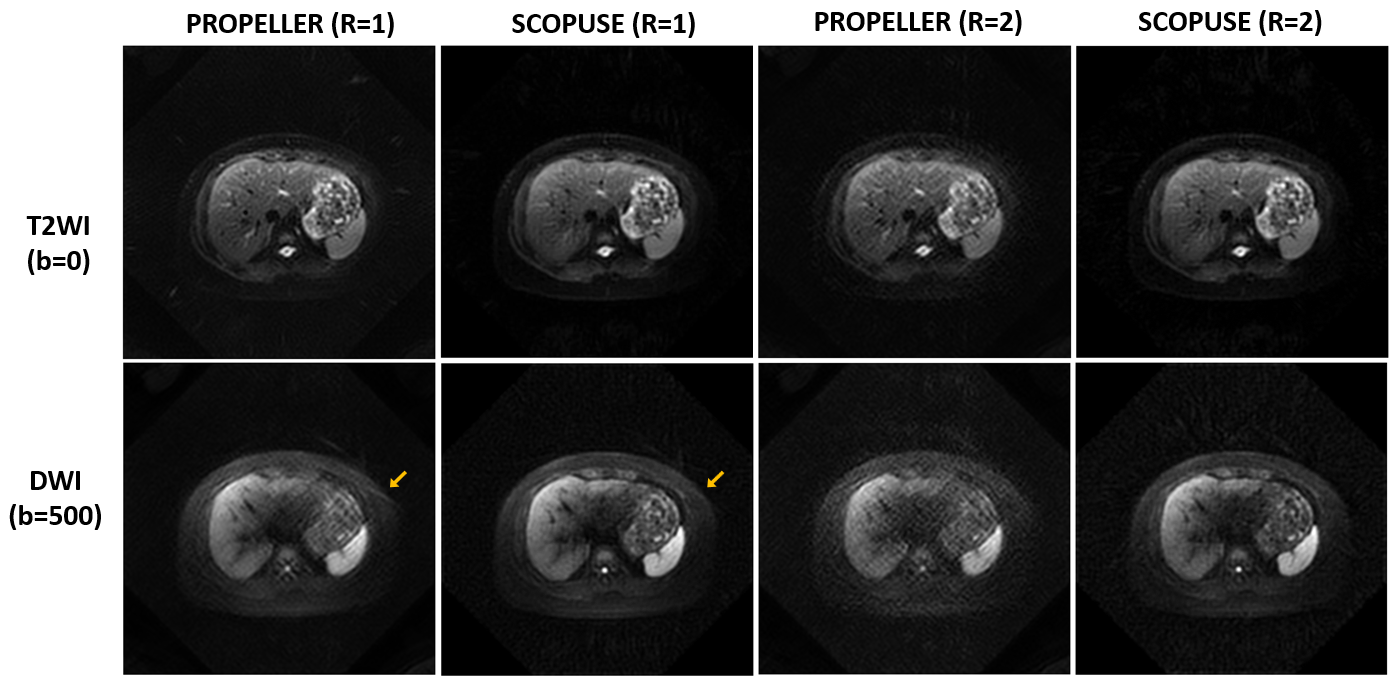
The BH liver DW-ROPELLER-EPI data was collected using a 17-channel body phase-array coil with the following scan parameters: TE/TR=93/1500ms, FOV=400mm, 8 blades with 45° rotation angle for 360° k-space coverage, slice thickness=8mm, number of slices=5, and BH time=29s for each diffusion direction. In addition, the scan acceleration with R=2 for each blade (i.e., selecting only one segment) was simulated to achieve an equivalent 15s BH time for each diffusion direction.
3) FB liver DWI
To further assess the robustness of the SCOPUSE for DW-PROPELLER-EPI reconstruction, five sets of FB liver DW-PROPELLER-EPI data were retrospectively collected from another study10 and then reconstructed with either conventional PROPELLER-EPI pipeline15 or proposed SCOPUES framework. The original scan parameters were as follows: TE/TR=66.4/4000ms, 24 blades with 15° rotation angle for 360° k-space coverage, blade size=128x32, slice thickness=8mm, number of slices=20, and scan time=6.5mins.
Results
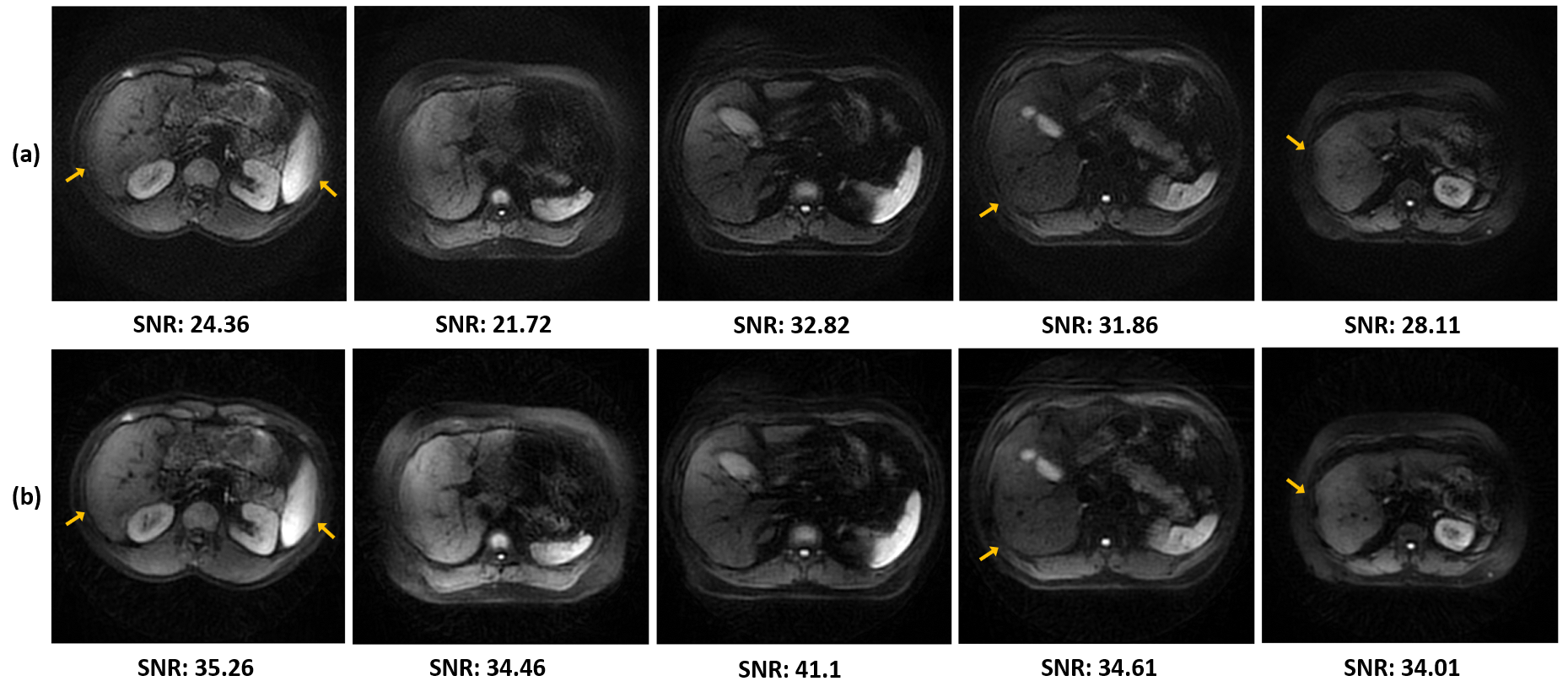
Fig.3 presents the phantom images reconstructed from data with different scan accelerations using either conventional PROPELLER-EPI or the proposed SCOPUSE method. Fig.4 compares BH liver DW-PROPELLER-EPI data reconstructed using either conventional PROPELLER-EPI or SCOPUSE method. Fig.5 shows the reconstruction results and SNR comparison for the retrospectively collected FB DW-PROPELLER-EPI data.Discussion
The proposed SCOPUES can accelerate the DW-PROPELLER-EPI acquisition by reconstructing the image with less blades, thereby enabling BH liver DWI with high geometric fidelity and robustness (Fig.4). Compared with conventional PROPELLER-EPI reconstruction, SCOPUES can eliminate streaking artifacts due to insufficient blades for 360° k-space coverage (Figs.3 & 4), and also improve the reconstruction performance through a joint iterative reconstruction framework. Although the SCOPUSE relies on the broadened blade size to sufficiently cover k-space with less blades for improving the performance, it can also improve the image quality of FB liver DW-PROPELLER-EPI without any scan acceleration (i.e., full 24 blades data presented in Fig.5). In conclusion, the proposed SCOPUSE can improve the feasibility of high-fidelity liver DW-PROPELLER-EPI with either FB or BH acquisition.Acknowledgements
The work was in part supported by grants from Hong Kong Research Grant Council (GRF17106820, GRF17125321, GRF14206723, and ECS24213522).References
1. Coenegrachts, K. et al. Improved focal liver lesion detection: Comparison of single-shot diffusion-weighted echoplanar and single-shot T2 weighted turbo spin echo techniques. Br. J. Radiol. 2007; 80: 524–531. https://doi.org/10.1259/bjr/33156643.
2. Shenoy-Bhangle A., Baliyan V., Kordbacheh H., Guimaraes A. R., Kambadakone A. Diffusion weighted magnetic resonance imaging of liver: Principles, clinical applications and recent updates. World J. Hepatol. 2017; 9: 1081-1091.
3. Chen N.K., Wyrwicz, A.M. Optimized distortion correction technique for echo planar imaging. Magnetic resonance in medicine. 2001; 45: 525–528.
4. Chen N.K, Wyrwicz, A.M. Removal of EPI Nyquist Ghost Artifacts With Two Dimensional Phase Correction, 2004.
5. Chang H.C., Chen N.K., Chuang T.C., Juan C.J., Wu M.L. and Chung H.W., “PROPELLER-EPI improved by 2D phase cycled reconstruction”, ISMRM, 20th Annual Meeting, Melbourne, Australia, May 2012.
6. Chen N.K, Guidon A, Chang H.C., Song A.W. A robust multi-shot scan strategy for high-resolution diffusion weighted MRI enabled by multiplexed sensitivity-encoding (MUSE). NeuroImage. 2013; 72: 41-47.
7. Liu X, Cui D, Dai E, Hui ES, Chan Q, and Chang HC, “Self-calibrated and Collaborative Propeller-EPI Reconstruction (SCOPER) for High-Quality Diffusion-Tensor Imaging”, International Society for Magnetic Resonance in Medicine, 27th Annual Meeting, Montreal, Canada, May 2019.
8. Shihui Chen, Mei-Lan Chu, Liyuan Liang, Yi-Jui Liu, Nan-Kuei Chen, He Wang, Chun-Jung Juan, Hing-Chiu Chang. Highly accelerated multi-shot intravoxel incoherent motion diffusion-weighted imaging in brain enabled by parametric POCS-based multiplexed sensitivity encoding. NMR in Biomedicine. 2023.
9. Kim Y.Y, Kim M.J, Gho S.M, Seo N. Comparison of multiplexed sensitivity encoding and single-shot echo-planar imaging for diffusion-weighted imaging of the liver, European Journal of Radiology, Volume 132, 2020.
10. Chang H.C., Wang L., Chen G.T., Liang L.Y., et al. Repeatability of Liver Apparent Diffusion Coefficient Measurement Using Free-Breathing Diffusion-Weighted Propeller Echo-Planar Imaging, ISMRM, Annual Meeting, May 2021.
11. Wang L., Li T., Cai J., and Chang H.C., Motion-Resolved Four-Dimensional Abdominal Diffusion-Weighted Imaging using Propeller Echo-Planar Imaging (4D-DW-Propeller-EPI), ISMRM, Annual Meeting, May 2021.
12. Chu M.L., Chang H.C., Chung H.W., et al. POCS‐based reconstruction of multiplexed sensitivity encoded MRI (POCSMUSE): a general algorithm for reducing motion‐related artifacts. Magnetic resonance in medicine. 2015; 74(5): 1336-1348.
13. Pruessmann K.P., Weiger M., Scheidegger M.B., Boesiger P., SENSE: sensitivity encoding for fast MRI. Magnetic resonance in medicine. 1999.
14. Pipe J.G. Motion correction with PROPELLER MRI: application to head motion and free-breathing cardiac imaging. Magn Reson Med 1999; 42: 963–969.
15. Wang F.N., Huang T.Y., Lin F.H., Chuang T.C., et al. PROPELLER EPI: An MRI technique suitable for diffusion tensor imaging at high field strength with reduced geometric distortions. Magnetic Resonance in Medicine. Nov 2005; 54(5): 1232-1240.
16. Chuang T.C., Huang T.Y., Lin F.H., et al. PROPELLER-EPI with parallel imaging using a circularly symmetric phased-array RF coil at 3.0 T: Application to high-resolution diffusion tensor imaging. Magnetic resonance in medicine. 2006; 56: 1352–1358.
Figures