4003
Predicting the Degree of Differentiation of Pancreatic Cancer: A Comparative Study of Four MRI Diffusion Models1Changzhou Medical Center, Nanjing Medical University, Changzhou, China, 2The Third Affiliated Hospital of Soochow University, Changzhou, China, 3MR Research Collaboration Team, Siemens Healthineers Ltd., Shanghai, China, 4Shanghai Key Laboratory of Magnetic Resonance, East China Normal University, Shanghai, China
Synopsis
Keywords: Pancreas, Diffusion/other diffusion imaging techniques, Pancreatic ductal adenocarcinoma; FROC;CTRW;Differentiation
Motivation: This study aims to determine the value of diffusion kurtosis imaging (DKI), intro-voxel incoherent movement (IVIM), continuous-time random walk (CTRW), and fractional order calculus (FROC) in assessing the degree of differentiation of pancreatic cancer.
Goal(s): The objective is to identify the most effective quantitative diffusion metrics for assessing the degree of differentiation in pancreatic cancer.
Approach: Four MRI diffusion models were generated using DXI technology to quantitatively evaluate the degree of differentiation in pancreatic cancer.
Results: Significant differences were observed between the low-grade PDAC group and the high-grade PDAC group in terms of these metrics (KDKI, αCTRW, μFROC).
Impact: These advanced diffusion models hold potential as noninvasive tools for predicting the degree of differentiation in pancreatic cancer, facilitating personalized management strategies for patients with pancreatic ductal adenocarcinoma (PDAC).
Introduction
Pancreatic cancer ranks among the most malignant tumors of the digestive system. Recent statistics reveal a 5-year survival rate of less than 11% for pancreatic cancer patients. Currently, radical resection remains the most critical and effective treatment for this disease1. By the year 2030, pancreatic cancer is projected to become the second leading cause of cancer-related mortality worldwide, following lung cancer in absolute mortality2.While numerous studies have employed DKI and IVIM models to evaluate pancreatic cancer differentiation, similar investigations using FROC and CTRW models have not been conducted on pancreatic cancer. This study employs four MRI diffusion models to quantitatively assess the degree of differentiation in pancreatic cancer, comparing eleven quantitative diffusion metrics across these four models.Methods
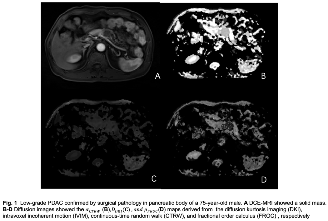
A retrospective analysis was conducted on 49 patients with PDAC from July 2022 to July 2023 at the First People’s Hospital of Changzhou. All participants were pathologically categorized into two groups: the low-grade PDAC group (n=24) and the high-grade PDAC group (n=25). All participants in our study underwent structural and diffusion magnetic resonance imaging (MRI) using a 3T scanner (MAGNETOM VIDA, Siemens Healthcare, Erlangen, Germany) equipped with a 32-channel abdomen coil. The diffusion MRI was conducted in the axial plane employing a comprehensive q-space Cartesian grid sampling technique, which included 13 different b-values ranging from 0 to 2500 s/mm². The acquisition time was 8 min 39 s. The resolution was 1.6×1.6×3.0 mm3. IVIM, DKI, FROC, and CTRW imaging were reconstructed from raw diffusion weighted data and eleven diffusion parameters, namely fIVIM, DIVIM, D*IVIM, DDKI, KDKI, αCTRW, βCTRW, DCTRW, βFROC, DFROC and μFROC of the primary tumor, were derived from diffusion models. The raw data from the diffusion MRI scans were converted into the NIFTI-1.1 format using MRIcron (https://www.nitrc.org/projects/mricron). Subsequently, the data underwent processing using the BodyLab software, which is based on DIPY (https://dipy.org/). The comparisons of diffusion metrics and clinicopathologic variables among the two groups were tested with the independent sample t-test and the chi-square test.Results
A total of 49 individuals with PDAC were included, with 24 participants in the low-grade PDAC group (mean age ± standard deviation, 66.46±9.94) and 25 participants in the high-grade PDAC group (mean age ± standard deviation, 67.64±7.82). Table 1 shows the comparisons of quantitative diffusion metrics between low-grade and high-grade PDAC. The KDKI (0.56±0.19× 10⁻³ mm²/s vs. 0.68±0.10⁻³mm²/s; p=0.008), αCTRW (0.77±0.17 vs. 0.86±0.10; p=0.035), and μFROC (3.03±1.12 mm vs. 3.77±0.70 mm; p=0.009) differed significantly between low-grade and high-grade PDAC. The KDKI , αCTRW and μFROC were notably lower in low-grade PDAC compared to their counterparts in high-grade PDAC (p=0.008 to p=0.035).Discussion
This study revealed differences in three diffusion metrics derived from DKI, FROC, and CTRW between low-grade and high-grade PDAC. These metrics may serve as imaging biomarkers for distinguishing between the two. Additionally, this finding suggests that FROC and CTRW can be considered as new diffusion models for PDAC, as previous studies have not explored their application in this context. However, it was a single-center study, and larger multi-center studies are needed to validate the discriminative performance of these diffusion-based metrics. Further research is warranted to investigate these models thoroughly.Conclusion
In conclusion, our preliminary study demonstrates that multiple diffusion metrics derived from DKI, FROC, and CTRW differ between low-grade and high-grade PDAC. The KDKI ,αCTRW and μFROC have the potential to serve as imaging biomarkers for identifying the degree of differentiation in pancreatic cancer, warranting further validation.Acknowledgements
No acknowledgement found.References
1 Park W, Chawla A, O Reilly EM. Pancreatic Cancer: A review. JAMA, 2021, 326(9):851-62.
2 Rahib L, Smith BD, Aizenberg R, ET al. Projecting cancer incidence and deaths to 2030: The unexpected burden of thyroid, liver, and pancreas cancers in the United States [J]. Cancer Res, 2014, 74(11): 2913-2921.
Figures
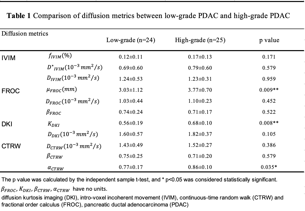
The p value was calculated by the independent sample t-test, and * p<0.05 was considered statistically significant.
βFROC, KDKI, βCTRW, αCTRW have no units.
diffusion kurtosis imaging (DKI), intro-voxel incoherent movement (IVIM), continuous-time random walk (CTRW) and fractional order calculus (FROC), pancreatic ductal adenocarcinoma (PDAC)