4001
Sensitivity and Specificity of Detecting Premalignant Pancreatic Lesions by Hyperpolarized Magnetic Resonance1Cancer System Imaging, UT MD Anderson Cancer Center, Houston, TX, United States, 2UT MD Anderson Cancer Center UT Health Science Center Houston Graduate School of Biomedical Sciences, Houston, TX, United States, 3Clinical Cancer Prevention, UT MD Anderson Cancer Center, Houston, TX, United States, 4Statistics, Rice University, Houston, TX, United States
Synopsis
Keywords: Pancreas, Hyperpolarized MR (Non-Gas), Pancreatic Cancer, Metabolic Imaging, Early Detection
Motivation: There is an unmet need for early diagnosis of pancreatic cancer due to the asymptomatic nature of the disease.
Goal(s): The goal is to detect the early stages of pancreatic cancer by monitoring the altered metabolism in premalignant pancreatic lesions in vivo with hyperpolarized metabolic imaging.
Approach: This is validated in two mechanistic pathways of cancer progression, (1) pancreatic intraepithelial neoplasia (PanIN), and (2) intraductal papillary mucinous neoplasm (IPMN).
Results: Increased lactate flux is observed with progression of the premalignant lesions in both the pathways. No increase in lactate flux is observed in pancreatitis, a known confounder demonstrating the specificity of this technique.
Impact: This research described has the potential for leading to practice-changing recommendations for non-invasively detecting and monitoring premalignant lesions and incipient pancreatic cancer, to prevent over-diagnosis and over-treatment of cysts that harbor indolent biology.
Introduction
Pancreatic cancer is one of the most aggressive types of cancers. It is difficult to detect due to its asymptomatic presentation at early stages. Therefore, there is an unmet need for non-invasive imaging markers that help identify the aggressive sub-type(s) in a pancreatic lesion at an early time point in pancreatic cancer. One of the most commonly used imaging biomarkers is the conversion of hyperpolarized pyruvate to lactate.1 It has been previously demonstrated that at early timepoints of pancreatic cancer the Warburg effect sets in and promotes the conversion to lactate. With metabolic HP-MR imaging, the conversion will be monitored between different premalignant models at different timepoints to determine sensitivity of the imaging technique. Concurrently, the specificity of HP MR of detecting premalignant lesions will be determined by imaging pancreatitis induced mice at different timepoints and compare them to the premalignant models.Methods
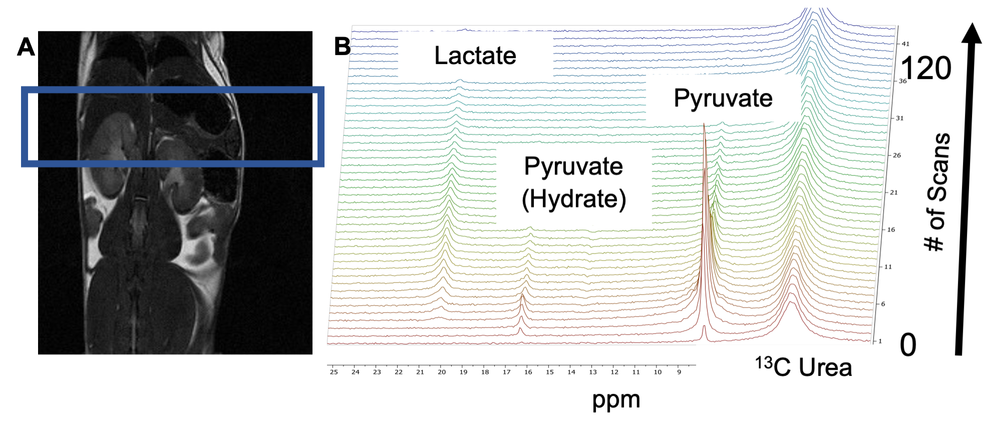
Hyperpolarized 1-13C Pyruvate MRS was employed to study the metabolic processes in tamoxifen inducible genetically engineered mouse (GEM) models (P48CreERT2;LSL-KrasG12D (iKC)) with pre-invasive pancreatic intraepithelial neoplasia (PanIN) precursor lesions, invasive pancreatic cancer model (P48CreERT2;LSL-KrasG12D; LSL-p53R172H (iKPC)) and control animals (P48CreERT2 (iC)) without pancreatic lesions. Similarly, inducible genetically engineered mouse models (p48-Cre; LSL-KrasG12D; Rosa26R-LSL-rtTA-TetO-GnasR201C) that develop premalignant cystic lesions (IPMN pathway) that progresses to pancreatic cancer on doxycycline diet (Dox+) for 15 weeks and mice on normal diet (Dox-) serving as control. The dissolution DNP (HyperSense, Oxford Instruments, UK) operating at 3T was utilized to hyperpolarize 1-13C pyruvate. The 13C magnetic resonance spectra of hyperpolarized 1-13C pyruvate were acquired at 7T Bruker MRI scanner.2 (Figure 1) The PanIN mice were imaged at different time points in their lifespan, before tamoxifen induction, 10-, 20-, and 30-weeks post induction. The IPMN mice were imaged after the 15 weeks of (Dox±) treatment. Simultaneously, wildtype, iC and iKC mice were treated with caerulein for three weeks for the development of pancreatitis. These mice were imaged 24 hours after the last caerulein injection.Results/Discussion
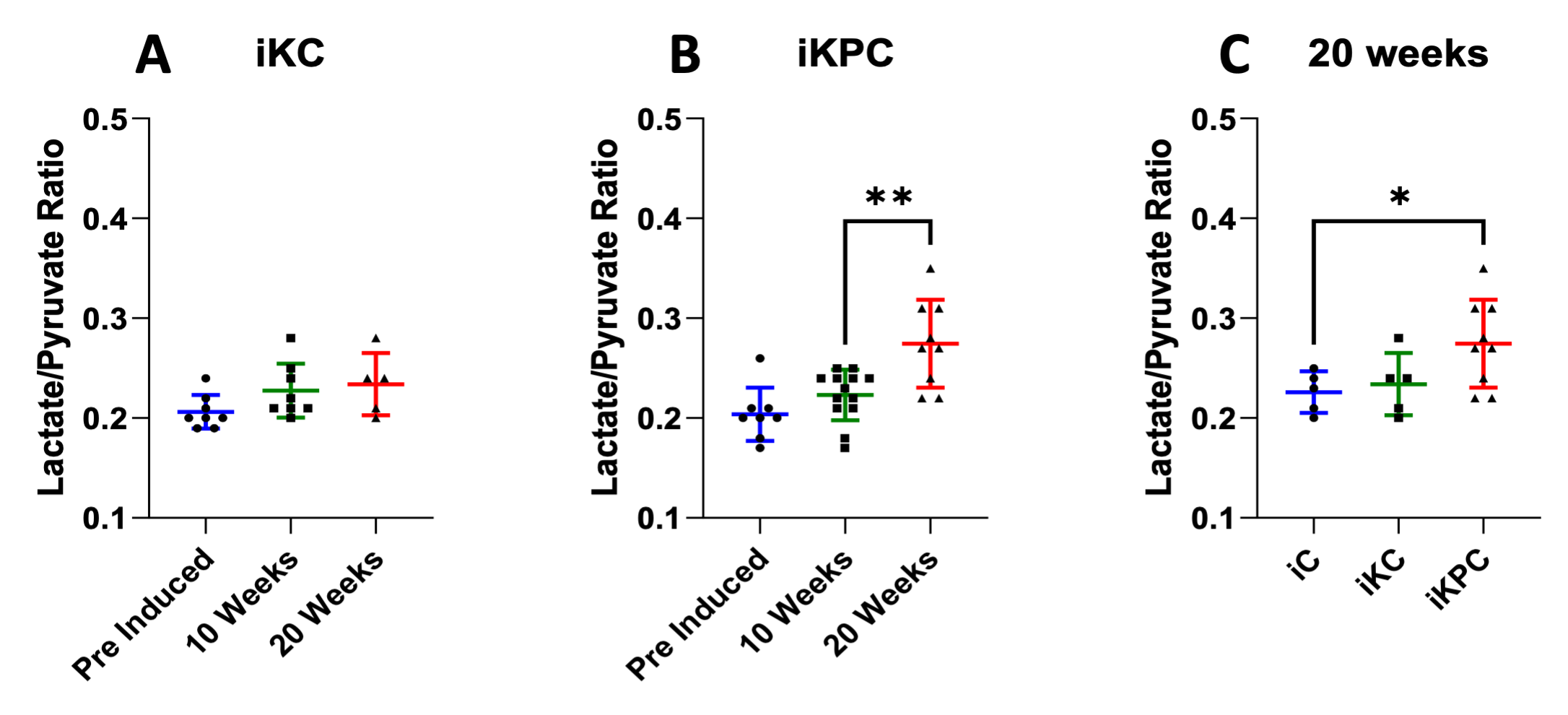
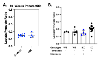
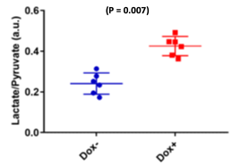
The lactate-to-pyruvate ratio increased in the pancreatic cancer models compared to the control model. These results demonstrate that there are significant alterations of LDH activities during the transformation from early to advanced PanINs lesions. As for the aggressive iKPC mouse model, at the 20-week post induction imaging there was a significant increase of the lactate-to-pyruvate ratio (0.28±0.04) compared to the 10-week time point after induction (0.22±0.03). The 20-week iKPC time point ratio compared to the iKC and control mouse models was significantly higher, (0.28±0.04 compared to 0.23±0.03 and 0.22±0.02 respectively) indicating the invasive nature of the cancer. Even in the iKC model there is a slight increase of the lactate-to-pyruvate ratio at 20-weeks post induction (0.23±0.03) compared to both previous time points, pre-induction (0.21±0.02) and 10-week (0.22±0.03). All this data is shown in Figure 2. At the same time, mice that were treated with caerulein developed pancreatitis as demonstrated by tissue histology. Surprisingly, the lactate-to-pyruvate ratio remained constant in both iC and iKC mice that developed pancreatitis, around 0.15±0.03 for both groups, even with an inflamed pancreas. (Figure 3) Even in wildtype mice that developed pancreatitis their ratio didn’t increase compared to wildtype mice that were not injected with caerulein, 0.16±0.01 and 0.19±0.05 respectively. Compared with mice developing premalignant lesions, the pancreatitis models exhibit lower ratios, indicating that the only factor increasing the lactate-to-pyruvate ratio is the progression towards pancreatic cancer. These results suggest that 1-13C Pyruvate HP-MR metabolic imaging can specifically employed to image pancreatic cancer progression and is not influenced by known confounders such as pancreatitis. IPMN mice feed with doxycycline showed increase lactate-to-pyruvate ratio (0.43±0.05) compared to mice on normal diet (0.24±0.05). We also plan to implement some Artificial Intelligence (AI) components to our metabolic profiles to further predict pancreatic cancer at even earlier stages.3Conclusion
Conclusion: 1-13C Pyruvate HP-MR can detect metabolic shift to lactate in two different models of pancreatic cancer premalignancy representing two different pathways of pancreatic cancer progression. This finding can be potentially translated to the clinic for detection of pancreatic premalignant lesion in high-risk populations and assist physicians determine malignant lesions before progression to pancreatic cancer.Acknowledgements
This research was funded in part by a grant from Pancreatic Cancer Action Network (PANCAN; 16-65-BHAT) (PB, FM); NCI PREVENT (PB, FM); NCI F99/K00 (1F99CA284365-01, JSE) Duncan Family Institute for Cancer Prevention and Risk Assessment Seed Funding; by grants from the US National Cancer Institute (U01 CA214263, U54 CA151668 and R21 CA185536, R01 CA218004; and 1P50 CA221707-01) and Department of Defense (PA220132). This work also was supported by the National Institutes of Health/NCI Cancer Center Support Grant under award number P30 CA016672.References
1. Dutta, P., Pando, S. C., Mascaro, M., et al. Early Detection of Pancreatic Intraepithelial Neoplasias (PanINs) in Transgenic Mouse Model by Hyperpolarized 13C Metabolic Magnetic Resonance Spectroscopy. International Journal of Molecular Sciences. 2020; 21(10), 3722.
2. Pudakalakatti, S., Raj, P., Salzillo, T. C., Enriquez, J. S., et al. Metabolic Imaging Using Hyperpolarization for Assessment of Premalignancy. In Cancer Immunoprevention. 2022; 169-180. Humana, New York, NY.
3. Enriquez, J.S., Chu, Y., Pudakalakatti, S., et al. Hyperpolarized magnetic resonance and artificial intelligence: Frontiers of imaging in pancreatic cancer. JMIR medical informatics. 2021; 9(6), p.e26601.
Figures