4000
Differentiation between inflammatory myofibroblastic tumor and colorectal liver metastasis on gadoxetic acid-enhanced MRI1The First Hospital of Lanzhou University, Lanzhou, China, 2Philips Healthcare, Beijing, China
Synopsis
Keywords: Hepatobiliary, Tumor, hepatic inflammatory myofibroblastic tumor
Motivation: Inflammatory myofibroblastic tumors (IMT) of the liver can mimic malignant tumors, yet, they lack definitive radiological characteristics.
Goal(s): To identify gadoxetate acid-enhanced MRI (Gd-EOB-MRI) features distinguishing hepatic inflammatory myofibroblastic tumor (IMT) from colorectal liver metastasis (CLM).
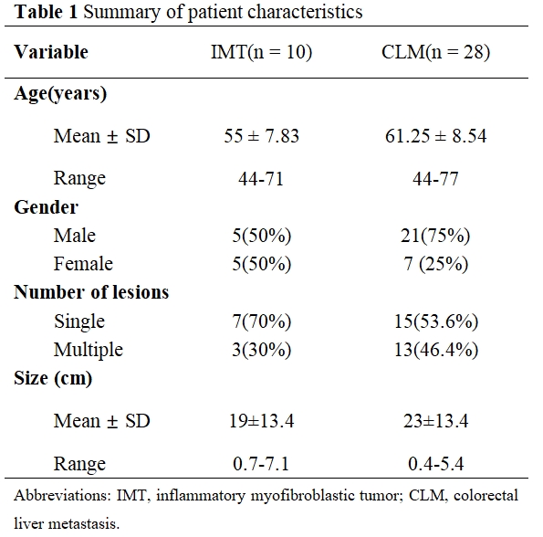
Approach: This retrospective study included 10 IMT (0.7-7.1 cm) and 28 CLM (0.4-5.4 cm) patients who underwent Gd-EOB-MRI. Two reviewers assessed tumor characteristics.
Results: Peripheral hypointense rim on T1WI, and arterial phase and/or portal venous phase, corresponding to the delayed enhanced rim on transitional phase, layered appearance on T2WI, and obscure hypointensity on hepatobiliary phase were commonly in IMTs, but rare in CLMs.
Impact: The distinctive imaging features on Gd-EOB-DTPA MRI such as early target appearance and delayed enhanced rim and obscure hypointensity on Hepatobiliary phase, are helpful in differential diagnosis of IMT from colorectal liver metastasis.
Motivation
Inflammatory myofibroblastic tumor (IMT) of the liver is a distinctive neoplasm of intermediate biological potential. IMT can mimic malignant tumors, such as liver metastasis, hepatocellular carcinoma, and intrahepatic cholangiocarcinoma, etc. However, there are no radiological features have been reported to be characteristic.Purpose
To identify gadoxetate acid-enhanced MRI (Gd-EOB-MRI) features distinguishing hepatic inflammatory myofibroblastic tumor (IMT) from colorectal liver metastasis (CLM).Methods
From August 2017 to August 2023, 10 patients (5 females and 5 males, median age 55 years, range 44-71 years) with 54 pathological confirmed IMTs (0.7–7.1 cm) and 28 patients (7 females and 21 males, median age 61.25 years, range 44-77 years) with 61 pathological confirmed CLMs (0.4–5.4 cm) who underwent Gd-EOB-MRI were enrolled. Two reviewers retrospectively assessed the morphology, boundary, signal intensity on T1WI, T2WI, DWI, and Gd-EOB-MRI imaging of tumors.Results
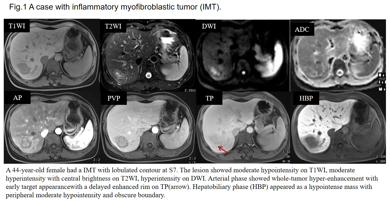
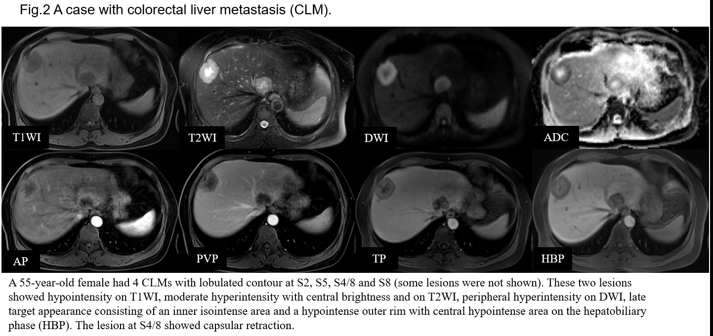
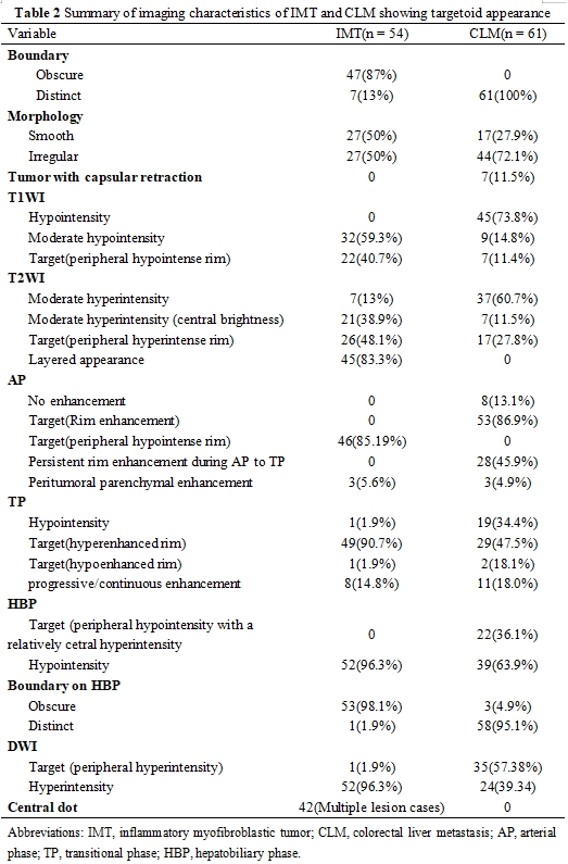
Tweenty-seven IMTs and 43 CLMs exhibited lobulated or wavy contours, and one IMT showed a patchy shape with bile duct dilatation, while the others were smooth. The following features were showed significantly higher frequencies in IMT than CLM: early target appearance characterized by a peripheral hypointense rim on T1WI, and arterial phase (AP) and/or portal venous phase (PVP), corresponding to the delayed enhanced rim on transitional phase (TP); layered appearance consisting of an inner isointense area and a hyperintense outer rim with tiny central dots with that appeared hypointense on T1WI and hyperintense on T2WI; obscure boundary and hypointensity on hepatobiliary phase (HBP) . While rim enhancement on AP, persistent rim enhancement during AP to TP, peripheral hyperintensity on DWI, late target appearance characterized by peripheral hypointensity with a relatively central hyperintensity and clear boundary on HBP were significantly more common in CLM than in IMT.Conclusion
Early target appearance and corresponding delayed enhanced rim on TP, layered appearance with a tiny central dot, obscure bondary and hypointensity on HBP may be reliable Gd-EOB-MRI features for distinguishing IMT from CLM.Acknowledgements
NoneReferences
1. Tang L, Lai EC, Cong WM, Li AJ, Fu SY, Pan ZY, Zhou WP, Lau WY, Wu MC. Inflammatory myofibroblastic tumor of the liver: a cohort study. World J Surg. 2010;34(2):309-313.
2. Elpek GÖ. Inflammatory Myofibroblastic Tumor of the Liver: A Diagnostic Challenge. J Clin Transl Hepatol. 2014;2(1):53-57.
3.Chang AI, Kim YK, Min JH, Lee J, Kim H, Lee SJ. Differentiation between inflammatory myofibroblastic tumor and cholangiocarcinoma manifesting as target appearance on gadoxetic acid-enhanced MRI. Abdom Radiol (NY). 2019;44(4):1395-1406.
Figures