3999
Diffusion–Based Virtual MR Elastography as a Potential Biomarker for Predicting Recurrence of Hepatocellular Carcinoma1Zhongshan hospital, Fudan University, Shanghai, China, 2MR Application Development, Siemens Shenzhen Magnetic Resonance Ltd., Shenzhen, China
Synopsis
Keywords: Liver, Liver
Motivation: Preoperative prediction of tumor recurrence is essential for surveillance and management of patients with hepatocellular carcinoma (HCC).
Goal(s): To explore the diagnostic performance of virtual magnetic resonance elastography (vMRE) derived from preoperative diffusion-weighted images in predicting HCC recurrence after hepatectomy.
Approach: Eighty patients who underwent magnetic resonance imaging with a dedicated diffusion-weighted imaging sequence were retrospectively recruited. The parameters derived from vMRE, together with image features, were used to predict tumor recurrence after hepatectomy.
Results: The μdiff values of vMRE and corona enhancement are potential biomarkers for the preoperative prediction of recurrence after hepatectomy in patients with HCC.
Impact: Our results revealed that preoperative diffusion–based virtual magnetic resonance elastography could be used for preoperative prediction of HCC recurrence without using additional hardware, which might help in deciding on treatment and formulating management strategies for patients with HCC.
Introduction
Hepatocellular carcinoma (HCC) is one of the most common malignant tumors worldwide, accounting for 75%-85% of primary liver cancer1. Despite advances achieved in early diagnosis and surgical treatment for HCC, approximately 50%-80% of patients experience recurrence within 5 years after hepatectomy2. Previous studies have reported that the magnetic resonance elastography (MRE)-based stiffness could be used as a potential biomarker for predicting HCC recurrence after surgical resection3,4. However, the application of MRE was limited by the need for an external mechanical setup and a dedicated magnetic resonance imaging (MRI) sequence. Recently, a novel technique named diffusion-weighted imaging (DWI)-based virtual elastography (vMRE) was proposed by Le Bihan et al.5 They revealed that the parameter obtained from DWI with b values of 200 and 1500 s/mm2, known as the shifted apparent diffusion coefficient (sADC), demonstrated equivalent efficiency to diagnose fibrosis stages compared with MRE6. Given that HCC lesions in patients with recurrence may exhibit with increased cellularity and stiffness, vMRE may have potential value in the tumor characterization of HCC without requiring additional hardware. vMRE has scarcely been applied to liver tumors yet.Methods
This study included 80 patients who underwent MRI including a dedicated DWI sequence. All MR images were obtained by using a 1.5T MR system (MAGNETOM Aera; Siemens Healthineers, Erlangen, Germany). The multiple-b-value DWI was acquired with a research single-shot spin-echo echo-planar DWI pulse sequence. The detailed parameters were as follows: repetition time (TR)/echo time: 8000 ms/63 ms; field of view, 380 × 308 mm2; matrix, 128 × 128; b-values: 0, 200, 500, and 1500 s/mm2; section thickness, 5 mm. The ADC maps from DWI with b values of 0 and 500 s/mm2 and sADC from DWI with b values of 200 and 1500 s/mm2 were generated using an in-house developed program based on MATLAB (Mathworks, MA, USA). The sADC was then converted into DWI-based virtual shear modulus (μdiff) according to the method proposed in previous studies5,6. The radiologists also evaluated the morphological MRI features of the tumor lesions. The Cox proportional-hazards model was used to identify the risk factors associated with tumor recurrence.Results
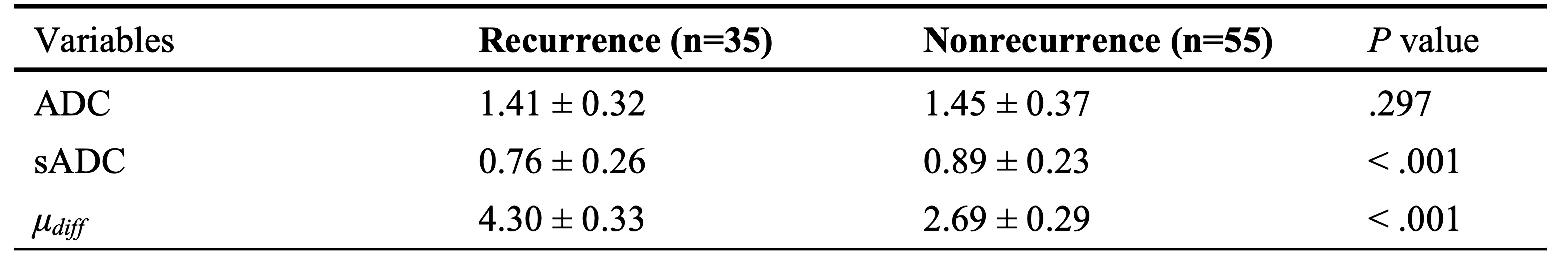
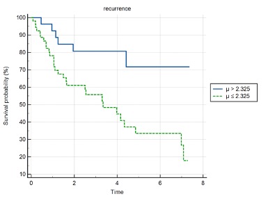
Thirty-five patients (43.8%) experienced tumor recurrence after hepatectomy. The sADC (P < .001) and μdiff (P < .001) in patients with HCC with and without recurrence demonstrated notable differences. However, the ADC (P = .297) showed no statistical differences in patients with HCC with and without recurrence (Figs 1, 2 and 3). In the multivariable Cox proportional-hazards model, μdiff > 2.325 kPa [hazard ratio (HR): 1.313, 95% CI: 1.113-1.548; P = .001] and corona enhancement (HR: 2.044, 95% CI: 1.017-4.108; P = .044) were independent risk factors for tumor recurrence. Patients with HCC with μdiff > 2.325 kPa had poorer 5-year disease-free survival after hepatectomy compared with patients with μdiff values ≤ 2.325 kPa (Fig. 4). The receiver operating characteristic (ROC) analysis demonstrated that the combination of μdiff values and corona enhancement yielded the best diagnostic efficiency for predicting a high risk of tumor recurrence with area under the curve (AUC) of 0.766 (Fig. 5).Acknowledgements
None declared.References
1 Sung, H. et al. Global cancer statistics 2020: GLOBOCAN estimates of incidence and mortality worldwide for 36 cancers in 185 countries. CA Cancer J Clin, doi:10.3322/caac.21660 (2021).
2 Forner, A., Reig, M. & Bruix, J. Hepatocellular carcinoma. Lancet 391, 1301-1314, doi:10.1016/s0140-6736(18)30010-2 (2018).
3 Zhang, L. et al. MR elastography as a biomarker for prediction of early and late recurrence in HBV-related hepatocellular carcinoma patients before hepatectomy. Eur J Radiol 152, 110340, doi:10.1016/j.ejrad.2022.110340 (2022).
4 Wang, J. et al. 3D MR Elastography of Hepatocellular Carcinomas as a Potential Biomarker for Predicting Tumor Recurrence. J Magn Reson Imaging 49, 719-730, doi:10.1002/jmri.26250 (2019).
5 Le Bihan, D., Ichikawa, S. & Motosugi, U. Diffusion and Intravoxel Incoherent Motion MR Imaging-based Virtual Elastography: A Hypothesis-generating Study in the Liver. Radiology 285, 609-619, doi:10.1148/radiol.2017170025 (2017).
6 Kromrey, M. L., Le Bihan, D., Ichikawa, S. & Motosugi, U. Diffusion-weighted MRI-based Virtual Elastography for the Assessment of Liver Fibrosis. Radiology 295, 127-135, doi:10.1148/radiol.2020191498 (2020).
7 Ota, T. et al. Diffusion-Based Virtual MR Elastography of the Liver: Can It Be Extended beyond Liver Fibrosis? J Clin Med 10, doi:10.3390/jcm10194553 (2021).
Figures
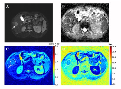
Representative MR images of a 47-year-old man with pathologically verified HCC. Tumor recurrence occurred 13 months after surgery.
(A) Axial T2-weighted image showing a hyperintensity mass (arrow) on segment VI of the liver. (B-D) ADC, sADC, and μdiff maps showing that the mean ADC, sADC, and μdiff values of the tumor (arrow) were 1.44 × 10⁻³ mm2/s, 0.70 × 10⁻³ mm2/s, and 5.10 kPa, respectively. ADC, apparent diffusion coefficient; sADC, shifted apparent diffusion coefficient; μdiff, DWI-based virtual shear modulus.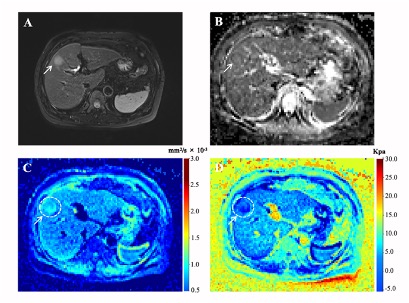
Representative MR images of a 60-year-old woman with pathologically verified HCC without recurrence during a 5-year follow-up.
(A) Axial T2-weighted image shows a hyperintensity mass (arrow) on segment VIII of liver. (B-D) ADC, sADC, and μdiff maps showing that the mean ADC, sADC, and μdiff values of the tumor (arrow) were 1.35 × 10⁻³ mm2/s, 0.94 × 10⁻³ mm2/s, and 1.99 kPa, respectively. ADC, apparent diffusion coefficient; sADC, shifted apparent diffusion coefficient; μdiff, DWI-based virtual shear modulus.
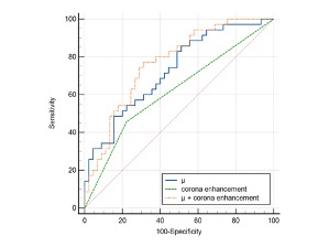
Graph indicating the ROC curves for μdiff, corona enhancement and the combination of μdiff and corona enhancement for predicting HCC recurrence after hepatectomy. The AUCs for the corresponding ROC curves were 0.728 (μdiff), 0.617 (corona enhancement), and 0.766 (μdiff + corona enhancement). AUC, Area under the curve; DWI, diffusion-weighted imaging; HCC, hepatocellular carcinoma; ROC, receiver operating characteristic; μdiff, DWI-based virtual shear modulus.