3997
The differential diagnostic value of APT combined with Gd-BOPTA in IMCC and SHHM.1School of Medical Imaging,Dalian Medical University, Dalian, China, 2Department of Radiology, The First Affiliated Hospital of Dalian Medical University, Dalian, China, 3Philips (China) Investment Co., Ltd, Shanghai, China
Synopsis
Keywords: Hepatobiliary, Precision & Accuracy, Differential diagnosis
Motivation: IMCC and SHHM have similarities in imaging manifestations, are prone to confusion, and the treatment methods are different, once misdiagnosed, it seriously affects the patient's treatment and prognosis.
Goal(s): This study aims to explore the differential diagnostic value of amide proton transfer (APT) combined with Gd-BOPTA in IMCC and SHHM.
Approach: Retrospective analysis, normality testing, logistic regression, ROC analysis, and other methods were used for the study.
Results: APT, LLR, and LSIR have significance in the differential diagnosis of IMCC and SHHM. When combined with LLR and LSIR, APT improves the differential efficiency.
Impact: This study helps to more accurately distinguish IMCC from SHHM, thus adopting the correct treatment method, which is of great help to the treatment and prognosis of patients.
Introduction
Massive intrahepatic cholangiocarcinoma (IMCC) and single hypovascularized hepatic metastases (SHHM) are common malignant tumors in the liver. The two have similar imaging manifestations and are easily confused. [1] This study aims to explore the differential diagnostic value of amide proton transfer (APT) combined with Gd-BOPTA in IMCC and SHHM.Methods
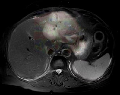
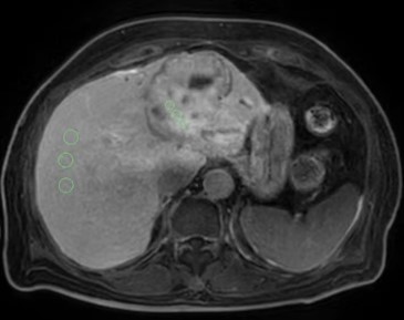
A retrospective analysis was conducted on the clinical and MRI data of patients with cholangiocarcinoma and liver metastasis confirmed by surgery pathology or clinical follow-up in our hospital from March 2021 to July 2023. Finally, 14 cases of IMCC and 17 cases of SHHM were included. Both IMCC and SHHM use the Philips post-processing platform to place three equally sized regions of interest (ROIs) at the maximum level of the tumor, record the average signal intensity (SI) value of the tumor APT, and place three equally sized ROIs at the maximum level of the tumor and at the same level of normal liver parenchyma during the hepatobiliary specific phase (HBP). Record the average SI value of the tumor and liver, Calculate the tumor lesion liver signal intensity ratio (LLR) and signal intensity ratio (LSIR) based on the mean. Using SPSS 27 statistical software, normality tests were used to evaluate the normality of each parameter, and independent sample t-tests or Mann Whitney U-tests were used to compare the differences in quantitative parameters between the two groups. Use logistic regression to establish prediction models with different parameter combinations. Evaluate diagnostic performance using receiver operating characteristics (ROC) analysis.Results
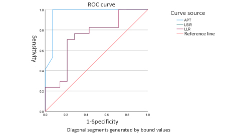
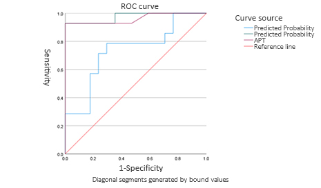
There was no statistically significant difference in SNR and CNR values between the IMCC group and the SHHM group (P>0.05). APT, LLR, and LSIR were significant in the differential diagnosis of IMCC and SHHM, and the statistical significance of APT (P<0.001) was much greater than that of LLR (P=0.042) and LSIR (P=0.042). The areas under the curve of APT, LLR, and LSIR were 0.962, 0.710, and 0.710, respectively, when LLR was combined with LSIR, and APT was combined with LLR and LSIR, Improved identification efficiency (AUC of 0.735 and 0.975, respectively).Conclusion
APT, LLR, and LSIR can effectively distinguish IMCC from SHHM, and the combined identification efficiency of APT, LLR, and LSIR is improved. This study contributes to a more accurate identification of IMCC and SHHM, thereby adopting correct treatment interventions to improve the treatment effectiveness and prognosis of patients.[2,3]Acknowledgements
References
[1]Qian, Haizhen*; Li, Shihong*; Ji, Ming; Lin, Guangwu. MRI characteristics for the differential diagnosis of benign and malignant small solitary hypovascular hepatic nodules. European Journal of Gastroenterology & Hepatology 28(7):p 749-756, July 2016. | DOI: 10.1097/MEG.0000000000000642
[2]徐壮. 肝内胆管细胞癌的预后影响因素分析[D].吉林大学,2023.DOI:10.27162/d.cnki.gjlin.2023.005123.
[3] Takahashi H, Berber E. Role of thermal ablation in the management of colorectal liver metastasis. Hepatobiliary Surg Nutr. 2020 Feb;9(1):49-58. doi: 10.21037/hbsn.2019.06.08. PMID: 32140478; PMCID: PMC7026789.
Figures