3992
the application value of automatic extraction of intratumoral susceptibility signal intensities in abdominal tumors1the First Affiliated Hospital of Dalian Medical University, Dalian, China, 2Dalian University of Technology, Dalian, China
Synopsis
Keywords: fMRI Analysis, Segmentation
Motivation: By studying the application of ITSS ratio values measured under different threshold conditions, we hope to be able to quickly identify and predict abdominal tumors preoperatively.
Goal(s): To explore the application value of automatic extraction of ITSS in abdominal tumors.
Approach: Friedman The rank sum test was used to compare the differences of ITSS measurements under different threshold conditions.
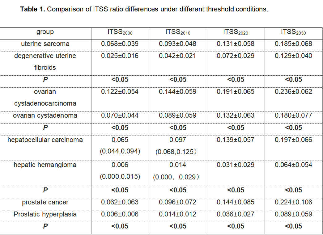
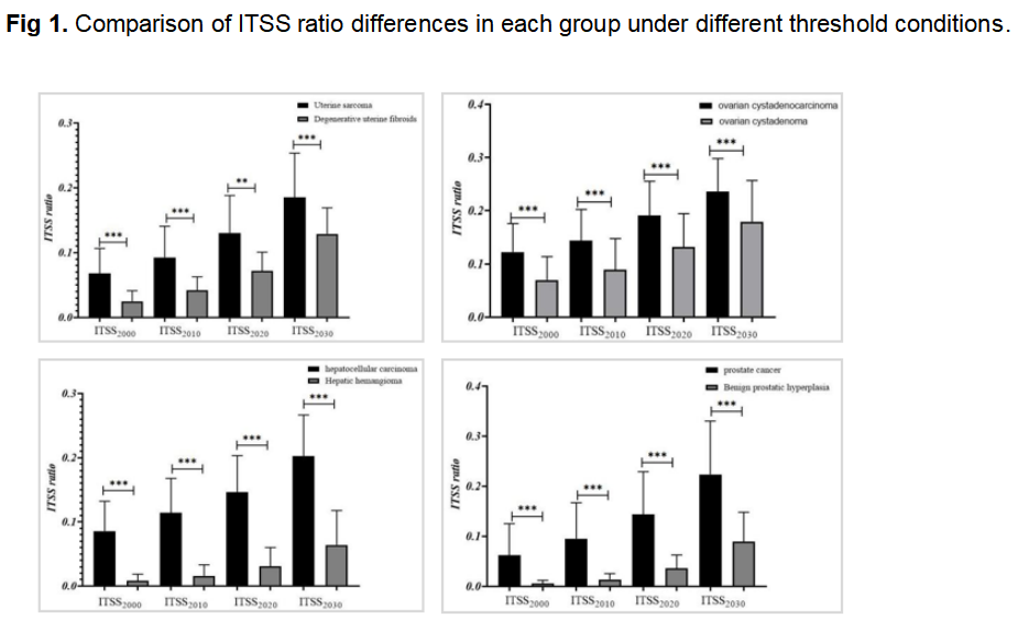
Results: Under different threshold conditions, the ITSS ratios of uterine sarcoma, ovarian cystadenocarcinoma, hepatic vascular carcinoma and prostate cancer were respectively higher than those of degenerative uterine fibroids, ovarian cystadenomas, hepatic hemangioma and prostate hyperplasia.
Impact: The automatic extraction of ITSS can effectively, quickly and subjectively predict the differential diagnosis of abdominal tumors under different threshold conditions, which has a good clinical application prospect.
Introduction
Intra-tumoral susceptibility signal intensities (ITSS) is consistent with the density and size of microvessels in histopathological samples [1]. It is a non-invasive imaging marker that can visually display the proliferation of blood vessels inside the tumor. At present, the ITSS measurement method mainly used is semi-quantitative, which has the problem of being unable to reflect local differences in lesions and making it difficult to identify. In addition, manual counting of ITSS requires a lot of time, effort, and is highly subjective. Therefore, this study is based on the automatic extraction of AS software to explore whether there are differences in the identification of abdominal tumors between ITSS ratio values measured under different threshold conditions, and whether the differential diagnosis and prediction of abdominal tumors can be effectively and quickly carried out.Methods
The clinical and imaging data of patients with abdominal tumors in our hospital were retrospectively analyzed. All patients underwent 1.5T magnetic resonance imaging two weeks before surgery, and the scanning sequences included T1WI, T2WI, ESWAN, etc. The ITSS ratio of lesions at different thresholds was automatically measured using Anatomy-Sketch software. The independent samples t test or Mann-Whitney U test was used to compare the differences in the ITSS ratio of each lesion under different threshold conditions; Friedman The rank sum test was used to compare the differences of ITSS measurements under different threshold conditions; the receiver operating characteristic (ROC) curve was used to evaluate the identification performance of the ITSS ratios of different threshold conditions; the Delong test was used to compare the area under the curve (AUC).Results
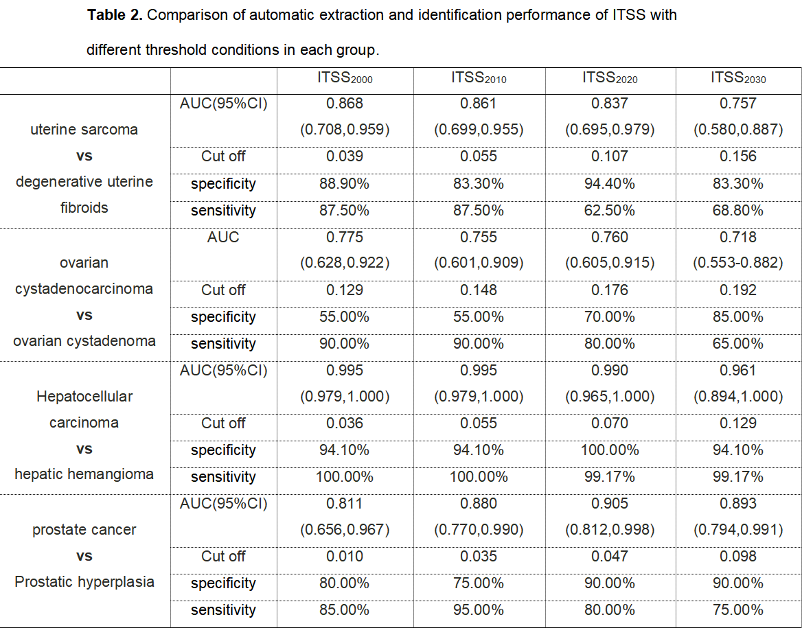
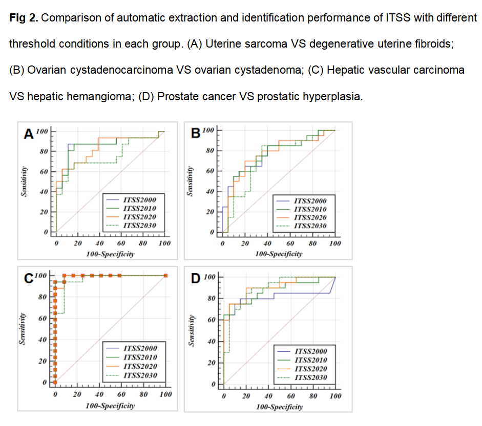
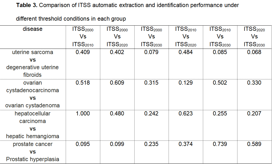
Under different threshold conditions, the ITSS ratios of uterine sarcoma, ovarian cystadenocarcinoma, hepatocellular carcinoma and prostate cancer were higher than those of degenerative uterine fibroids, ovarian cystadenomas, hepatic hemangioma and prostate hyperplasia, respectively, and the differences were statistically significant (Table 1, Fig 1). Furthermore, the ITSS ratio has high efficiency in identifying benign and malignant tumors in each group, with AUC values ranging from 0.718 to 0.995 (Table 2, Fig 2). The Delong test results showed that there was no statistically significant differences in AUC under different threshold conditions (Table 3).Discussion
In this study, the ITSS automatic extraction software does not require pre-screening of images with ITSS visible to the eye, and is more suitable for abdominal organs that are prone to motion artifacts under the premise of using phase map artifact removal; and the ITSS automatic extraction software can be used under different threshold conditions. To effectively and quickly subjectively predict the differential diagnosis of abdominal tumors, in order to improve the detection rate of tumor microvessels and microbleeds, this study finally concluded that the optimal threshold is ITSS2020. Compared with semi-quantitative or other quantitative ITSS extraction methods, it has higher specificity and sensitivity, and can provide certain help for the examination of tumor neovascularization and microbleeding. However, the number of patients included in each disease group in this study was small, and the sample size needs to be further increased to verify the results of this study.Conclusion
The automatic extraction of ITSS can effectively, quickly and subjectively predict the differential diagnosis of abdominal tumors under different threshold conditions, which can assist clinicians in formulating more effective treatment strategies.Acknowledgements
No acknowledgement found.References
[1]. Christoforidis GA, Kangarlu A, Abduljalil AM, Schmalbrock P, Chaudhry A, Yates A, Chakeres DW: Susceptibility-based imaging of glioblastoma microvascularity at 8 T: correlation of MR imaging and postmortem pathology. AJNR American journal of neuroradiology 2004, 25(5):756-760.Figures