3988
A comparative study of dynamic contrast-enhanced MRI and voxel-incoherent motion in evaluating microcirculation in rectal cancer1Shuguang Hospital Affiliated to Shanghai University of Traditional Chinese Medicine, Shanghai, China, 2Diagnostic Imaging, Siemens Healthineers Ltd, Shanghai, China
Synopsis
Keywords: Cancer, Digestive
Motivation: For the preoperative assessment of rectal cancer microcirculation use DCE ang IVIM.
Goal(s): To analyze the correlation between rectal cancer microvascular density, as indicated by CD34 immunohistochemistry, and quantitative parameters obtained from DCE and IVIM MRI.
Approach: Assess the correlation between quantitative imaging parameters from DCE and IVIM, and microvascular density.
Results: Significant differences in Ktrans and D values among different degrees of rectal cancer differentiation; furthermore, Ktrans, Kep, and D* values exhibited significant correlations with rectal cancer microvascular density, while Ve, D, and f did not show significant correlations.
Impact: The validation of the utility of non-invasive MRI techniques, particularly DCE and IVIM, assesses rectal cancer microcirculation, which can enhance early diagnosis and treatment planning for patients, ultimately improving the prognosis and management of rectal cancer.
Abstract
Introduction: Recurrence and metastasis are the primary factors contributing to the mortality of rectal cancer patients [1]. Previous research has indicated a significant correlation between the local tumor microcirculation status and the recurrence and metastasis of tumors [2]. Accurate assessment of the rectal cancer microcirculation status is crucial for early diagnosis and treatment. Conventional evaluation of rectal cancer tumor microcirculation status typically requires biopsy and postoperative pathology. Biopsy cannot provide a comprehensive assessment, while postoperative pathology loses its significance in preoperative diagnosis and surgical guidance. Therefore, there is a need to explore a non-invasive and visual quantitative magnetic resonance imaging (MRI) technique for preoperative tumor microenvironment assessment.Purpose: This study aims to analyze the correlation between rectal cancer microvascular density reflected by immunohistochemistry CD34 and the quantitative parameters of IVIM and DCE, investigating the value of both methods in reflecting tumor microcirculation.
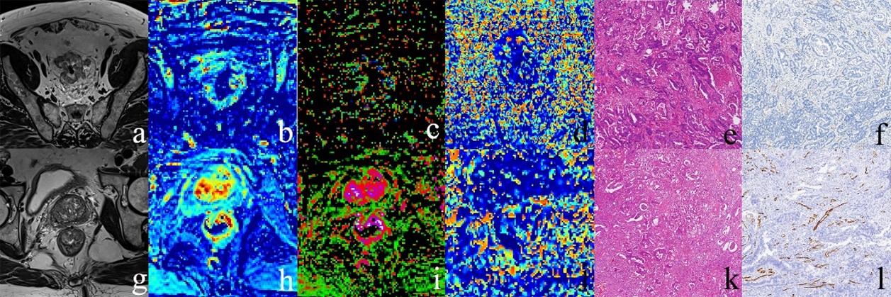
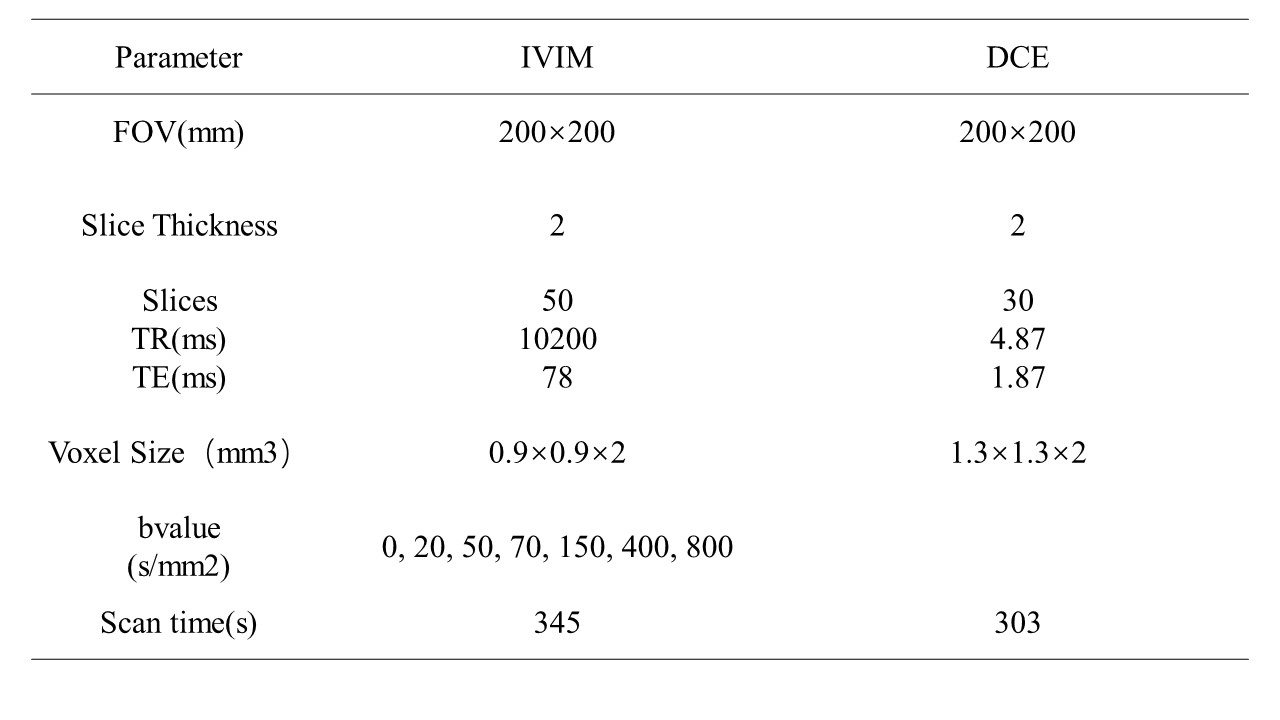
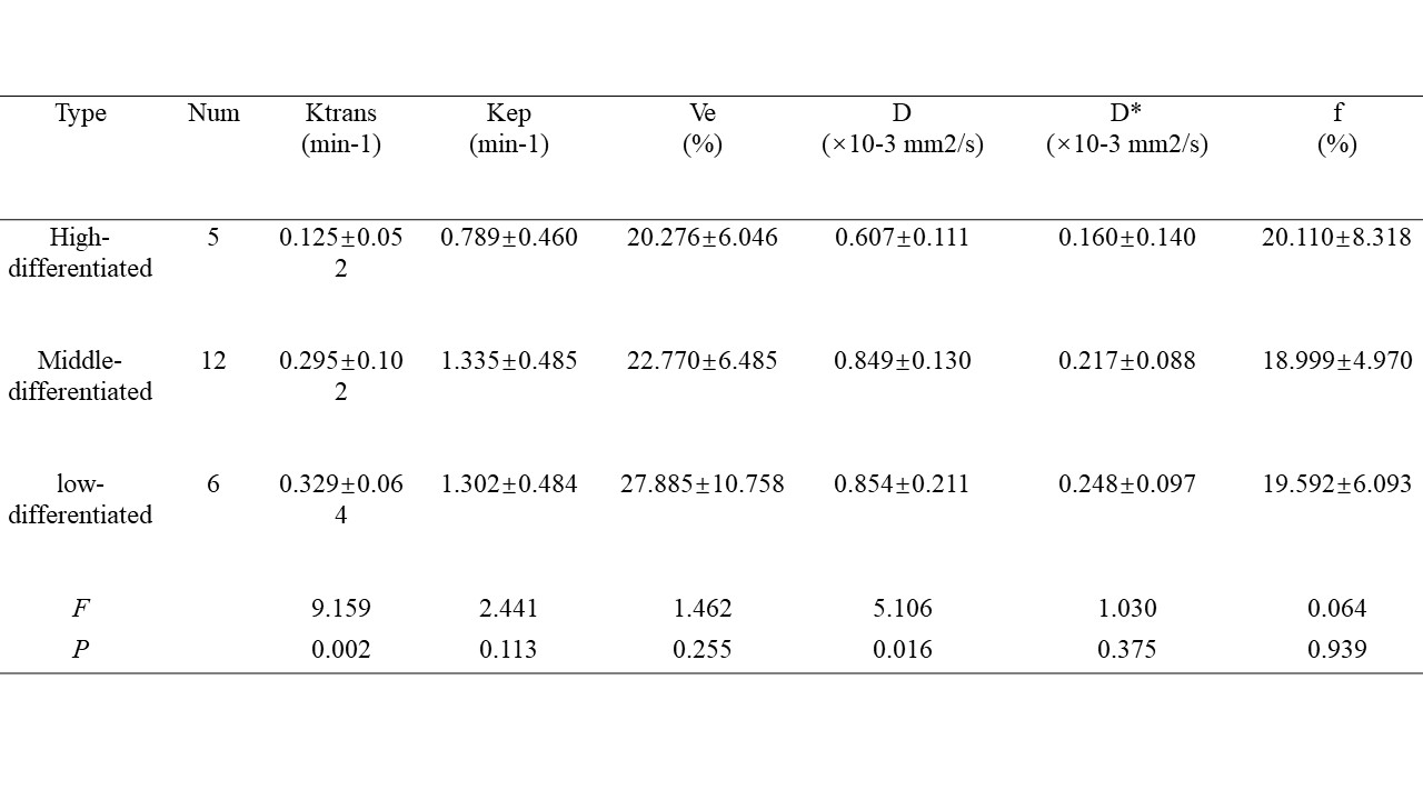
Methods: Prospective data were collected from 23 rectal cancer patients (10 males and 13 females) between December 2019 and December 2022, with an average age of 64.00±12.47 years (range: 35-82 years). All patients underwent routine high-resolution MRI, IVIM, and DCE sequences of the rectum. Inclusion criteria were age over 18, good general condition, biopsy-confirmed rectal adenocarcinoma, no distant metastases, and eligibility for surgical resection without prior radiotherapy or chemotherapy. Exclusion criteria included the presence of pacemakers, artificial heart valves, inability to undergo MRI due to other underlying diseases, concurrent malignancies, claustrophobia, and an MRI-surgery interval of more than 1 week. Informed consent was obtained from all participants. This study was approved by the medical ethics committee (Ethics Committee of Shuguang Hospital, Shanghai University of Traditional Chinese Medicine, 2019-750-105-01). The scans were performed using a 3T Siemens MAGNETOM Skyra MRI scanner. Before the MRI examination, patients emptied their bowels. An 18-channel phased-array surface coil was used for imaging, with the positioning marker at the upper margin of the pubic symphysis. The scan covered the entire rectal region. The IVIM and DCE parameters were listed in table1. A high-pressure injector was used for contrast agent injection into the median cubital vein, with a contrast agent dose of 0.2 ml/kg and an injection rate of 2 ml/s. All post-processing of DCE-MRI images was performed using Tissu4D software (Siemens Healthcare) with the Tofts pharmacokinetic model. Diffusion Image were processed by MITK-Diffusion(MITK, DKFZ). An area of interest (ROI) was delineated in the deepest infiltrated plane of rectal cancer, avoiding hemorrhagic, necrotic, cystic areas, bowel contents, and mesentery. The ROI area was not less than 1 cm2. HE staining and CD34 staining were performed on the transverse sections of the deepest infiltrated layer of rectal cancer. Data were analyzed using SPSS 19.0(SPSS Inc., Chicago, IL, USA) statistical software. Kolmogorov-Smirnov tests were initially used to assess the normality of DCE-MRI and IVIM parameters within each group. One-way analysis of variance (ANOVA) was used to compare differences in vascular density, DCE, and IVIM parameters among different degrees of rectal cancer differentiation. Spearman's correlation analysis was employed to assess the correlation between DCE and IVIM quantitative parameters and vascular density. A significance level of P<0.05 was considered statistically significant.
Results: Among the patients, 6 had poorly differentiated adenocarcinoma, 12 had moderately differentiated adenocarcinoma, and 5 had highly differentiated adenocarcinoma. There were significant statistical differences in Ktrans and D values among the three groups (P<0.05), while Kep, Ve, D*, and f showed no statistical differences (P>0.05), as shown in Table 2. The microvascular densities for highly, moderately, and poorly differentiated rectal cancer were 2.549%±1.04%, 3.625%±1.629%, and 3.716%±1.221%, respectively. No significant statistical differences were observed among the three groups (F=1.175, P=0.329). Ktrans, Kep, and D* values exhibited significant statistical differences in their correlation with rectal cancer microvascular density (r=0.734, P=0.000; r=0.617, P=0.002; r=0.456, P=0.029), while Ve, D, and f showed no statistical differences (r=0.101, P=0.647; r=0.199, P=0.362; r=-0.239, P=0.272), as shown in Table 2 and Figures 1 and 2.
Conclusion: DCE-MRI, employing a pharmacokinetic model to analyze the dynamic distribution of contrast agents, reflects the microcirculation status of biological tissues. IVIM-DWI can also reflect microcirculation and provides additional information on water molecule diffusion. This study suggests that both DCE-MRI and IVIM-DWI can non-invasively evaluate the microcirculation status of rectal cancer. For assessing rectal cancer microcirculation perfusion, DCE-MRI outperforms IVIM-DWI. Clinical preference should be given to DCE-MRI for evaluating rectal microcirculation status. However, for patients who cannot receive intravenous contrast agents, IVIM-DWI can serve as an effective alternative.
Acknowledgements
NoneReferences
[1] Augestad KM, Keller DS, Bakaki PM, Rose J, Koroukian SM, Øresland T, Delaney CP. The impact of rectal cancer tumor height on recurrence rates and metastatic location: A competing risk analysis of a national database. Cancer Epidemiol. 2018;53:56-64.
[2] Matsumoto K, Nakayama Y, Inoue Y, Minagawa N, Katsuki T, Shibao K, Tsurudome Y, Hirata K, Nagata N, Itoh H. Lymphatic microvessel density is an independent prognostic factor in colorectal cancer. Dis Colon Rectum. 2007;50(3):308-14.
Figures