3986
Added Value of Amide Proton Transfer Weighted MRI in the Evaluation of treatment Response to Neoadjuvant Therapy of Locally Advanced Rectal Cancer1Union Hospital, Tongji Medical College, Huazhong University of Science and Technology, Wuhan, China, 2Philips Healthcare, Beijing, China
Synopsis
Keywords: Treatment Response, Tumor, complete response
Motivation: The existing evaluation system remains inadequate for assessing complete response(CR) to neoadjuvant therapy (NAT) for locally advanced rectal cancer (LARC). There is a pressing need for more precise imaging evaluation methods.
Goal(s): To investigate the added value of Amide Proton Transfer weighted (APTw) MRI in the evaluation of CR to NAT in patients with LARC.
Approach: Diagnosis performance of conventional assessment and APTw-added combined assessment was assessed by receiver operating characteristic curve (ROC) analysis.
Results: Diagnostic efficiency in the evaluation of CR was significantly improved when APTw imaging was added to conventional evaluation, with AUC improved from 0.706 to 0.969
Impact: Superimposing the advantage of noninvasiveness, APTw imaging may have great application value in solving the clinical problem of diagnosing CR to NAT and provide additional valuable information for clinical decision making.
Purpose
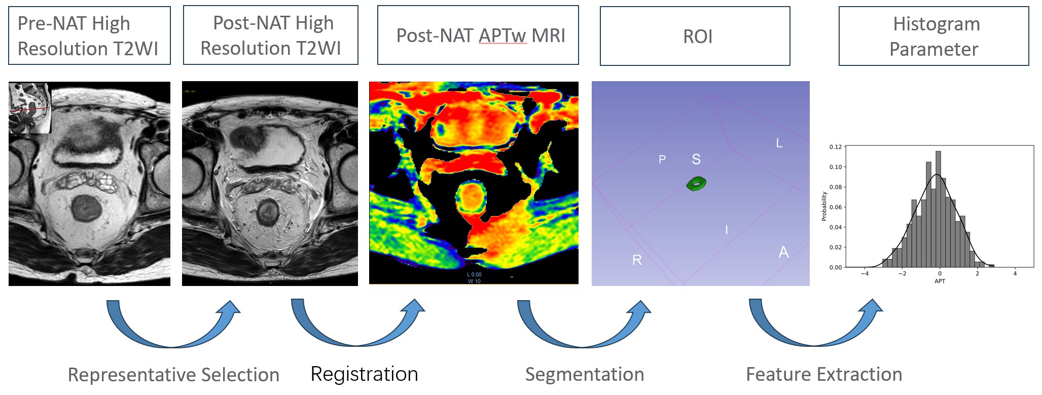
To investigate the added value of Amide Proton Transfer weighted (APTw) magnetic resonance imaging (MRI) in the evaluation of CR to NAT in patients with LARCMaterials and Methods
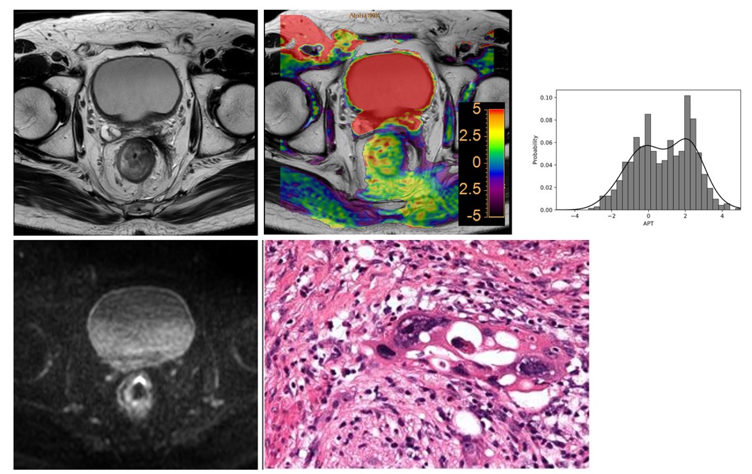
In this approved single-center prospective study, patients with LARC (T3-4N0M0 or T1-4N+M0) who underwent NAT and subsequent surgery, with adequate MRI quality were enrolled. All patients underwent conventional MRI and APTw imaging before and after NAT. Conventional evaluation of CR to NAT based on post-NAT T2WI and DWI, tumor segmentation and histogram analysis based on post-NAT APTw images, were performed by two experienced radiologists independently. Pathologic complete response (pCR) served as the reference standard. Feature selection of histogram parameters was according to significant difference between-groups and low Pearson correlation coefficients between variables. Diagnosis performance of conventional assessment and APTw-added combined assessment was assessed by receiver operating characteristic curve (ROC) analysis. The AUCs were compared using the Delong method.Results
64 participants were evaluated: 21 with pCR and 43 with non-pCR. The accuracy, sensitivity, and specificity of the conventional assessment based on T2WI and DWI in discriminating CR to NAT were 0.703 (95% CI: 0.697,0.710), 0.698 (95% CI: 0.560,0.835), and 0.714 (95% CI: 0.521,0.908), respectively. Four variables, namely. 90percentile, totalenergy, range, and entropy, were selected for further multivariate logistic regression analysis and finally totalenergy and entropy entered into the APTw-added combined model (combination of APT histogram parameters, T2WI and DWI). The accuracy, sensitivity, and specificity of the APTw-added combined model in discriminating CR to NAT were 0.953 (95% CI: 0.952,0.954), 0.930 (95% CI: 0.854,1.000), and 1.000 (95% CI: 1.000,1.000), respectively. Diagnostic efficiency in the evaluation of CR was significantly improved when APTw imaging was added to conventional evaluation based on T2WI and DWI, with AUC improved from 0.706 to 0.969. Using the APTw-added combined model, the accuracy, sensitivity, and specificity for evaluation CR in test set were 0.900 (95% CI: 0.882,0.918), 0.857 (95% CI: 0.598,1.000), and 1.000 (95% CI: 1.000,1.000), respectively (Table 3), and the AUC was 0.952 (95% CI: 0.820,1).Conclusion
Use of additional APTw imaging yields significantly better diagnostic accuracy than does use of conventional T2WI and DWI in the evaluation of CR to NAT in patients with LARC. Superimposing the advantage of noninvasiveness, APTw imaging may have great application value in solving the clinical problem of diagnosing CR to NAT and provide additional valuable information for clinical decision making.Acknowledgements
No acknowledgement found.References
1. Benson AB, Venook AP, Al-Hawary MM, Azad N, Chen YJ, Ciombor KK, Cohen S, Cooper HS, Deming D, Garrido-Laguna I, Grem JL, Gunn A, Hecht JR, Hoffe S, Hubbard J, Hunt S, Jeck W, Johung KL, Kirilcuk N, Krishnamurthi S, Maratt JK, Messersmith WA, Meyerhardt J, Miller ED, Mulcahy MF, Nurkin S, Overman MJ, Parikh A, Patel H, Pedersen K, Saltz L, Schneider C, Shibata D, Skibber JM, Sofocleous CT, Stotsky-Himelfarb E, Tavakkoli A, Willett CG, Gregory K, Gurski L. Rectal Cancer, Version 2.2022, NCCN Clinical Practice Guidelines in Oncology. J Natl Compr Canc Netw 2022;20(10):1139-1167. doi: 10.6004/jnccn.2022.0051
2. European Society of Coloproctology collaborating g. Evaluating the incidence of pathological complete response in current international rectal cancer practice: the barriers to widespread safe deferral of surgery. Colorectal Dis 2018;20 Suppl 6(1463-1318 (Electronic)):58-68. doi: 10.1111/codi.14361
3. Riesco-Martinez MC, Fernandez-Martos C, Gravalos-Castro C, Espinosa-Olarte P, La Salvia A, Robles-Diaz L, Modrego-Sanchez A, Garcia-Carbonero R. Impact of Total Neoadjuvant Therapy vs. Standard Chemoradiotherapy in Locally Advanced Rectal Cancer: A Systematic Review and Meta-Analysis of Randomized Trials. Cancers (Basel) 2020;12(12). doi: 10.3390/cancers12123655
4. Lin Z, Cai M, Zhang P, Li G, Liu T, Li X, Cai K, Nie X, Wang J, Liu J, Liu H, Zhang W, Gao J, Wu C, Wang L, Fan J, Zhang L, Wang Z, Hou Z, Ma C, Yang K, Wu G, Tao K, Zhang T. Phase II, single-arm trial of preoperative short-course radiotherapy followed by chemotherapy and camrelizumab in locally advanced rectal cancer. J Immunother Cancer 2021;9(11). doi: 10.1136/jitc-2021-003554
5. Rullier E, Vendrely V, Asselineau J, Rouanet P, Tuech JJ, Valverde A, de Chaisemartin C, Rivoire M, Trilling B, Jafari M, Portier G, Meunier B, Sieleznieff I, Bertrand M, Marchal F, Dubois A, Pocard M, Rullier A, Smith D, Frulio N, Frison E, Denost Q. Organ preservation with chemoradiotherapy plus local excision for rectal cancer: 5-year results of the GRECCAR 2 randomised trial. Lancet Gastroenterol Hepatol 2020;5(5):465-474. doi: 10.1016/S2468-1253(19)30410-8
6. Dossa F, Chesney TR, Acuna SA, Baxter NN. A watch-and-wait approach for locally advanced rectal cancer after a clinical complete response following neoadjuvant chemoradiation: a systematic review and meta-analysis. Lancet Gastroenterol Hepatol 2017;2(7):501-513. doi: 10.1016/S2468-1253(17)30074-2
7. Duldulao MP, Lee W, Streja L, Chu P, Li W, Chen Z, Kim J, Garcia-Aguilar J. Distribution of residual cancer cells in the bowel wall after neoadjuvant chemoradiation in patients with rectal cancer. Dis Colon Rectum 2013;56(2):142-149. doi: 10.1097/DCR.0b013e31827541e2
8. Maas M, Lambregts DM, Nelemans PJ, Heijnen LA, Martens MH, Leijtens JW, Sosef M, Hulsewe KW, Hoff C, Breukink SO, Stassen L, Beets-Tan RG, Beets GL. Assessment of Clinical Complete Response After Chemoradiation for Rectal Cancer with Digital Rectal Examination, Endoscopy, and MRI: Selection for Organ-Saving Treatment. Ann Surg Oncol 2015;22(12):3873-3880. doi: 10.1245/s10434-015-4687-9
9. Horvat N, Carlos Tavares Rocha C, Clemente Oliveira B, Petkovska I, Gollub MJ. MRI of Rectal Cancer: Tumor Staging, Imaging Techniques, and Management. Radiographics 2019;39(2):367-387. doi: 10.1148/rg.2019180114
10. Park SH, Cho SH, Choi SH, Jang JK, Kim MJ, Kim SH, Lim JS, Moon SK, Park JH, Seo N, Korean Society of Abdominal Radiology Study Group for Rectal C. MRI Assessment of Complete Response to Preoperative Chemoradiation Therapy for Rectal Cancer: 2020 Guide for Practice from the Korean Society of Abdominal Radiology. Korean J Radiol 2020;21(7):812-828. doi: 10.3348/kjr.2020.0483
11. Beets-Tan RGH, Lambregts DMJ, Maas M, Bipat S, Barbaro B, Curvo-Semedo L, Fenlon HM, Gollub MJ, Gourtsoyianni S, Halligan S, Hoeffel C, Kim SH, Laghi A, Maier A, Rafaelsen SR, Stoker J, Taylor SA, Torkzad MR, Blomqvist L. Magnetic resonance imaging for clinical management of rectal cancer: Updated recommendations from the 2016 European Society of Gastrointestinal and Abdominal Radiology (ESGAR) consensus meeting. Eur Radiol 2018;28(4):1465-1475. doi: 10.1007/s00330-017-5026-2
12. Patel UB, Blomqvist LK, Taylor F, George C, Guthrie A, Bees N, Brown G. MRI after treatment of locally advanced rectal cancer: how to report tumor response--the MERCURY experience. AJR Am J Roentgenol 2012;199(4):W486-495. doi: 10.2214/AJR.11.8210
13. Delli Pizzi A, Basilico R, Cianci R, Seccia B, Timpani M, Tavoletta A, Caposiena D, Faricelli B, Gabrielli D, Caulo M. Rectal cancer MRI: protocols, signs and future perspectives radiologists should consider in everyday clinical practice. Insights Imaging 2018;9(4):405-412. doi: 10.1007/s13244-018-0606-5
14. Seo N, Kim H, Cho MS, Lim JS. Response Assessment with MRI after Chemoradiotherapy in Rectal Cancer: Current Evidences. Korean J Radiol 2019;20(7):1003-1018. doi: 10.3348/kjr.2018.0611
15. Jia X, Zhang Y, Wang Y, Feng C, Shen D, Ye Y, Hong N. MRI for Restaging Locally Advanced Rectal Cancer: Detailed Analysis of Discrepancies With the Pathologic Reference Standard. AJR Am J Roentgenol 2019;213(5):1081-1090. doi: 10.2214/AJR.19.21383
16. Lambregts DMJ, Boellaard TN, Beets-Tan RGH. Response evaluation after neoadjuvant treatment for rectal cancer using modern MR imaging: a pictorial review. Insights Imaging 2019;10(1):15. doi: 10.1186/s13244-019-0706-x
17. Kim SH, Lee JM, Hong SH, Kim GH, Lee JY, Han JK, Choi BI. Locally advanced rectal cancer: added value of diffusion-weighted MR imaging in the evaluation of tumor response to neoadjuvant chemo- and radiation therapy. Radiology 2009;253(1):116-125. doi: 10.1148/radiol.2532090027
18. Foti PV, Privitera G, Piana S, Palmucci S, Spatola C, Bevilacqua R, Raffaele L, Salamone V, Caltabiano R, Magro G, Li Destri G, Milone P, Ettorre GC. Locally advanced rectal cancer: Qualitative and quantitative evaluation of diffusion-weighted MR imaging in the response assessment after neoadjuvant chemo-radiotherapy. Eur J Radiol Open 2016;3(2352-0477 (Print)):145-152. doi: 10.1016/j.ejro.2016.06.003
19. Lambregts DMJ, van Heeswijk MM, Delli Pizzi A, van Elderen SGC, Andrade L, Peters N, Kint PAM, Osinga-de Jong M, Bipat S, Ooms R, Lahaye MJ, Maas M, Beets GL, Bakers FCH, Beets-Tan RGH. Diffusion-weighted MRI to assess response to chemoradiotherapy in rectal cancer: main interpretation pitfalls and their use for teaching. Eur Radiol 2017;27(10):4445-4454. doi: 10.1007/s00330-017-4830-z
20. Geng Z, Zhang Y, Yin S, Lian S, He H, Li H, Xie C, Dai Y. Preoperatively Grading Rectal Cancer with the Combination of Intravoxel Incoherent Motions Imaging and Diffusion Kurtosis Imaging. Contrast Media Mol Imaging 2020;2020:2164509. doi: 10.1155/2020/2164509
21. Zhang XY, Wang L, Zhu HT, Li ZW, Ye M, Li XT, Shi YJ, Zhu HC, Sun YS. Predicting Rectal Cancer Response to Neoadjuvant Chemoradiotherapy Using Deep Learning of Diffusion Kurtosis MRI. Radiology 2020;296(1):56-64. doi: 10.1148/radiol.2020190936
22. Horvat N, Veeraraghavan H, Khan M, Blazic I, Zheng J, Capanu M, Sala E, Garcia-Aguilar J, Gollub MJ, Petkovska I. MR Imaging of Rectal Cancer: Radiomics Analysis to Assess Treatment Response after Neoadjuvant Therapy. Radiology 2018;287(3):833-843. doi: 10.1148/radiol.2018172300
23. Ryan JE, Warrier SK, Lynch AC, Heriot AG. Assessing pathological complete response to neoadjuvant chemoradiotherapy in locally advanced rectal cancer: a systematic review. Colorectal Disease 2015;17(10):849-861. doi: https://doi.org/10.1111/codi.13081
24. Zhou J, Payen JF, Wilson DA, Traystman RJ, van Zijl PC. Using the amide proton signals of intracellular proteins and peptides to detect pH effects in MRI. Nat Med 2003;9(8):1085-1090. doi: 10.1038/nm907
25. Zhou J, Heo HY, Knutsson L, van Zijl PCM, Jiang S. APT-weighted MRI: Techniques, current neuro applications, and challenging issues. J Magn Reson Imaging 2019;50(2):347-364. doi: 10.1002/jmri.26645
26. Zhou J, Zaiss M, Knutsson L, Sun PZ, Ahn SS, Aime S, Bachert P, Blakeley JO, Cai K, Chappell MA, Chen M, Gochberg DF, Goerke S, Heo HY, Jiang S, Jin T, Kim SG, Laterra J, Paech D, Pagel MD, Park JE, Reddy R, Sakata A, Sartoretti-Schefer S, Sherry AD, Smith SA, Stanisz GJ, Sundgren PC, Togao O, Vandsburger M, Wen Z, Wu Y, Zhang Y, Zhu W, Zu Z, van Zijl PCM. Review and consensus recommendations on clinical APT-weighted imaging approaches at 3T: Application to brain tumors. Magn Reson Med 2022;88(2):546-574. doi: 10.1002/mrm.29241
27. Law BKH, King AD, Ai QY, Poon DMC, Chen W, Bhatia KS, Ahuja AT, Ma BB, Ka-Wai Yeung D, Fai Mo FK, Wang YX, Yuan J. Head and Neck Tumors: Amide Proton Transfer MRI. Radiology 2018;288(3):782-790. doi: 10.1148/radiol.2018171528
28. Ma B, Blakeley JO, Hong X, Zhang H, Jiang S, Blair L, Zhang Y, Heo HY, Zhang M, van Zijl PC, Zhou J. Applying amide proton transfer-weighted MRI to distinguish pseudoprogression from true progression in malignant gliomas. J Magn Reson Imaging 2016;44(2):456-462. doi: 10.1002/jmri.25159
29. Nishie A, Asayama Y, Ishigami K, Ushijima Y, Takayama Y, Okamoto D, Fujita N, Tsurumaru D, Togao O, Sagiyama K, Manabe T, Oki E, Kubo Y, Hida T, Hirahashi-Fujiwara M, Keupp J, Honda H. Amide proton transfer imaging to predict tumor response to neoadjuvant chemotherapy in locally advanced rectal cancer. J Gastroenterol Hepatol 2019;34(1):140-146. doi: 10.1111/jgh.14315
30. Chen W, Mao L, Li L, Wei Q, Hu S, Ye Y, Feng J, Liu B, Liu X. Predicting Treatment Response of Neoadjuvant Chemoradiotherapy in Locally Advanced Rectal Cancer Using Amide Proton Transfer MRI Combined With Diffusion-Weighted Imaging. Front Oncol 2021;11:698427. doi: 10.3389/fonc.2021.698427
Figures